Original Article
Expression of STING and MIF in tumor infiltration
lymphocytes as prognostic factors in patients with ESCC
Zhi-Chao Wang
1*, Lin Zhang
2*, Ze-Lei Li
3, Jing He
3, Ting-Ting Cai
4, Da-Jun Yang
4,5, De-Rong Xie
1,61Department of Medical Oncology, Zengcheng District People’s Hospital of Guangzhou, Guangzhou, China; 2Department of Clinical Laboratory, Sun Yat-Sen University Cancer Center, Guangzhou, China; 3Department of
Medical Oncology, Ganzhou People’s Hospital, Jiangxi Province, China; 4Experimental Research, Sun Yat-Sen
University Cancer Center, State Key Laboratory of Oncology in South China, Guangzhou, China; 5Collaborative
Innovation Center for Cancer Medicine, Guangzhou, China; 6Department of Medical Oncology, Sun Yat-Sen
Memorial Hospital, Sun Yat-Sen University, Guangzhou, China. *Equal contributors.
Received August 20, 2016; Accepted August 28, 2016; Epub September 1, 2017; Published September 15, 2017
Abstract: STING and MIF are Tumor-immune related proteins act as immune regulating roles that effect the progres-sion of cancer. In these studies, we aimed to detect the expresprogres-sion levels of STING and MIF in tumor cells and in lymphocytes in tumor microenvironments and their association with survivals of patients diagnosed with esopha-geal squamous cell carcinoma (ESCC). The expression levels of STING and MIF were accessed by immunochemistry staining in tumor tissues from 112 resected ESCC. Correlation analyses and independent prognostic outcomes were determined using Pearson’s chi-square test. Independent prognostic outcomes were measured by Cox
regres-sion analysis. We found that STING high expresregres-sion in TILs or MIF high expresregres-sion in tumor cells or in tumor infiltrat
-ing lymphocytes (TILs) was significantly related to reduced disease-free survival (DFS) and overall survival (OS) of
ESCC patients (P<0.05). Multivariate analysis indicated that the expression of STING and MIF in TILs were adverse independent prognostic factors in the whole cohort of patients (P<0.05). The expression of MIF in tumor cells or in
TILs was significantly positively correlated with STING in TILs (P<0.05). The combined STING and MIF expression in TILs was strongly related to reduced DFS and OS of ESCC patients (P<0.05). Our studies indicated the expression
levels of STING and MIF in TILs were independent predictive factors of survivals in patients with ESCC.
Keywords: STING, MIF, esophageal squamous cell carcinoma, tumor microenvironment
Introduction
Esophageal squamous cell carcinoma (ESCC)
is a prevalent malignancy leading cause of
can-cer-related death worldwide [1]. ESCC is one of
the malignant tumors with a five-year survival
rate less than 30% [2]. Expect for the
tradition-al prognostic factors such as TNM stage and
cell differentiation, molecular markers such as
IL-17, Foxp3 in tumor micro environments have
been studied for the prognoses of ESCC
patients in recent studies [3-8]. However, novel
prognostic markers in ESCC remain to be
explored.
Stimulator of interferon genes (STING) a protein
that is expressed in various cell types such as
epithelial cells, T cells, macrophages and
den-dritic cells [9, 10]. Early studies found STING as
a DNA sensor that is critical for activating type I
interferon in response to invading DNA viruses
or bacteria [11-14]. Recently, the role of STING
in cancer generation and progression was
investigated. In these studies, STING acts as a
double-edged sword in favor or against tumor
progression [15-18]. So far, the role of STING in
ESCC has not been studied. Macrophage
migra-tion inhibitory factor (MIF) is a cytokine
com-monly expressed in diverse cell types including
lymphocytes and endothelial cells [19]
.
Over-expression of MIF can be detected in many
pathological conditions, such as autoimmunity
and cancer [20, 21]. In many kinds of tumors,
MIF was found to be associated with
tumorigen-esis, tumor metastasis and tumor
anginogene-sis [22-24]. MIF is considered as a link between
In our study, we investigated the expression of
STING and MIF protein in tumor cells and TILs
in 112 ESCC tissues by immunohistochemical
staining. The correlations between the
expres-sion levels of STING and MIF in different cell
types include tumor cells and TILs in tumor
microenvironment and their prognostic values
were assessed.
Materials and methods
Patients and tumor tissue samples
A total of 112 ESCC patients who underwent
surgery at Sun Yat-Sen University Cancer Cen-
ter in China from November of 2000 to
December of 2002 were involved in this study.
No patient had received any antitumor
treat-ment prior to surgery. All patients had
histologi-cally confirmed primary ESCC. The follow-up
data from the 112 patients with ESCC in this
study were available and complete. OS was
defined as the time interval from the date of
surgery to the date of cancer-related death or
the end of follow-up (December 2011). DFS
was defined as the time interval from the date
of surgery to the date of tumor progression. The
study was approved by the Research Ethics
Committee of the Sun Yat-Sen University Can-
cer Center.
Reagents and antibodies
Primary antibodies: mouse anti-human MIF
(ab55445; Abcam, USA), rabbit anti-human
STING (ab198951; Abcam, USA), and
horserad-ish peroxidase-labeled antibody against a
mouse/rabbit IgG (Envision; Dako, Glostrup,
Denmark).
Immunohistochemistry and evaluation of
im-munohistochemical staining
The formalin-fixed paraffin-embedded tumor
tissues were cut continuously into 4-μm sec
-tions. The antigens were retrieved by heating
the tissue slices in a pressure cooker for 8 min
in EDTA (1 mmol/L, pH 8.0) solution. The
sec-tions were then immersed in a 0.3% hydrogen
peroxide solution for 30 min. Slices were
incu-bated with anti-MIF, anti-STING antibodies at
4°C overnight. A negative control was
incubat-ed with a normal murine IgG antibody. The
sec-tions were then incubated with a secondary
antibody at room temperature for 30 min. Then
the tissue sections were stained with DAB. Two
independent observers blinded to the
clinico-pathological information scored the STING and
MIF expression levels in tumor cells by
assess-ing (a) the percentage of positive cells: (0, <5%;
1, 6 to 25%; 2, 26 to 50%; 3, 51 to 75%; 4,
>75%) and (b) the staining intensity: (0,
nega-tive; 1, light yellow; 2, yellow; 3, brown). The
score was the product of a × b. The levels of
STING and MIF expression in lymphocytes were
measured by counting the positively and
nega-tively stained lymphocytes by a 400 ×
high-power microscopic for 5 fields and then calcu
-lating the mean positive percentage among the
total lymphocytes per field.
Statistical analysis
Statistical analyses were performed with SPSS
16.0 software (SPSS Inc, Chicago, IL, USA). We
divided the patients into two groups (a
high-level group and a low-high-level group) based on the
median values of different immunohistoche-
mical variables. Pearson’s chi-square test
was applied to analyze the correlation between
the patients’ clinicopathological characteristics
and immunohistochemical variants in different
cell types. Clinical prognosis including
disease-free survival and overall survival was analyzed
by Kaplan-Meier analysis using the log-rank
test according to the expression levels of STING
and MIF examined in tumor cells and in TILs.
Independent prognostic factors were identified
by univariate and multivariate analyses using
the Cox regression model. The correlations
among the expression levels of STING and MIF
in both tumor cells and TILs were tested by
Pearson’s correlation coefficient and linear
regression analyses. In this study, a two-tailed
P
-value <0.05 was considered statistically
significant.
Results
Expression patterns of STING and MIF in ESCC
and their correlations with clinicopathological
parameters
In this study, the median age of the 112
patients was 62 years, range from 35 to 90
years; 94 (83.9%) of the patients were males
and 18 (16.1%) were females. There were 58
(51.8%) cases of Stage I and II tumors and 54
(48.2%) cases of Stage III and IV tumors based
on the International Union against Cancer 2002
83 (74.1%) had died, 86 (76.8%) had suffered
the relapse or progression of the disease. The
patients’ clinical characteristics are listed in
Additional file 1:
Table S1.
The expression levels of STING and MIF were
detected in tumor tissues from 112 patients
with ESCC. STING and MIF was mainly expre-
ssed in the cytoplasm of tumor cells and TILs
(
Figure 1
). The mean percentage and the range
of the percentage for MIF or STING expression
in TILs per field were 33% (range, 0 to 92%) and
31% (range, 0 to 85%), respectively (Additional
[image:3.612.90.523.71.311.2]file 1:
Table S2).
Figure 1. Immunohistochemical staining for STING and MIF in the tumor tissue samples of human esophageal
carci-noma. Our data showed low expression
levels of STING (A) and MIF (C) (× 400) and high expression levels of STING (B) and MIF (D) (× 400) in tumor tissues from patients with ESCC compared with the negative control (E) (× 400). The sol-id arrows point to the positive staining of TILs. The dotted arrows point to the positive staining of tumor cells.
Table 1.
Association of the expression of STING, MIF and clinicopathologic parameters in 112
pa-tients with ESCC
Clinicopathologic parameter Total case
Expression in tumor cells Expression in lymphocytes High level
expression of STING (%) P
High level expression
of MIF (%) P
High level expression of STING (%) P
High level expression
of MIF (%) P Age
≤62 (y) 56 31 (55.4%) 0.345 26 (46.4%) 0.450 31 (55.4%) 0.257 26 (46.4%) 0.450 >62 (y) 56 26 (46.4%) 30 (53.6%) 25 (44.6%) 30 (53.6%) Gender
Female 18 9 (50.0%) 0.934 6 (33.3%) 0.123 7 (38.9%) 0.303 8 (51.1%) 0.607
Male 94 48 (50.9%) 50 (53.2%) 49 (52.1%) 48 (44.4%)
T status
T1-2 32 14 (43.8%) 0.339 14 (43.8%) 0.403 15 (46.8%) 0.676 11 (34.4%) 0.036*
T3-4 80 43 (53.8%) 42 (52.5%) 41 (51.3%) 45 (56.3%)
N status
N0 52 28 (53.8%) 0.561 23 (44.2%) 0.256 25 (48.1%) 0.705 22 (42.3%) 0.130
N1 60 29 (48.3%) 33 (55.0%) 31 (51.7%) 34 (56.7%)
Clinical stage
I-II 58 31 (53.4%) 0.575 28 (48.3%) 0.705 30 (51.7%) 0.705 27 (46.6%) 0.449
III-IV 54 26 (48.1%) 28 (51.9%) 26 (48.1%) 29 (53.7%)
[image:3.612.94.522.357.603.2]Associations between clinicopathological
pa-rameters and immunohistochemical variables
The associations between clinicopathological
parameters and immunohistochemical
vari-ables in different cell types of 112 ESCC
[image:4.612.94.522.71.589.2]whereas the expression levels of MIF in tumor
cells or the expression levels of STING in tumor
cells and in TILs were not related to any of the
clinicopathological parameters.
Association between STING and MIF
expres-sion and clinical outcome
The median survival time of the 112 patients
was 26 months (range: 0 to 133 months). The
cumulative five-year OS rate and DFS rate of
the patients in this study were 30% and 29%,
respectively (data not shown). The statistical
analysis showed a significant negative correla
-tion between DFS, OS and the expression lev
-els of MIF in tumor cells and TILs. Negative
cor-relation between DFS, OS and the expression
levels of STING in TILs was also detected
(
P<
0.05,
Figure 2
).
Univariate and multivariate analyses of STING
and MIF expression level as prognostic factors
The univariate analysis indicated that high ex-
pression level of MIF (
P=
0.034 and
P=
0.032)
in tumor cells was significantly correlated with
reduced DFS and OS. High expression level of
MIF (
P=
0.001 and
P=
0.001) in TILs was also
significantly associated with decreased DFS
and OS. High expression level of STING
(
P=
0.003 and
P=
0.004) in TILs was also
nota-bly correlated with decreased DFS and OS,
whereas the expression levels of STING in TILs
were not significantly correlated with reduced
DFS and OS (
Table 2
). Clinicopathological
parameters such as gender, Tumor status,
nodal status and TNM stage were also of
prog-nostic value in univariate analysis. Furthermore,
according to the multivariate Cox model
analy-sis, we observed that the expression levels of
MIF or STING in TILs were independent
predic-tors of DFS and OS (
Table 3
).
Correlation between STING and MIF
expres-sion
Pearson’s correlation coefficient and a linear
regression analysis were used to evaluate the
correlations between the expression levels of
STING and MIF in tumor cells and TILs. STING
[image:5.612.90.528.84.233.2]expression levels in TILs were significantly posi
-tively correlated with MIF expression levels in
tumor cells and in TILs (
P=
0.022, R=0.217 and
P=
0.016, R=0.226, respectively). Besides, the
expression levels of MIF in tumor cells were
positively correlated with MIF expression levels
Table 2.
Univariate analysis of DFS and OS in 112 patients with ESCC
Variables DFS (n=136) OS (n=136)
HR (95% CI) P HR (95% CI) P
Age, years (≤62/>62) 0.754 (0.492-1.154) 0.193 0.822 (0.534-1.266) 0.373 Gender (male/female) 0.502 (0.265-0.951) 0.034* 0.406 (0.203-0.812) 0.011* Tumor (T) status (1-2/3-4) 1.710 (1.040-2.812) 0.035* 1.739 (1.039-2.909) 0.035* Nodal (N) status (0/1) 2.270 (1.455-3.540) <0.001* 2.141 (1.365-3.357) 0.001* TNM stage (I-II/III-IV) 2.081 (1.352-3.202) 0.001* 1.297 (1.044-1.613) 0.019* MIF in tumor cells (low/high) 1.570 (1.026-2.402) 0.038* 1.594 (1.034-2.457) 0.035* STING in tumor cells (low/high) 1.079 (0.706-1.651) 0.724 1.098 (0.713-1.690) 0.672 MIF in lymphocytes (low/high) 2.081 (1.352-3.202) 0.001* 2.000 (1.290-3.102) 0.002* STING in lymphocytes (low/high) 1.883 (1.223-2.900) 0.004* 1.868 (1.205-2.896) 0.005*
Note: p value is determined by log-rank test. *P<0.05.
Table 3.
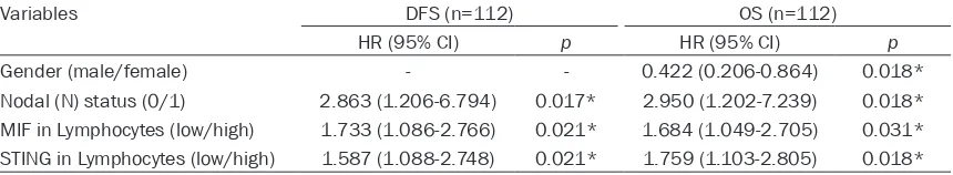
Multivariate Cox analyses for DFS and OS of 112 patients with ESCC
Variables DFS (n=112) OS (n=112)
HR (95% CI) p HR (95% CI) p
Gender (male/female) - - 0.422 (0.206-0.864) 0.018*
Nodal (N) status (0/1) 2.863 (1.206-6.794) 0.017* 2.950 (1.202-7.239) 0.018* MIF in Lymphocytes (low/high) 1.733 (1.086-2.766) 0.021* 1.684 (1.049-2.705) 0.031* STING in Lymphocytes (low/high) 1.587 (1.088-2.748) 0.021* 1.759 (1.103-2.805) 0.018*
Note: The Cox proportional hazards regression model contained the significantly different factors in univariate analysis, includ
[image:5.612.93.523.278.357.2]in TILs (
P
<0.001, R=0.475), as shown in
Fig-
ures 3A
and
4B
. Furthermore, our study showed
that the combined high expression of STING
and MIF in TILs was strongly associated with
reduced DFS and OS (
Figure 4
).
Discussion
Immune cells in tumor microenvironment play
an important role in the generation and
devel-opment of cancer [26]. The roles of STING in
tumor microenvironment are Contradictory. In
some studies, STING was found to be a
protec-tive factor that prevented the generation and
progression of cancer. The activation of STING
in phagocytes was reported to leading to T cell
responses and function as an antitumor role
[27]. STING mediates protection against colon
cancer by recognizing intestinal DNA damage
and promoting wound repair in the colon [28,
29]. Radiation-induced cancer cell death
stimu-lated STING-dependent cytosolic DNA sensing
resulted in type I interferon-dependent
antitu-mor responses [30]. STING agonists were
capa-ble to cause tumor regression and showed
potent antitumor therapeutic effects [31]. In
other reports, the negative role of STING has
been uncovered in the prevention of inflamma
-tion-induced cancer. DNA leakage in the cy-
toplasm caused by DNA damage activates
STING-mediated inflammation and finally leads
to skin carcinogenesis [32]. Besides, studies
found STING is capable to influence the func
-tion of immunosuppressive cells include T
regu-latory cells and Myeloid-derived Suppressor
Cells (MDSCs) in tumor microenvironment. A
study on tongue squamous cell carcinoma
indi-Figure 3. Scatter dot plots and correlation analysis between the STING and MIF expressions in different cellpopula-tions. A: The expression of MIF in tumor cells were significantly positively correlated with STING expression in TILs
(P=0.022, R=0.217). B: The expression levels of MIF in TILs were significantly positively correlated with STING ex -pression in TILs (P=0.016, R=0.226). C: The expression of MIF in tumor cells were significantly positively correlated
[image:6.612.90.524.73.202.2]with MIF expression in TILs (P<0.001, R=0.475).
Figure 4. Survival curves for ESCC patients according to their expression levels of STING and MIF in tumor cells. A
and B: DFS and OS curves for patients according to the combined low expression level, single high expression level
[image:6.612.94.522.284.440.2]cated that activated STING promoted the
gen-eration of several immunosuppressive
cyto-kines including IL-10, IDO and CCL22, and
enhanced the recruitment of Foxp3
+regulatory
T cells (Tregs) via the c-jun/CCL22 signaling
[33, 34]. Moreover, a study on STING ligand
c-di-GMP revealed that, Low doses of c-di-GMP
increased the production of IL12 by MDSCs,
whereas a high dose of c-di-GMP killed the
tumor cells directly [35].
Here, we discussed about the
immunosuppres-sive potential of STING in tumor
microenviron-ment via Tregs and MDSCs. Interestingly, MIF is
also considered as an immunosuppressive
fac-tor in cancer in variety of studies. In some
stud-ies, MIF promotes tumor progression by incre-
asing the prevalence of MDSCs in tumor
micro-environment [36]. MIF can also inhibit the
dif-ferentiation of MDSCs to normal monocytes
and promote the immunosuppressive function
of MDSCs [37, 38]. Moreover, a study on
tumor-bearing mice indicates that MIF promotes
tumor growth by promoting the generation of
Tregs generation through the regulation of IL-2
production
[39]
. Our data confirmed the roles
of STING and MIF in TILs in facilitating the
carci-nogenesis and cancer progression. We also
investigated the expression of STING and MIF
in tumor cells. But no significant difference was
found in the survival of patients in high and low
expression of STING in tumor cells. It may be
explained by the contradictory role of STING in
tumorigenesis. Further investigations are
need-ed to uncover the function of STING in tumor
cells and the molecular mechanisms of STING
in tumor induced immunity.
Our data provides novel prognostic indicators
of STING and MIF in TILs in predicting the
sur-vival of patients with ESCC. The expression
pat-tern of STING in TILs in ESCC was firstly
described. Our results indicate that STING has
different biological functions in tumor cells and
in TILs. Importantly, the expression levels of
STING and MIF in tumor cells and in TILs were
positively associated (
Figure 3
). Our results
showed for the first time that combined high
expression of STING and MIF in TILs strongly
indicate a reduced DFS and OS (
Figure 4
). The
relationship between STING and MIF is still
unclear and waiting to be explored. Besides,
the immune regulatory function of STING and
MIF in tumor microenvironment is an
interest-ing project that deserves further investigation.
Acknowledgements
This work was supported by Guangdong Eso-
phageal Cancer Research Institute Project (No.
Q201405).
Disclosure of conflict of interest
None.
Address correspondence to: Dr. De-Rong Xie, De-
partment of Medical Oncology, Zengcheng District
People’s Hospital of Guangzhou; Department of
Medical Oncology, Sun Yat-Sen Memorial Hospital,
Sun Yat-Sen University, 107 Yanjiang West Road, Guangzhou 510120, Guangdong Province, China. Tel: +86-20-81332616; E-mail: derongxie@126. com; Dr. Da-Jun Yang, Experimental Research, Sun Yat-Sen University Cancer Center, State Key Labo-
ratory of Oncology in South China; Collaborative
Innovation Center for Cancer Medicine, 651 Dong- feng East Road, Guangzhou 510060, China. Tel: +86-20-87342335; E-mail: yangdj@sysucc.org.cn
References
[1] Torre LA, Bray F, Siegel RL, Ferlay J, Lortet-Tieulent J, Jemal A. Global cancer statistics, 2012. CA Cancer J Clin 2015; 65: 87-108. [2] Yan W, Wistuba II, Emmert-Buck MR, Erickson
HS. Squamous Cell Carcinoma - Similarities and Differences among Anatomical Sites. Am J Cancer Res 2011; 1: 275-300.
[3] Su XD, Zhang DK, Zhang X, Lin P, Long H, Rong TH. Prognostic factors in patients with recur-rence after complete resection of esophageal squamous cell carcinoma. J Thorac Dis 2014; 6: 949-957.
[4] Chen MQ, Xu BH, Zhang YY. Analysis of prog-nostic factors for esophageal squamous cell carcinoma with distant organ metastasis at initial diagnosis. J Chin Med Assoc 2014; 77: 562-566.
[5] Okumura H, Uchikado Y, Matsumoto M, Owaki T, Kita Y, Omoto I, Sasaki K, Sakurai T,
Setoyama T, Nabeki B, Matsushita D, Ishigami S, Hiraki Y, Nakajo M, Natsugoe S. Prognostic factors in esophageal squamous cell carcino-ma patients treated with neoadjuvant
chemo-radiation therapy. Int J Clin Oncol 2013; 18:
329-334.
squa-mous cell carcinoma. J Thorac Oncol 2013; 8:
495-501.
[7] Lv L, Pan K, Li XD, She KL, Zhao JJ, Wang W, Chen JG, Chen YB, Yun JP, Xia JC. The
accumu-lation and prognosis value of tumor infiltrating
IL-17 producing cells in esophageal squamous
cell carcinoma. PLoS One 2011; 6: e18219.
[8] Huang C, Fu ZX. Localization of IL-17+Foxp3+ T cells in esophageal cancer. Immunol Invest 2011; 40: 400-412.
[9] Ishikawa H, Barber GN. STING is an endoplas-mic reticulum adaptor that facilitates innate immune signalling. Nature 2008; 455: 674-678.
[10] Ishikawa H, Ma Z, Barber GN. STING regulates intracellular DNA-mediated, type I interferon-dependent innate immunity. Nature 2009; 461: 788-792.
[11] Ishikawa H, Barber GN. STING is an endoplas-mic reticulum adaptor that facilitates innate immune signalling. Nature 2008; 455: 674-678.
[12] Ishikawa H, Ma Z, Barber GN. STING regulates intracellular DNA-mediated, type I interferon-dependent innate immunity. Nature 2009; 461: 788-792.
[13] Burdette DL, Monroe KM, Sotelo-Troha K, Iwig JS, Eckert B, Hyodo M, Hayakawa Y, Vance RE. STING is a direct innate immune sensor of cy-clic di-GMP. Nature 2011; 478: 515-518. [14] Cai X, Chiu YH, Chen ZJ. The
cGAS-cGAMP-STING pathway of cytosolic DNA sensing and signaling. Mol Cell 2014; 54: 289-296. [15] Ahn J, Ruiz P, Barber GN. Intrinsic self-DNA
triggers inflammatory disease dependent on
STING. J Immunol 2014; 193: 4634-4642. [16] Ahn J, Xia T, Konno H, Konno K, Ruiz P, Barber
GN. Inflammation-driven carcinogenesis is me -diated through STING. Nat Commun 2014; 5: 5166.
[17] Zhu Q, Man SM, Gurung P, Liu Z, Vogel P,
Lamkanfi M, Kanneganti TD. Cutting edge:
STING mediates protection against colorectal tumorigenesis by governing the magnitude of
intestinal inflammation. J Immunol 2014; 193:
4779-4782.
[18] Gajewski TF, Schreiber H, Fu YX. Innate and adaptive immune cells in the tumor microenvi-ronment. Nat Immunol 2013; 14: 1014-1022. [19] Calandra T, Roger T. Macrophage migration
in-hibitory factor: a regulator of innate immunity. Nat Rev Immunol 2003; 3: 791-800.
[20] Greven D, Leng L, Bucala R. Autoimmune
dis-eases: MIF as a therapeutic target. Expert Opin
Ther Targets 2010; 14: 253-264.
[21] Plant BJ, Gallagher CG, Bucala R, Baugh JA,
Chappell S, Morgan L, O’Connor CM, Morgan K, Donnelly SC. Cystic fibrosis, disease severi -ty, and a macrophage migration inhibitory
fac-tor polymorphism. Am J Respir Crit Care Med 2005; 172: 1412-1415.
[22] Gordon-Weeks AN, Lim SY, Yuzhalin AE, Jones K, Muschel R. Macrophage migration inhibito-ry factor: a key cytokine and therapeutic target in colon cancer. Cytokine Growth Factor Rev 2015; 26: 451-461.
[23] Tomiyasu M, Yoshino I, Suemitsu R, Okamoto T, Sugimachi K. Quantification of macrophage
migration inhibitory factor mRNA expression in non-small cell lung cancer tissues and its
clini-cal significance. Clin Cancer Res 2002; 8:
3755-3760.
[24] Meyer-Siegler KL, Bellino MA, Tannenbaum M. Macrophage migration inhibitory factor eva-
luation compared with prostate specific anti -gen as a biomarker in patients with prostate carcinoma. Cancer-Am Cancer Soc 2002; 94: 1449-1456.
[25] Bucala R, Donnelly SC. Macrophage migration inhibitory factor: a probable link between
in-flammation and cancer. Immunity 2007; 26:
281-285.
[26] Sheu BC, Chang WC, Cheng CY, Lin HH, Chang DY, Huang SC. Cytokine regulation networks in the cancer microenvironment. Front Biosci 2008; 13: 6255-6268.
[27] Woo SR, Fuertes MB, Corrales L, Spranger S, Furdyna MJ, Leung MY, Duggan R, Wang Y, Barber GN, Fitzgerald KA, Alegre ML, Gajewski TF. STING-dependent cytosolic DNA sensing mediates innate immune recognition of immu-nogenic tumors. Immunity 2014; 41: 830-842. [28] Zhu Q, Man SM, Gurung P, Liu Z, Vogel P,
Lamkanfi M, Kanneganti TD. Cutting edge:
STING mediates protection against colorectal tumorigenesis by governing the magnitude of
intestinal inflammation. J Immunol 2014; 193:
4779-4782.
[29] Ahn J, Konno H, Barber GN. Diverse roles of STING-dependent signaling on the
develop-ment of cancer. Oncogene 2015; 34:
5302-5308.
[30] Deng L, Liang H, Xu M, Yang X, Burnette B, Arina A, Li XD, Mauceri H, Beckett M, Darga T, Huang X, Gajewski TF, Chen ZJ, Fu YX, Weichselbaum RR. STING-Dependent Cytoso- lic DNA Sensing Promotes Radiation-Induced Type I Interferon-Dependent Antitumor Im- munity in Immunogenic Tumors. Immunity 2014; 41: 843-852.
[32] Ahn J, Xia T, Konno H, Konno K, Ruiz P, Barber
GN. Inflammation-driven carcinogenesis is me -diated through STING. Nat Commun 2014; 5: 5166.
[33] Liang D, Xiao-Feng H, Guan-Jun D, Er-Ling H, Sheng C, Ting-Ting W, Qin-Gang H, Yan-Hong N,
Ya-Yi H. Activated STING enhances Tregs infil -tration in the HPV-related carcinogenesis of tongue squamous cells via the c-jun/CCL22 signal. Biochim Biophys Acta 2015; 1852: 2494-2503.
[34] Lemos H, Mohamed E, Huang L, Ou R,
Pacholczyk G, Arbab AS, Munn D, Mellor AL. STING Promotes the Growth of Tumors
Characterized by Low Antigenicity via IDO
Activation. Cancer Res 2016; 76: 2076-2081. [35] Chandra D, Quispe-Tintaya W, Jahangir A,
Asafu-Adjei D, Ramos I, Sintim HO, Zhou J,
Hayakawa Y, Karaolis DK, Gravekamp C. STING ligand c-di-GMP improves cancer vaccination against metastatic breast cancer. Cancer Immunol Res 2014; 2: 901-910.
[36] Simpson KD, Templeton DJ, Cross JV. Macrophage migration inhibitory factor pro-motes tumor growth and metastasis by induc-ing myeloid-derived suppressor cells in the tu-mor microenvironment. J Immunol 2012; 189: 5533-5540.
[37] Waigel S, Rendon BE, Lamont G, Richie J, Mitchell RA, Yaddanapudi K. MIF inhibition
re-verts the gene expression profile of human
melanoma cell line-induced MDSCs to normal monocytes. Genom Data 2016; 7: 240-242. [38] Yaddanapudi K, Rendon BE, Lamont G, Kim EJ,
Al RN, Richie J, Albeituni S, Waigel S, Wise A, Mitchell RA. MIF Is Necessary for Late-Stage Melanoma Patient MDSC Immune Suppression and Differentiation. Cancer Immunol Res 2016; 4: 101-112.
Table S1.
Clinical characteristics of the 112 patients
With ESCC
Characteristic No. (%)
Total cases 112 (100%)
Age
Median 62
Range 35-90
Gender
Male 94 (83.9%)
Female 18 (16.1%)
Tumor (T) status
T1 4 (3.6%)
T2 28 (25.0%)
T3 77 (68.8%)
T4 3 (2.7%)
Lymphoid Nodal (N) status
N0 52 (46.4%)
N1 60 (53.6%)
Distant metastasis (M) status
M0 107 (95.5%)
M01 5 (4.5%)
TNM stage
I 3 (2.7%)
IIa-IIb 55 (49.1%)
III 49 (43.8%)
IV 5 (4.5%)
Progression or Relapse
No 26 (23.2%)
Yes 86 (76.8%)
Death
No 29 (25.9%)
Yes 83 (74.1%)
Table S2.
Descriptive statistics of
immunohisto-chemical variables in 112 patients
Variable In tumor cells In TILs Low
expression level (%)
High expression
level (%)
Mean percentage
(%)
Range of percentages
(%)
MIF 56 (50%) 56 (50%) 33 0-92
[image:10.612.192.420.543.620.2]