Original Article
C-erbB-2 expression is related with pathological
progression of gastric cancer: results of a
non-radioactive in situ hybridization
Keqiang Wang1*, Jiangang Liu2*, Yanchao Duan3, Jiafeng Wu1, Shengyi Dongye4, Yiren Wang5, Zhenzhong Liu2, Guoxin Han2
Departments of 1Clinical Laboratory, 2general surgery, 3Hematology, 4Pathology, Affiliated Hospital of Taishan Medical University, Tai’an, Shandong Province, China; 5Medical Laboratory Class, Grade 2016, Medical College, Qingdao University, Qingdao, Shandong Province, China. *Equal contributors.
Received June 7, 2017; Accepted August 23, 2017; Epub September 1, 2017; Published September 15, 2017
Abstract: Objective: To study the relationship of c-erbB-2 oncogene expression with major pathological character-istics of gastric cancer (GC) progression. Methods: Eighty-one GC specimens were studied for c-erbB-2 oncogene
amplification using non-radioactive in situ hybridization method. The c-erbB-2 overexpression status was correlated with tumor differentiation, tumor invasion and lymph node metastasis. Results: Among the 81 pathology confirmed
GC patients, 41 (50.6%) were found to have c-erbB-2 overexpression in cancer tissues. The rate of c-erbB-2
over-expression was significantly higher in those with poor tumor differentiation (63.0%, 29/46) than in those with
well differentiated tumor (34.3%, 12/35) (χ2=6.576, P<0.001); significantly higher in those that invaded into deep muscle and beyond (55.7%, 39/70) than in those with tumors limited to the superficial muscle (18.2%, 2/11)
(χ2=5.357, P<0.025); and significantly higher in those with lymph node metastases (59.6%, 34/57) than in those
without lymph node involvement (29.2%, 7/24) (χ2=6.278, P<0.025). Conclusions: c-erbB-2 oncogene
overexpres-sion may indicate a more aggressive biological behavior of the tumor and could be used as a predictive marker for GC pathological progression.
Keywords: Gastric cancer, c-erbB-2 gene, in situ hybridization
Introduction
Proto-oncogene c-erbB-2 (also called neu of HER-2) located on human chromosome 17q21 belongs to a subfamily of type I receptor tyro-sine protein kinase, which encodes a 185 kDa transmembrane growth factor glycoprotein (also called P185 protein) that contains an extracellular ligand-binding domain and intra-cellular tyrosine kinase activity. The extracellu-lar region is structurally simiextracellu-lar to that of epider-mal-growth-factor receptor [1]. Like epidermal
growth factor, c-erbB-2 expression reflects an
increase in the proliferative activity of a tumor [2]. c-erbB-2 expression is observed only in low levels in epithelial cells of most organs in nor-mal human tissues and at slightly higher levels in fetal tissues. Overexpression is observed in many different malignancies such as breast, ovarian, gastric, lung, prostate, colonic, and
other cancers [3-6]. In this paper, we studied
c-erbB-2 expression profile in GC tissues using in situ hybridization, and explored the signifi -cance of this gene in GC pathological progression.
materials and methods
Clinical specimens
Eight-one pathology-confirmed gastric carcino -ma specimens were collected from Jan 2016 to
Jun 2017 from the Affiliated Hospital of Taishan
Lauren classification, there were 33 cases with
intestinal type carcinoma and 48 cases with
diffused type carcinoma. Thirty-five cases were
with well-differentiated carcinoma and 46 were poorly differentiated. In 11 cases cancer
invad-ed superficial muscle layer and in the rest can -cer invaded beyond deep muscle. Fifty-seven of these cases had lymph node metastases and 24 had no lymph node metastases. After surgi-cal removal, the specimens were snap frozen in liquid nitrogen and cryo-sectioned at -260C,
with thickness between 6 to 10 µm, which were mounted on aminopropyltriethoxysilane (APES, Sigma, St Louis, Ohio, USA) treated slides.
In situ hybridization
c-erbB-2 cDNA probe was a kind gift of Professor Yamamoto from Tokyo University, Japan. It is the product of c-erbB-2 cDNA degra-dation by endonucleases Dra Iand Sma I, with the length of 461 base pairs (bp). Non-radioactive digoxigenin-11-UTP labeling was performed using random priming method, with reagent kit from Boehringer mannheims (pur-chased from Sino-American Biotechnology Company, No. 007, Sanshan Road, National Hi-Tech Industry Development Zone, Luoyang, Henan Province, China). The probe concentra-tion was 15 µg/ml and sensitivity was 0.1 pg DNA. After treatment with 0.1 N HCl, RNase A, protease K and 0.1% Glycin, the slides were pre-hybridized for one hour at room tempera-ture in pre-hybridization solution (4×SSC, 50% formamide, 1× Denhardt solution, 0.5% PEG and 0.5 mg/ml ssDNA). After washing the hybridization solution (probe concentration 0.2 µg/ml) was added, the slides were sealed with cover glass and treated in formamide chamber at 95°C for 10-15 min. Then the slides were hybridized overnight at 42°C in a humid cham-ber, after which the cover glass was removed in 2×SSC solutions, and alkaline phosphatase labeled anti-DIG antibody (1:500 dilution) was added and incubated for 2 h. After washing, 5-bromo-4-chloro-3-indoxyl phosphate disodi-um (BCIP)/nitroblu-tetrazolidisodi-um (NBT) color de- velopment substrate was added and kept in the dark for color reaction, which was terminated by TE buffer washing when color reaction was
sufficient. The slides were counter-stained with
eosin, and sealed. Normal gastric tissues from the same specimens were treated in the same fashion as negative controls.
Slides interpretation
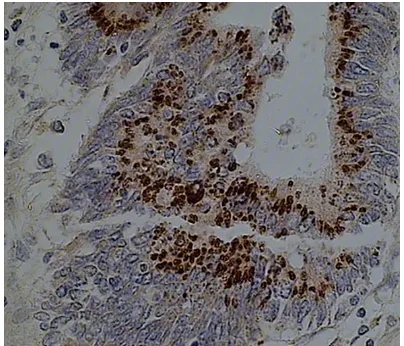
[image:2.612.88.290.71.244.2]When the slides were viewed under microscope (400×), dark brown-blue granules and particles could be found in positively stained cells, and the background was stained red by eosin. Positive and negative stained specimens were recorded respectively. Data on conventional pathology of the specimens were collected from routine pathological report based on gross pathology and hematoxylin and eosin (HE) stained tissue slides.
Figure 1. c-erbB-2 expression in gastric cancer tis-sues detected by non-radioactive labeled in situ hy-bridization. Frozen section gastric cancer slides were hybridized with digoxigenin-11-UTP labeled c-erbB-2 probe, developed by BCIP/NBT and counterstained by eosin. Positive stain was shown as dark brown particles in gastric cancer cell nests (400×).
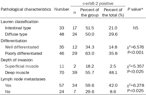
[image:2.612.89.289.346.497.2]Table 1. c-erbB-2 expression and the pathological characteristics of the GC studied
Pathological characteristics Number
c-erbB-2 positive
P value* n Percent of the group the total (%)Percent of
Lauren classification
Intestinal type 33 17 51.5 21.0 NS Diffuse type 48 24 50.0 29.6
Differentiation
Well differentiated 35 12 34.3 14.8 χ2=6.576 P<0.001 Poorly differentiated 46 29 63.0 35.8
Depth of invasion
Superficial muscle 11 2 18.2 2.5 χ2=5.357 P<0.025 Deep muscle 70 39 55.7 48.1
Lymph node metastases
Yes 57 34 59.6 42.0 χ2=6.278
P<0.025
No 24 7 29.6 8.6
NS: not significant. *Chi-square test.
or, accounting for 14.8% (12/81) of the total speci-mens studied and 34.3% (12/35) of the well differ-entiated group, and 29 were poorly differentia- ted, accounting for 35.8% (29/81) of the total and 63.0% (29/46) of the poorly differentiated gro- up (χ2=6.576, P<0.001) (Figure 3).
In terms of tumor inva-sion, there were 11 cases (13.6%, 11/81) with tumo- rs invasion limited to the
superficial muscle layer
and 70 cases (86.4%, 70/81) with tumors invad-ing beyond the deep mus-statistical analysis
Comparisons between c-erbB-2 positive rates in different pathological subgroups were ana-lyzed using Chi-square test, with P<0.05 as
sta-tistical significance. results
c-erbB-2 expression in gC tissues
Of the 81 GC specimens, 41 (50.6%) were found to have c-erbB-2 overexpression, which showed dark-brown granules in cancer tissue (Figure 1), negative results are shown in Figure 2. The relationship of c-erbB-2 overexpression and the pathologic characteristics of tumors were summarized in Table 1.
Correlation between c-erbB-2 overexpression and major pathological characteristics pertain -ing to cancer progression
Three major pathological parameters related to cancer progression-tumor differentiation, depth of tissue invasion and lymph node metastasis-were particularly analyzed in this study to explore their correlation with c-erbB-2 expression.
In terms of tumor differentiation, there were 35 (43.2%, 35/81) cases with well differentiat-ed tumors and 46 (56.8%. 46/81) poorly dif-ferentiated tumors. Among the 41 c-erbB-2 po- sitive cases, 12 were well differentiated tum-
cle layer. In cases with superficial muscle layer
invasion, c-erbB-2 overexpression was found in 2 cases, accounting for 18.2% (2/11) of the group or 2.5% (2/81) of the total specimens studied. On the other hand, in cases with deep muscle invasion, c-erbB-2 overexpression was observed in 39 cases, accounting for 55.7% (39/70) of the group or 48.1% (39/81) of the total specimens studied. The difference in c-erbB-2 expression between these two groups
were statistically very significant (χ2=5.357, P<0.005) (Figure 3).
In terms of lymph node metastases, there were 57 cases (70.4%, 57/81) with lymph node metastases and 24 cases (29.6%, 24/81) with-out lymph node metastases. Of the lymph node positive group, c-erbB-2 overexpression was found in 34 cases, which was 59.6% (34/57) of the group or 42.0% (34/81) of the total speci-mens studied. In contrast, in the lymph node negative group, c-erbB-2 overexpression was found in 7 cases, accounting for 21.2% (7/24) of the group or 8.6% (7/81) of the total. The dif-ferences between these two groups reached
statistically significance (χ2=6.278, P<0.001) (Figure 3).
Discussion
GC depends mainly on the stage of the disease, with early GC having a 5 year survival of 90-100% and advanced tumors a 5 year sur-vival of 15-25%. The role of other prognostic factors in these tumors is still under investiga-tion [7]. Conveninvestiga-tional pathology demonstrated that certain types of GC usually showed much more aggressive biological behavior than other types, and such pathological features include tumor differentiation, depth of tumor invasion and lymph node metastasis [8]. With the devel-opment of molecular biology, it has been found that there are many genes involved in GC devel-opment and pathological progression, includ-ing the plasminogen activator (uPA) and its inhibitor PAI-1 (plasminogen activator inhibitor type 1), the cell-cycle regulator cyclin E, epider-mal growth factor (EGF), growth factor recep-tors c-erbB-2, the apoptosis inhibitor bcl-2, the cell adhesion molecule E-cadherin, and the multifunctional protein beta-catenin [9]. The re-
those reported by other investigators. Secon- dly, we used in situ hybridization method to detect c-erbB-2 overexpression at mRNA level on freshly obtained GC tissues snap frozen in liquid nitrogen immediately after resection. This method can preserve the gene product to the maximum. On the other hand, most previ-ous study used immunohistochemical method to study c-erbB-2 protein expression on
paraf-fin embedded tissue blocks fixed in various fixa -tives. This method has limited sensitivity as
demonstrated by a study that confirmed fixa
-tion and paraffin wax embedding affect the
results of immunohistochemical demonstra-tion of c-erbB-2 in GC [14].
[image:4.612.93.373.74.328.2]Our results suggested that c-erbB-2 overex-pression was related with GC pathological pro-gression. The percentage of c-erbB-2 overex-pression was higher in poorly differentiated GC (35.8%) than in well differentiated GC Figure 3. Relationship between c-erbB-2 overexpression and pathological
characteristics in gastric cancer. Well = well differentiated, poor = poorly
dif-ferentiated; S = tumor invading superficial muscle layer, D = tumor invading
deep muscle layer; LN- = lymph node metastasis negative, LN+ = lymph node metastasis positive. The percentage of c-erbB-2 overexpression was much higher in poorly differentiated tumors (35.8%) than in well differentiated tumors (14.8%) (P<0.001), much higher in tumors invading deep muscle
(48.1%) than those invading superficial muscle (2.5%) (P<0.001), and much
higher in tumors with lymph node metastases (42.0%) than in those without lymph node metastases (8.6%) (Chi-square test).
lationship between these mo- lecular markers and the con-ventional pathological param-eters is currently under inten-sive investigation, as elucida- tion of these molecular
pro-files may provide more infor -mation on biological behavior of GC.
Using highly sensitive in situ hybridization method, we found 50.6% (41/81) of the gastric tumors had c-erbB-2 overexpression in the present work. Previous studies of c-erbB-2 in GC have shown that the frequency of its over-expression varies from 9 to 38% [10-13]. Two major rea-sons may account for the fact that c-erbB-2 overexpression was higher in our series than those reported previously. First, most of our patients were late stage GC. Among the 81 tumors, 57 (70.3%) had lymph node metastasis and 70 (86.4%) had tumors spreading beyond the deep muscle layer, as examined by
(14.8%), higher in tumors invading beyond deep
muscle 48.1% than in those limited to superfi -cial muscle (2.5%), and higher in tumors with lymph node metastases (42.0%) than those without lymph node metastases. These results
are in consistency with previous findings sug -gesting that c-erbB-2 overexpression may denote a group of highly aggressive GC with poor prognosis [7, 9, 13]. As c-erbB-2 may play an important role to predict more aggressive biological behavior of gastric cancer, this mol-ecule could be used as a valuable marker for GC prognosis.
Acknowledgements
Shandong science and technology develop-ment plan (No. 2013YD21013); Shandong medical and health science and technology development plan (No. 2011HW083).
Disclosure of conflict of interest None.
Address correspondence to: Drs. Guoxin Han and Zhenzhong Liu, Department of General Surgery,
Affiliated Hospital of Taishan Medical University,
Tai’an 27100, Shandong Province, China. Tel: +86-538-6236422; Fax: +86-538-8420042; E-mail: tahgx@163.com (GXH); Tel: +86-538-6230822; Fax: +86-538-8420042; E-mail: lzhzhtsh@126.com (ZZL)
references
[1] Lupu R, Colomer R, Kannan B, Lippman ME. Characterization of a growth factor that binds exclusively to the erbB-2 receptor and induces cellular responses. Proc Natl Acad Sci U S A 1992; 89: 2287-2291.
[2] Lordick F, Al-Batran SE, Dietel M, Gaiser T, Hofheinz RD, Kirchner T, Kreipe HH, Lorenzen S, Möhler M, Quaas A, Röcken C, Rüschoff J, Tannapfel A, Thuss-Patience P, Baretton G. HER2 testing in gastric cancer: results of a German expert meeting. J Cancer Res Clin On-col 2017; 143: 835-841.
[3] Hurvitz SA, Gelmon KA, Tolaney SM. Optimal management of early and advanced HER2 breast cancer. Am Soc Clin Oncol Educ Book 2017; 37: 76-92.
[4] Bhaumik S, Ahmad F, Das BR. Somatic mutati-on analysis of KRAS, BRAF, HER2 and PTEN in EGFR mutation-negative non-small cell lung carcinoma: determination of frequency,
distri-bution pattern and identification of novel dele -tion in HER2 gene from Indian patients. Med Oncol 2016; 33: 117.
[5] Murray NP, Reyes E, Fuentealba C, Jacob O, Orellana N. possible role of her-2 in the pro-gression of prostate cancer from primary tu-mor to androgen independence. Asian Pac J Cancer Prev 2015; 16: 6615-9.
[6] Fanotto V, Ongaro E, Rihawi K, Avallone A, Sil-vestris N, Fornaro L, Vasile E, Antonuzzo L, Le-one F, Rosati G, Giuliani F, Bordonaro R, Scar-tozzi M, De Maglio G, Negri FV, Fasola G, Aprile G. HER-2 inhibition in gastric and colorectal cancers: tangible achievements, novel acquisi-tions and future perspectives. Oncotarget 2016; 7: 69060-69074.
[7] Kim HJ, Karpeh MS. Surgical approaches and outcomes in the treatment of gastric cancer. Semin Radiat Oncol 2002; 12: 162-169. [8] Strand MS, Lockhart AC, Fields RC. Genetics of
gastric cancer. Surg Clin North Am 2017; 97: 345-370.
[9] Ertao Z, Jianhui C, Kang W, Zhijun Y, Hui W, Chuangqi C, Changjiang Q, Sile C, Yulong H, Shirong C. Prognostic value of mixed lineage kinase domain-like protein expression in the survival of patients with gastric caner. Tumour Biol 2016; 37: 13679-13685.
[10] Ross JS, McKenna BJ. The HER-2/neu onco-gene in tumors of the gastrointestinal tract. Cancer Investig 2001; 19: 554-568.
[11] Woo CG, Ho WJ, Park YS, Park SR, Ryu MH, Jung HY, Kang YK. A potential pitfall in evalua-ting HER2 immunohistochemistry for gastric signet ring cell carcinomas. Pathology 2017; 49: 38-43.
[12] Babu TM, Rammohan A, Baki VB, Devi S, Gu-nasekar D, Rajendra W. Development of novel HER2 inhibitors against gastric cancer derived
from flavonoid source of Syzygium alternifo -lium through molecular dynamics and pharma-cophore-based screening. Drug Des Devel Ther 2016; 10: 3611-3632.
[13] Wu H, Cai Z, Lu G, Cao S, Huang H, Jiang Y, Sun W. Impact of c-erbB-2 protein on 5-year survi-val rate of gastric cancer patients after surge-ry: a cohort study and meta-analysis. Tumori 2017; 103: 249-254.