Original Article
MicroRNA-153 regulated AKT1 expression and
suppressed cell proliferation of epithelial
ovarian cancer cells
Weizhen Li1*, Mengjie Wang2,3*, Bi Meng2,3, Jingwen Yu2,4, Qiaoyun Chen5, Hao Li1, Yangchen Liu3
Departments of 1Clinical Laboratory, 3Radiotherapy, 4Obstetrics and Gynecology, The Taixing People’s Hospital, Taixing, Jiangsu, China; 2Bengbu Medical School, Bengbu, Anhui, China; 5Department of Central Laboratory, The Affiliated People’s Hospital of Jiangsu University, Zhenjiang, Jiangsu, China. *Equal contributors and co-first authors.
Received September 4, 2016; Accepted March 17, 2017; Epub July 1, 2017; Published July 15, 2017
Abstract: Epithelial ovarian cancer (EOC) is the most fatal malignancies in females worldwide, with increasing inci-dence recently in China. MiR-153 was reported to be dysregulated in some human cancers, including EOC. In this study, we explored the roles of miR-153 and its target AKT1 in regulating growth and migration in EOC. Cell prolif-eration was measured with a CCK-8 assay. Real-time quantitative RT-PCR was performed to investigate expression levels of miR-153. Cell cycle features were analyzed by Flow cytometry system. The direct target gene was confirmed by dual-luciferase reporter assay. We found the expression levels of miR-153 were generally lower in the EOC tis-sues than in the matched normal tistis-sues. The miR-153 mimics caused significant G0/G1 arrest in A2780 cells. Overexpression of miR-153 suppressed cell proliferation and migration in ovarian cancer. Results of dual-luciferase reporter assay suggested that AKT1 was a direct target of miR-153 in ovarian cancer cells. Overexpression of AKT1 reverses the inhibition effect of miR-153 on cell proliferation. Introduction of miR-153 into EOC cell lines leaded to inhibition of cell proliferation and migration by directly targeting AKT1. MiR-153 may have prognostic or therapeutic value for the future management of ovarian cancer patients.
Keywords: Epithelial ovarian cancer, microRNA-153, AKT1
Introduction
Ovarian cancer (OC) represents the most fatal malignancies in females worldwide, with in- creasing incidence recently in China [1, 2]. As the most common types of ovarian cancer, epi-thelial ovarian cancer (EOC), accounts for about 90% of all cases of ovarian cancer. Despite improvements in chemotherapy and surgical techniques, the relative survival rate at 5 years in advanced stage EOC patients is rarely >30% [3, 4]. However, the molecular basis of EOC is still largely unknown. Therefore, there is conse-quently an urgent need to define the molecular mechanisms underlying EOC and to develop more specific and more effective treatment modalities.
MicroRNAs (miRNAs) are small noncoding RNA gene products about 22 nt long that negatively
regulate protein expression of target mRNA by either translational inhibition or mRNAs degra-dation. Growing evidence has suggested that miRNAs are associated with various biological and pathological processes, such as prolifera-tion, differentiaprolifera-tion, and carcinogenesis [5, 6]. Accumulating evidence has demonstrated mi- RNA mutations or abnormal expression in many human cancers, including ovarian cancer [7, 8]. Increasing evidences have shown that miRNAs may function as oncogenes or tumor suppres-sors in the occurrence of various human can-cers [9, 10]. However, the mechanisms of miR-NAs in tumorigenesis remains largely unclear, and further studies are needed to identify their exact roles in occurrence and development of tumors.
However, the function and mechanism of miR-153 in EOC remains unknown. In this study, we
observed that miR-153 is significantly
down-regulated in human EOC tissue compared with adjacent-normal tissue. Next, we explored the roles of miR-153 and its target AKT1 in regulat-ing growth and migration in EOC. Our results show that microRNA-153 functions as a tumor suppressor in epithelial ovarian cancer.
Materials and methods
Human tissue samples
The 20 EOC tissue specimens and paired non-cancerous tissues used in the present study were obtained from the Department of Ob-
stetrics and Gynecology of the Taixing People’s
Hospital after surgical resection. The fresh tis-sue specimens were immediately placed in liq-uid nitrogen after surgery and stored at -80°C until the isolation of RNA. All patients recruited to this study did not receive any preoperative radiotherapy, chemotherapy, or hormonal ther-apy. This study was approved by The Human
Ethics Committee of the Taixing People’s
Hospital and written informed consent was obtained from each participant.
Cell lines and cell culture
Four ovarian carcinoma cell lines (A2780, SKOV3, OVCAR3, SW626) were purchased from Shanghai Institute of Cell Biology, China Aca- demy of Sciences (Shanghai, China). Human Ovarian Surface Epithelial Cells (HOSEpiC) was stored in our laboratory. EOC Cells were
cul-tured in Dulbecco’s modified eagle’s medium
supplemented with 10% fetal bovine serum (Sijiqing Company, Hangzhou, China), 100 units mL-1 penicillin and 100 μg mL-1 streptomycin
(Beyotime, Haimen, China) in a humidified
atmosphere of 5% CO2 at 37°C. The complete media were refreshed every 3 days.
MiRNA-153 mimic transfection
MiR-153 mimic or the nonspecific control (NC) was synthesized Gene Pharma (Shanghai,
China). A2780 cells (1 × 10⁵) were seeded in
six-well plates and transfected with miR-153 mimic or the NC using lipofectamine 2000 reagent (Invitrogen, CA, USA) according to the
manufacturer’s instructions. The tranfected
cells were subjected to further analysis as pre-sented in the results section.
pcDNA3.1-AKT1 vectors transfection
The CDS sequence of AKT1 was synthesized by Lanjing Biotech (Zhenjiang, China) and cloned into pcDNA3.1-vector (Promega, Madison, WI, USA) between the HindIII and BamHI sites. Ovarian cancer cells A2780 were cultured at 5 × 10⁵ cells/well in 6-well plates until they rea-
ched 75% confluence. Subsequently, the cells
were transiently transfected with miR-153 mim-ics (genepharma, Shanghai, China) and the pcDNA3.1-AKT1 vectors or empty vector using Lipofectamine 2000 (Invitrogen, CA, USA)
according to the manufacturer’s protocol.
Real-time quantitative PCR for miR-153
The expression levels of miR-153 in 20 self-paired tissues and A2780 cells were deter-mined by Real-time quantitative PCR. Total RNA was isolated using TRIzol® reagent (Invitrogen)
from each sample according to the
manufac-turer’s directions. For miR-153 analysis, the stem-loop RT primer was 5’-GTCGTATCCAG-TGCAGGGTCCGAGGTATTCGCACTGGATACGAC GATCAC-3’ and the amplifying primers were as follows: sense, 5’-TTGCATAGTCACAAAA-3’ and antisense, 5’-GTGCAGGGTCCGAGGT-3’. U6 was
used as an internal control. Primers for
qRT-PCR of RNU6B were: RT 5’-CGCTTCACGAAT-TTGCGTGTCAT-3’; sense, 5’-GCTTCGGCAGCAC-ATATACTAAAAT-3’ and antisense, 5’-CGCTTCA-CGAATTTGCGTGTCAT-3’. The reactions of quan -titative PCR were incubated in 96-well optical plates at 95°C for 5 min, followed by 35 cycles of 95°C for 15 sec and 60°C for 30 sec and were performed in a Bio-Rad CFX-96 PCR sys-tem. Fold change was calculated by the 2-ΔΔCt method. For each sample, all experiments were performed in triplicate.
Dual-luciferase reporter assay
The luciferase reporter gene construct contain-ing 2 adjacent miR-153 targetcontain-ing sites from the
3’-UTR of AKT1 mRNA was created by cloning a 41-bp fragment (5’-AACCUGGUGGCACCAGAU-GCAACCUCACUAUGGUAUGCUGG-3’) into the pGL3 luciferase reporter vector (Promega) as
described previously [11]. The corresponding mutant constructs were created by replacing the seed regions of the miR-153 family binding
sites with 5’-UAUGC-3’. All sequences were
con-firmed by sequencing. The reporter constructs
2000 (Invitrogen, Grand Island, NY, USA). The luciferase activities were then determined using the dual-luciferase reporter gene assay kit (Promega, USA) as described previously [12]. Each experiment was repeated three times independently.
Cell proliferation assay
Cell proliferation was measured with a CCK-8 (Beyotime; Haimen, China) assay. Twenty-four hours after transfection, cells were seeded at a density of 4 × 10³/well into 96-well plates. Cells
were incubated in 10% CCK-8 that was diluted in normal culture medium at 37°C. Cell viability was determined at 48 hours after transfection using an enzyme-linked immunosorbent assay reader (Multiskan FC, ThermoFisher).
Colony formation assay
Colony formation assays were performed as described by Nyhan et al. [13]. Adherent cells were trypsinised and used to generate single-cell suspensions (1 × 10⁴cells/mL). Aliquots of
0.2 ml of cell suspension (containing 200 via-ble cells) were reseeded in fresh media into a well of a six well plate, which was incubated at
37°C for 14 days. The clones were fixed with
3.7% methanol and stained with 0.1% crystal violet(Beyotime; Haimen, China) for 5 min. The cell clones contained >50 cells were counted under a microscope.
Transwell migration assay
Briefly, A2780 cells were washed and resus -pended in serum-free DMEM medium at 48 h after treatment and 1 × 10⁴ cells were added to
the upper transwell chamber. Then, 500 μl
DMEM containing 10% serum was added to the lower chamber and used as the chemoattrac-tant. After 24 h at 37°C, non-migratory cells in the top chamber were gently wiped off with cot-ton swabs. Migrated cells at the bottom of the
membrane were fixed with 95% ethanol and
then stained with 0.2% crystal violet. The num-ber of migrated cells was counted to assess the migration ability of cells.
Cell cycle assay
The 48 h after the transfection, ovarian cancer cells were harvested and washed with cold PBS
and fixed with 75% ethanol at -20°C. After 24 h
fixation, the cells were washed with PBS again and stained with propidium iodide using the cell cycle analysis kit (Beyotime, Haimen, China) for 30 min. Cell cycle features were analyzed by BD FACSCanto™ II Flow cytometry system. The data were analyzed by ModFit LT software.
Western blot
Cells were washed twice with ice-cold PBS and then lysed in Lysis Reagent (Beyotime, Haimen,
China). Cell lysates (approximately 30 μg of pro
-tein) were loaded on an 8% SDS-PAGE gel and
subsequently transferred to a polyvinylidene
difluoride membrane (PVDF; Millipore).
Mono-clonal antibodies against AKT1 were purchased from Santa Cruz Biotechnology. Antibodies to
AKT1 or the endogenous control GAPDH were
incubated with the PVDF membrane overnight at 4°C in the TBST buffer. Band detection via a commercial enzyme-linked chemiluminescence kit was performed according to the
manufac-turer’s recommendation (Beyotime, Haimen,
China). The protein bands were normalized to
GAPDH.
Tumor formation in BALB/c nude mice
Healthy, male, infertile, 18-20 g, 5-week old nude mice were purchased from Shanghai SLAC Laboratory Animals Co., Ltd. (Shanghai, China). All animal experiments were performed in accordance with the Animal Studies Ethics Committee of Jiangsu University. To establish lung cancer xenograft model, 1 × 10⁷ A2780
cells were suspended in 200 μL
phosphate-buffered saline and subcutaneously injected
into the right flank of nude mice. After 7 days,
the transplanted nude mice were randomly divided into two groups (n = 10 each). miR-153
mimics or NC mimics (Genepharma Co., Ltd,
Shanghai, China) was directly injected into the
implanted tumor at the dose of 1 nmol (in 20 μL
phosphate-buffered saline) per mouse every 2 days for 14 times. The tumor size was moni-tored by measuring the length (L) and width (W) with calipers every 4 day, and the volumes were monitored by measuring the length (L) and width (W) with calipers. The volumes were cal-culated using the formula: (L × W2)/2.
Statistical analysis
Version 12.0 (SPSS 12.0). All results were shown as the mean ± SD from three separate experiments. Differences between groups were assessed by two-tailed Student’s t test. Sta- tistical significance was considered when P<0.05.
Results
miR-153 was downregulated in ovarian cancer
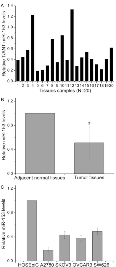
In order to investigate the role of miR-153 in ovarian cancer, the expression levels of miR-153 were first determined in 20 pairs of human ovarian cancer tissues and adjacent normal tis-sues using Real-time quantitative PCR. The results of RT-PCR shown that miR-153 levels was decreased in the 18 patients (18/20, 90.0%) (Figure 1A). The results also indicated that the expression levels of miR-153 were sig-nificantly lower in the EOC tissues than in the adjacent normal tissues (Figure 1B). Furth- ermore, we detected the expression levels of miR-153 in human normal epithelial ovarian cell line HOSEpiC and human ovarian carcino-ma cell lines. We found the expression level of miR-153 was significantly lower in the EOC cells than that in HOSEpiC cells (Figure 1C). In fur-ther experiments, the A2780 cell line was selected for the research object because the level of miR-153 in this cell line is the lowest. These findings indicate that the expression of miR-153 is down-regulated in EOC tissues and cell lines.
Overexpression of miR-153 attenuates the proliferative capacity of ovarian cancer cells
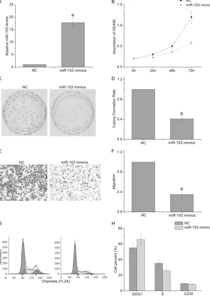
To further confirm the effect of miR-153 on the EOC cell proliferation, we used the miR-153 mimics to infect A2780 cells. The results of RT-PCR confirmed that miR-153 expression level was significantly increased in A2780 cells transfected with miR-153 mimic compared to cells transfected with NC (Figure 2A). Further, the CCK-8 and colony formation assays showed that the overexpression of miR-153 significant-ly attenuates the proliferative capacity of A2780 cells, compared with cells transfected with NC (Figure 2B-D). In the transwell assay, we observed that the overexpression of miR-153 led to a decrease in the number of cells that had migrated (Figure 2E, 2F). The cell cycle assay showed that the A2780 cells transfected with the miR-153 mimics had a significant increase in the G1/G0 phase cell population at 72 h post-infection, compared with negative control transfected cells (Figure 2G, 2H). Meanwhile, a concomitant decrease in the S phase cell population at 72 h post-infection in A2780 cells also occurred. These results
sug-Figure 1. Identification of the expression of miR-153
in human ovarian cancer tissues and cell lines. A, B: The expression level of miR-153 in 20 pairs of
[image:4.612.91.287.68.520.2]gest that the overexpression of miR-153 inhib-ited proliferation and induced cell cycle arrest of EOC cells.
AKT1 is a direct target of miR-153 in ovarian cancer cells
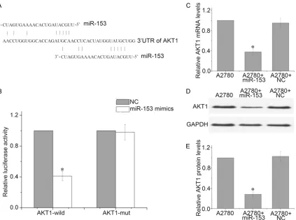
To determine the molecular mechanism by which miR-153 induces cell growth arrest and senescence, we used an open-target predic-tion program (TargetScanHuman 6.2, http:// www.targetscan.org/) to predict the targets of 153. We found AKT1 was a potential miR-153 target (Figure 3A). To determine whether AKT1 is the direct target gene for miR-153, a dual-luciferase reporter assay was performed. The results indicated that the luciferase activi-ty of the reporter containing the AKT1 gene’s wide-type 3’-UTR decreased (52%) following
treatment with miR-153 mimics. By contrast, the inhibitory effect of the miR-153 mimics was abolished in the mutated construct (Figure 3B). In addition, qRT-PCR and western blot analysis revealed that the expression of AKT1 mRNA and protein was inhibited by treatment with miR-153 mimics in A2780 cells (Figure 3C-E). Taken together, these data suggest that miR-153 reduces AKT1 expression by inhibiting translation and/or causing mRNA instability.
Overexpression of AKT1 reverses the inhibition effect of miR-153 on cell proliferation
[image:6.612.97.520.71.386.2]structed to increase endogenous AKT1 expres-sion (Figure 4A). The results of the CCK-8 and colony formation assays shown that the prolif-eration suppressing effect of miR-153 was reversed by AKT1 in A2780 cells (Figure 4B-D). Additionally, the transwell assay indicated that AKT1 significantly reversed the effect of miR-153 on the migration capability of A2780 cells (Figure 4E, 4F). Furthermore, cell cycle analysis demonstrated that BTG1 decreased the pro-portion of cells in G0/G1 phase, and inhibited the effect of miR-153 on cell cycle progression (Figure 4G and 4H). These findings suggested
that the overexpression of AKT1 could reverse the effect of miR-153 on the proliferation and cycle progression of ovarian cancer cells.
miR-153 suppresses ovarian cancer growth in vivo
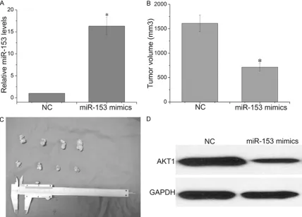
[image:8.612.93.521.174.481.2]To confirm the tumor suppressor role of miR-153, we established a BALB/c nude mouse xenograft model using A2780 cells. The A2780 cells were pre-treated with miR-153 or NC and injected into BALB/c athymic nude mice. RT-PCR analyses demonstrated the increased Figure 4. Overexpression of AKT1 reverses the Inhibition Effect of miR-153 on cell proliferation. A: The mRNA levels were detected by RT-PCR in A2780 cells cotransfected with pcDNA3.1-AKT1 and miR-153 mimics. B: The CCK-8 assay was performed every 24 h until 72 h after co-transfection with miR-153 mimics and pcDNA3.1-AKT1 or with miR-153 and control vector. C, D: Representative micrographs and quantifications of crystal violet stained colonies formed by the A2780 cells co-transfected with miR-153 mimics and pcDNA3.1-AKT1 or with miR-153 and control vector. E, F: Cell migration of A2780 cells that were treated as indicated in a transwell cell migration assay, as shown by the quantification and representative images. G, H: PI staining and flow cytometry were used to analyze the cell cycle distribution of A2780 cells that were treated as indicated. *P<0.05.
expression of miR-153 in tumors developed from miR-153-treated cells relative to NC- treated cells (Figure 5A). The tumor volume was measured every 4 days until day 28. The tumor volume of the A2780 cells treated with miR-153 mimics was significantly reduced rela-tive to NC (Figure 5B, 5C). We also performed Western blot assay to determine the expres-sion of AKT1 in xenografted tumor tissues (Figure 5D). We found that overexpression of miR-153 significantly inhibited the expression of AKT1. These results indicated that overex-pression of miR-153 significantly inhibits the tumorigenicity of A2780 cells in vivo.
Discussion
Although several miRNAs have been reported to play a role in the carcinogenesis of EOC, the function of miR-153 in EOC remains unclear. In this study, we observed that the levels of miR-153 were remarkably decreased in EOC tissues or EOC cell lines in comparison with paired adjacent normal tissues and normal epithelial ovarian cells. Furthermore, we found that over-expression of miR-153 can inhibit A2780 cells proliferation and induce cell cycle arrest. We also confirmed that AKT1 is a direct target of miR-153. Our findings suggested that miR-153 may attenuate the proliferative capacity of ovarian cancer cells.
MicroRNAs (miRNAs) are a class of endogenous small RNA molecules of approximately 18-24 nucleotides that modulate the post-transcrip-tional level of target gene mRNAs by binding to 3’-UTR directly [14, 15]. Recently, growing evi-dences indicated that miRNAs are involved in biological and pathological process regulation, such as cell proliferation, migration and apop-tosis [16]. Some of the miRNAs have been reported that they act as oncogenes genes or tumor suppressor in ovarian cancer. For exam-ple, Liu et al. reported that MiR-383 was over-expressed in both human EOC tumors and EOC cell lines [17]. Xiaohong Z. et al. confirmed the oncogenic effects of miR-203 on the prolifera-tion of ovarian cancer cells [18]. Recently sev-eral miRNAs have been proven to inhibit the growth and to enhance the drug sensitivity of EOC cells [19, 20]. An increasing body of evi-dence indicated that miR-153 expression was downregulated in multiple malignancies [21, 22]. However, to our knowledge the biological role and underlying molecular mechanism of
miR-153 in EOC remains unclear. Here, our find-ings demonstrated that ectopic expression of miR-153 inhibited proliferation, colony forma-tion, and migration by repressing AKT1, sug-gesting that miR-153 might act as tumor sup-pressor in EOC.
AKT, a serine/threonine-protein kinase, also known as protein kinase B (PKB), is involved in various signaling pathways, such as those of cell proliferation, survival, metabolism [23, 24]. Overactivation of AKT can affect many down-stream effectors and mediate multiple path-ways including cell proliferation and survival and thus favor tumorigenesis. Lee et al. report-ed that overexpression of phosphorylatreport-ed Akt (pAkt) in histologic specimens from EOC pa- tients was shown to be associated with poor prognostic [25]. Yin et al. found Wip1 modulate ovarian cancer metastasis through negative regulation of p-Akt [26]. Blocking of PI3K/AKT pathway has been reported to be negatively correlated with EOC tumor growth [27]. Con- sistently, we found that in the present study p-AKT levels were increased in the EOC tissues compared to paired noncancerous tissues. Moreover, overexpression of AKT1 reverses the inhibition effect of miR-153 on EOC cell prolif-eration. We also confirmed that AKT1 is the direct target of miR-153 in EOC cells. Our find-ings indicated that microRNA-153 suppressed EOC cell proliferation via regulating AKT1 expression.
In summary, we have found that miR-153 is mostly downregulated in ovarian cancer tis-sues. Transfection of miR-153 into EOC cell lines leads to inhibition of cell proliferation and induces cell cycle arrest by directly targeting AKT1. Hence, our data suggest that miR-153 and AKT1 may become promising therapeutic candidates and targets for EOC treatment. Disclosure of conflict of interest
None.
Address correspondence to:Hao Li, Department of Clinical Laboratory, The Taixing People’s Hospital, Taixing, Jiangsu, China. E-mail: lihao0523@outlook. com
References
and 485-5p and its clinicopathological rele-vance in ovarian epithelial tumours. Histopa-thology 2010; 57: 734-743.
[2] Siegel R, Naishadham D and Jemal A. Cancer statistics, 2013. CA Cancer J Clin 2013; 63: 11-30.
[3] Tinelli A, Vergara D, Martignago R, Leo G, Pi-sano M and Malvasi A. An outlook on ovarian cancer and borderline ovarian tumors: focus on genomic and proteomic findings. Curr Ge-nomics 2009; 10: 240-249.
[4] Coleman RL, Monk BJ, Sood AK and Herzog TJ. Latest research and treatment of advanced-stage epithelial ovarian cancer. Nat Rev Clin Oncol 2013; 10: 211-224.
[5] Wu F, Yang Z and Li G. Role of specific microR-NAs for endothelial function and angiogenesis. Biochem Biophys Res Commun 2009; 386: 549-553.
[6] Zhang W, Dahlberg JE and Tam W. MicroRNAs in tumorigenesis: a primer. Am J Pathol 2007; 171: 728-738.
[7] Wan WN, Zhang YQ, Wang XM, Liu YJ, Zhang YX, Que YH, Zhao WJ and Li P. Down-regulated miR-22 as predictive biomarkers for prognosis of epithelial ovarian cancer. Diagn Pathol 2014; 9: 178.
[8] Gao N, Tian JX, Shang YH, Zhao DY and Wu T. Catalpol suppresses proliferation and facili-tates apoptosis of OVCAR-3 ovarian cancer cells through upregulating microRNA-200 and downregulating MMP-2 expression. Int J Mol Sci 2014; 15: 19394-19405.
[9] Zhang ZC, Li YY, Wang HY, Fu S, Wang XP, Zeng MS, Zeng YX and Shao JY. Knockdown of miR-214 promotes apoptosis and inhibits cell pro-liferation in nasopharyngeal carcinoma. PLoS One 2014; 9: e86149.
[10] Shen W, Song M, Liu J, Qiu G, Li T, Hu Y and Liu H. MiR-26a promotes ovarian cancer prolifera-tion and tumorigenesis. PLoS One 2014; 9: e86871.
[11] Jin Y, Tymen SD, Chen D, Fang ZJ, Zhao Y, Dra-gas D, Dai Y, Marucha PT and Zhou X. MicroR-NA-99 family targets AKT/mTOR signaling pathway in dermal wound healing. PLoS One 2013; 8: e64434.
[12] Lei BX, Liu ZH, Li ZJ, Li C and Deng YF. miR-21 induces cell proliferation and suppresses the chemosensitivity in glioblastoma cells via downregulation of FOXO1. Int J Clin Exp Med 2014; 7: 2060-2066.
[13] Nyhan MJ, O’Donovan TR, Boersma AW, Wiemer EA and McKenna SL. MiR-193b pro-motes autophagy and non-apoptotic cell death in oesophageal cancer cells. BMC Cancer 2016; 16: 101.
[14] Bartel DP. MicroRNAs: genomics, biogenesis, mechanism, and function. Cell 2004; 116: 281-297.
[15] Ambros V. The functions of animal microRNAs. Nature 2004; 431: 350-355.
[16] Chang W, Liu M, Xu J, Fu H, Zhou B, Yuan T and Chen P. MiR-377 inhibits the proliferation of pancreatic cancer by targeting Pim-3. Tumour Biol 2016; 37: 14813-14824.
[17] Liu J, Dou Y and Sheng M. Inhibition of microR-NA-383 has tumor suppressive effect in hu-man epithelial ovarian cancer through the ac-tion on caspase-2 gene. Biomed Pharmacother 2016; 83: 1286-1294.
[18] Xiaohong Z, Lichun F, Na X, Kejian Z, Xiaolan X and Shaosheng W. MiR-203 promotes the growth and migration of ovarian cancer cells by enhancing glycolytic pathway. Tumour Biol 2016; 37: 14989-14997.
[19] Zhou F, Chen J and Wang H. MicroRNA-298 in-hibits malignant phenotypes of epithelial ovar-ian cancer by regulating the expression of EZH2. Oncol Lett 2016; 12: 3926-3932. [20] Tian S, Zhang M, Chen X, Liu Y and Lou G.
Mi-croRNA-595 sensitizes ovarian cancer cells to cisplatin by targeting ABCB1. Oncotarget 2016; 7: 87091-87099.
[21] Zuo J, Wang D, Shen H, Liu F, Han J and Zhang X. MicroRNA-153 inhibits tumor progression in esophageal squamous cell carcinoma by tar-geting SNAI1. Tumour Biol 2016; [Epub ahead of print].
[22] Wu X, Li L, Li Y and Liu Z. MiR-153 promotes breast cancer cell apoptosis by targeting HECTD3. Am J Cancer Res 2016; 6: 1563-1571.
[23] Hennessy BT, Smith DL, Ram PT, Lu Y and Mills GB. Exploiting the PI3K/AKT pathway for can-cer drug discovery. Nat Rev Drug Discov 2005; 4: 988-1004.
[24] Kumar A, Rajendran V, Sethumadhavan R and Purohit R. AKT kinase pathway: a leading tar-get in cancer research. ScientificWorldJournal 2013; 2013: 756134.
[25] Lee YK, Chung HH, Kim JW, Song YS and Park NH. Expression of phosphorylated Akt and hTERT is associated with prognosis of epitheli-al ovarian carcinoma. Int J Clin Exp Pathol 2015; 8: 14971-14976.
[26] Yin S, Wang P, Yang L, Liu Y, Wang Y, Liu M, Qi Z, Meng J, Shi TY, Yang G and Zang R. Wip1 suppresses ovarian cancer metastasis through the ATM/AKT/Snail mediated signaling. Onco-target 2016; 7: 29359-29370.