Original Article
Peroxiredoxin 4 as an independent prognostic marker
for survival in patients with early-stage
lung squamous cell carcinoma
Ji An Hwang1*, Joon Seon Song2*, Dae Yeul Yu3,4, Hyeong Ryul Kim5, Hye Jin Park2, Young Soo Park2, Woo
Sung Kim1, Chang Min Choi1
Departments of 1Pulmonary and Critical Care Medicine, 2Pathology, 5Thoracic Surgery, Asan Medical Center, University of Ulsan College of Medicine, Seoul, Korea; 3Disease Model Research Laboratory, Aging Research Center, Korea Research Institute of Bioscience and Biotechnology, Daejeon, Korea; 4Department of Functional Genomics, University of Science and Technology, Daejeon, Korea. *Equal contributors.
Received March 16, 2015; Accepted May 17, 2015; Epub June 1, 2015; Published June 15, 2015
Abstract: Objectives: Peroxiredoxin 4 (Prx 4) is a newly emerging antioxidant protein that has been studied in several human cancers. Recently, it was revealed that Prx 4 is highly expressed in human lung cancer and is needed for the promotion of lung cancer progression in vitro. However, there are no clinical data regarding the association of Prx 4 and prognosis in lung cancer. Materials and methods: The Prx 4 expression state as a prognostic indicator was assessed by immunohistochemical staining in 142 patients with stage II non-small cell lung cancer (NSCLC) who had undergone curative surgery between 2006 and 2010. The association between the degree of Prx 4 expression and several clinicopathologic parameters was then evaluated by statistical analyses. Results: The degree of Prx 4 expression was associated with histology and recurrence in the overall NSCLC patient group, with the proportion of patients with positive Prx 4 expression significantly higher for the adenocarcinoma subtype (39/70, 56%) than the squamous cell carcinoma subtype (23/72, 32%) (P = 0.004). However, when subgroup analyses according to histo-pathology were performed in terms of recurrence, positive Prx 4 expression was significantly correlated with higher recurrence rates (P = 0.003) and shorter disease-free survival (DFS) (P = 0.003, hazard ratio = 3.910) in patients with squamous cell carcinoma. In contrast, no meaningful relationship was observed between the level of Prx 4 ex-pression and DFS in the adenocarcinoma subgroup. Conclusion: Positive Prx 4 exex-pression is significantly correlated with recurrence and shorter DFS in patients with early-stage lung squamous cell carcinoma.
Keywords : Lung cancer, peroxiredoxin, immunohistochemistry
Introduction
Lung cancer is the most common cause of can-cer death worldwide, with an estimated 228,190 newly diagnosed cases in the United states in 2013 [1]. Approximately 85% to 90% of lung cancer cases are of non-small cell lung cancer (NSCLC), of which the main types are adenocarcinoma and squamous cell carcino-ma. The prognosis is poor, with an overall 5-year survival rate for NSCLC (all stages com-bined) of less than 15% [2]. Although early-stage NSCLC may be cured by surgical resec-tion, the recurrence rates are high, possibly due to early metastasis [3, 4]. Hence, it is important to identify biological markers that
are predictive of poor outcomes for early perti-nent treatment.
mamma-hyperoxidized and loses its antioxi-dant properties, which can only be reduced by sulfiredoxin (Srx) [13-15]. Then, instead, it may function as a molecular chaperone that facil-itates protein folding [16, 17]. Recently, Wei et al [18]. revealed that Srx and Prx 4 are highly expressed in human lung cancer cells from squamous cell carcinoma and adenocarcinoma and that Srx preferentially binds to Prx 4, so that Srx-Prx 4 complex significantly con-tributes to the maintenance of the tumor cell phenotype (anchorage-independent colony formation, cell migration, and invasion of lung can-cer cells) in vitro and the formation of metastases in vivo (mouse xeno-graft) through positive regulation of specific phosphokinase signaling cascades including the AP-1/MMP 9 axis, CREB, and MAPK pathways. Furthermore, many researches have been in progress recently con-cerning the associations between Prx 4 and cancer progression in various cancers, including ovarian cancer, oral cavity squamous cell carcinoma, and breast cancer [19-21]. However, there are no clinical data on the correlation between Prx 4 and prognosis in lung cancer. We therefore examined the associ-ation between the level of Prx 4 expression and prognosis in pa- tients with early-stage NSCLC who underwent curative surgical resec- tion.
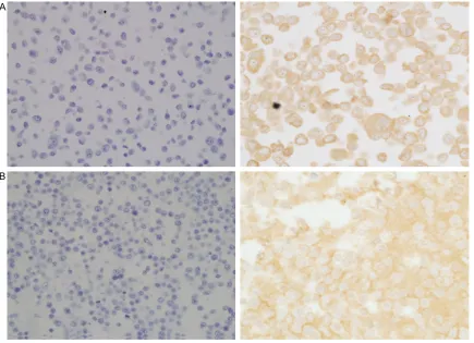
Figure 1. Representative photomicrographs of Prx 4 immunohistochemical staining in arrayed NSCLC tissues. The left and right columns show samples of adenocarcinoma and squamous cell carcinoma representing (A) 0, nega-tive, (B) 1+, weak, (C) 2+, moderate, (D) 3+, strong immunoreactivity in tumor cells, respectively. Original magnifi-cation: ×200. Prx 4 was not expressed in normal alveolar or bronchial epithelial cells. Normal bronchial epithelial cell image (Figure S1), negative and positive control cell images (Figure S2) were provided in supplementary data. NSCLC = non-small cell lung cancer; Prx 4 = peroxiredoxin 4.
lian cells, Prx 1 to Prx 6. Of these, Prx 4 is an antioxidant enzyme mainly localized in the endoplasmic reticulum (ER) within cells [11, 12]. Its antioxidant function is via the cysteine residue, which is oxidized to sulfenic acid form, and then, reduced by thioredoxin. However, under oxidative stress conditions, Prx 4 can be
Materials and methods
Patients and clinicopathologic data
[image:3.612.91.354.178.523.2]Institutional Review Board approval was ob- tained to use the relevant human archival tis-sues to investigate the immunohistochemical
Table 1. Clinicopathologic characteristics of the patients with stage II NSCLC who underwent complete resection
Variablesa No. of subjects (n = 142)
Sex
Male 108 (76.1%) Female 34 (23.9%) Smoking
Nonsmoker 40 (28.2%) Ex-smoker 73 (51.4%) Current smoker 29 (20.4%) Age (median, years) 63 (range, 32-85) Tumor size (median, cm) 3.0 (range, 1.0-7.0) T stageb
T1 73 (51.4%)
T2 69 (48.6%)
Type of surgical resection
Lobectomy 113 (79.6%) Bilobectomy 15 (10.6%) Pneumonectomy 14 (9.8%) Adjuvant therapy
Not done 52 (36.6%)
Done 90 (63.4%)
Median follow-up period (median, months) 42.9 (range, 3.4-88.4) Recurrence cases (recurrence rate) 59 (41.5%) Death cases (death rate) 48 (33.8%) Immunohistochemistry of Prx 4c
Negative 80 (56.4%) Positive 62 (43.6%)
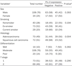
Table 2. Correlation of Prx 4 expression with clinicopathologic variables in patients with resectable stage II NSCLC
Variablesa Total number Prx 4 expression P-valueb
Negative Positive Sex
Male 108 (76) 63 (58) 45 (42) 0.393 Female 34 (24) 17 (50) 17 (50)
Smoking
Nonsmoker 40 (28) 18 (45) 22 (55) 0.194 Ex-smoker 73 (52) 43 (59) 30 (41) Current smoker 29 (20) 19 (66) 10 (34) Histology
Adenocarcinoma 70 (49) 31 (44) 39 (56) 0.004 Squamous cell carcinoma 72 (51) 49 (68) 23 (32) Differentiation
Well 14 (10) 7 (50) 7 (50) 0.392 Moderate 108 (76) 59 (55) 49 (45) Poor 20 (14) 14 (70) 6 (30) T stage
1 73 (51) 38 (52) 35 (48) 0.290 2 69 (49) 42 (61) 27 (39)
NSCLC = non-small cell lung cancer; Prx 4 = peroxiredoxin 4. aThe percentages for the variables are shown within parentheses. bThe significance was tested with chi-square or Fisher’s exact tests (P).
Department of Pathology and tissue arrays were prepared. All available histological slides, which had been routinely stained with hematoxylin and eosin (H&E), were reviewed. All information about staging or clinicopathologic characteris-tics was complete and tissue specimens were available for all 142 patients. Data was col-lected on patient demograph-ics, smoking history, type of surgery, histopathologic diag-nosis, grade of differentiation, tumor stage, date of diagnosis, date of recurrence, date of death (or last follow-up), and whether each patient received adjuvant therapy (chemothera-py and/or radiothera(chemothera-py) or not. In the process, cases of the patients who had experienced non-lung cancer related death were excluded. Tumor stages were assessed according to the AJCC Cancer Staging Man- ual, 7th edition [22].
Specimen preparation and IHC staining
IHC staining was performed on formalin-fixed, paraffin-embed-ded tissue sections using an automated IHC staining device (Benchmark XT; Ventana Me- dical Systems, Tucson, AZ) (Figure 1). Briefly, 4 um-thick whole tissue sections were transferred onto poly-L-lysine-coated adhesive slides and dried at 74°C for 30 min. After standard heat epitope retrieval for 1 h in ethylene diamine tet-raacetic acid (EDTA), pH 8.0, in the autostainer, samples were incubated with antibodies ag- ainst human Prx 4 antibody (IHC) expression of Prx 4. A total of 142 cases
of patients with stage II NSCLC who had under-gone complete surgical resection at Asan Medical Center (Seoul, Korea) between 2006 and 2010 were chosen from the archives of the
(1:100 dilution, rabbit polyclonal, AbFrontier, Seoul, South Korea). Sections were subsequ- ently incubated with an OptiView Universal DAB kit (Ventana Medical Systems). Slides were counterstained with Harris hematoxylin. They
Table 3. Correlation of Prx 4 expression with recurrence or deatha
in patients with resectable stage II NSCLC
Total number Prx 4 expression P-valueb
Negative Positive Recurrence
No 83 (59) 54 (65) 29 (35) 0.013 Yes 59 (41) 26 (44) 33 (56)
Recurrence in patients with adenocarcinoma
No 32 (46) 14 (44) 18 (56) 0.934 Yes 38 (54) 17 (45) 21 (55)
Recurrence in patients with squamous cell carcinoma
No 51 (71) 40 (78) 11 (22) 0.003 Yes 21 (29) 9 (43) 12 (57)
Death
No 94 (66) 52 (55) 42 (45) 0.732 Yes 48 (34) 28 (58) 20 (42)
[image:4.612.92.369.426.613.2]were evaluated separately by 2 independent experienced pathologists (J.S., Y.P.) with good agreement (Cohen’s ĸ = 0.696, P < 0.001).
The expression of Prx 4 was semiquantitatively evaluated using the method of Conklin et al. [23]. All individual tissue microarray cores were scored according to the cytoplasmic pattern intensity from 0 to 3 as follows: 0, no reactivity; 1+, faint/weak; 2+, moderate; 3+, strong reac-tivity in tumor cells. The expression levels of Prx 4 were categorized into two groups: cases with score 0 or 1 were considered negative and cases with score 2 or 3 were considered positive.
Statistical analysis
Statistical analyses were performed using SPSS software (version 18.0). The association between the Prx 4 immunoreactivity index and other clinicopathologic factors was analyzed by using the Pearson’s chi-square test or Fisher’s exact test. The Kaplan-Meier method and log-rank test were applied to estimate survival rates and compare the survival curves between groups. The Cox proportional hazard regression model was used for the analyses of the rela-tionship between disease-free survival (DFS) and Prx 4 expression or other clinicopathologic variables. All results were considered statisti-cally significant at P-value < 0.05.
Results
[image:5.612.90.286.68.489.2]The clinicopathologic characteristics of the patients with stage II NSCLC who had under-gone complete surgical resection are summa-rized in Table 1. Of the 142 patients, 108 (76%) were male. 40 (28%) of the patients had never smoked, whereas 102 (72%) were current or past smokers. The median patient age was 63 (range, 32-85). All patients had undergone curative surgery by lobectomy (79.6%), bilobec-tomy (10.6%), or pneumonecbilobec-tomy (9.8%). In the pathologic review after surgery, all patients had pathologic N1 node-positive disease. 73 patients (51%) were in the T1 stage and 69 (49%) were in T2. Ninety patients (63%) had received adjuvant therapy. The median follow-up period was 42.9 months (range, 3.4-88.4 months). During the follow-up period after sur-gery, 59 patients (41.5%) experienced recur-rence and 48 (33.8%) expired. In the IHC stain-ing of the resected tumor, 80 tissue samples (56.4%) showed negative Prx 4 expression and 62 (43.6%) showed positive Prx 4 immuno- reactivity.
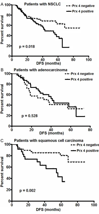
currence in the over-all NSCLC population (P = 0.013) and in pa- tients with squamous cell carcinoma (P = 0.003), but not in patients with adeno-carcinoma (P = 0.9- 34) (Table 3). In accordance with the- se results, Kaplan- Meier curves and log-rank tests showed that positive Prx 4 expression was sig-nificantly correlated with shorter DFS in all NSCLC patients (P = 0.018) and in pa- tients with the squa-mous cell carcinoma subtype (P = 0.002). However, in the ade-nocarcinoma sutype group, there was no meaningful diffeence between positive and negative Prx 4 expre- ssion state with re- gard to DFS (P = 0.5- 28 by log-rank test) (Figure 2). Meanwhi- le, as to overall sur-vival, no statistically significant difference was found according
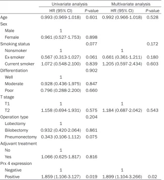
Table 4. Cox proportional hazard regression model analysis between DFS and Prx 4 expression or clinicopathologic variables in patients with NSCLC (n = 142)
Univariate analysis Multivariate analysis HR (95% CI) P-value HR (95% CI) P-value Age 0.993 (0.969-1.018) 0.601 0.992 (0.966-1.018) 0.528 Sex
Male 1
Female 0.961 (0.527-1.753) 0.898
Smoking status 0.077 0.172
Nonsmoker 1 1
Ex-smoker 0.567 (0.313-1.027) 0.061 0.661 (0.361-1.211) 0.180 Current smoker 1.072 (0.548-2.100) 0.839 1.205 (0.597-2.434) 0.603 Differentiation 0.902
Well 1
Moderate 0.928 (0.436-1.975) 0.847 Poor 0.796 (0.288-2.200) 0.660 T stage
T1 1 1
T2 1.158 (0.694-1.931) 0.575 1.184 (0.687-2.042) 0.543 Operation type 0.204
Lobectomy 1
Bilobectomy 0.932 (0.420-2.064) 0.861 Pneumonectomy 0.343 (0.106-1.112) 0.075 Adjuvant treatment
No 1
Yes 1.066 (0.625-1.817) 0.816 Prx 4 expression
Negative 1 1
Positive 1.859 (1.106-3.127) 0.019 1.899 (1.104-3.266) 0.02
HR = hazard ratio; CI = confidence interval; DFS = disease-free survival; NSCLC = non-small cell lung cancer; Prx 4 = peroxiredoxin 4.
The association between Prx 4 expression and various clinicopathologic factors that might be predictive of prognosis was evaluated in all 142 patients. It turned out that only histology was significantly associated with the degree of Prx 4 expression. Concretely, the proportion of patients with positive Prx 4 immunoreactivity was significantly higher in the adenocarcinoma subtype (39 out of 70, 56%) than in the squa-mous cell carcinoma subtype (23 out of 72, 32%) (P = 0.004). However, Prx 4 expression was not correlated with sex (P = 0.393), smok-ing status (P = 0.194), pathologic differentia-tion (P = 0.392), or pathologic stage (P = 0.290) (Table 2).
In terms of clinical outcome, Prx 4 expre- ssion level was meaningfully related to re-
to Prx 4 expression level in the overall NSCLC group or in each subtype group.
cells derived from a patient with lung ad- enocarcinoma caus- es a reduction or acceleration, respe- ctively, of tumor gr- owth and lung meta- stasis formation in mouse xenografts in vivo. However, there has been no human data reported to da- te about how com-patible these results are with the real clin-ical situation or ab- out the correlation between the level of Prx 4 expression and prognosis in pa- tients with NSCLC. In our present study, in IHC staining of stage II NSCLC tis-sues, Prx 4 expres-sion state was found to be associated with histology and recurrence rate. Th- at is, the proportion of patients with posi-tive Prx 4 expression
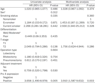
Table 5. Cox proportional hazard regression model analysis between DFS and Prx 4 expression or clinicopathologic variables in patients with the lung squamous cell carcinoma subtype (n = 72)
Univariate analysis Multivariate analysis HR (95% CI) P-value HR (95% CI) P-value Age 1.020 (0.965-1.077) 0.486 1.028 (0.967-1.092) 0.379
Sexa 0.312
Smoking status 0.335 0.360
Nonsmoker 1 1
Ex-smoker 1.184 (0.153-9.172) 0.871 1.453 (0.187-11.269) 0.720 Current smoker 2.266 (0.281-18.281) 0.442 2.930 (0.340-25.212) 0.328 Differentiation
Well/Moderateb 1
Poor 0.449 (0.06-3.353) 0.435 T stage
T1 1 1
T2 2.049 (0.794-5.286) 0.138 1.756 (0.624-4.944) 0.286 Operation type 0.664
Lobectomy 1
Bilobectomy 1.180 (0.419-3.324) 0.754 Pneumonectomy 0.611 (0.170-2.197) 0.451 Adjuvant treatment
No 1
Yes 0.759 (0.320-1.798) 0.530 Prx 4 expression
Negative 1
Positive 3.658 (1.490-8.978) 0.005 3.910 (1.587-9.632) 0.003
HR= hazard ratio; CI = confidence interval; DFS = disease-free survival; Prx 4 = peroxiredoxin 4. aThe significance (P-value) in the correlation between sex and recurrence in the squamous cell carcinoma subgroup was tested with a Fisher’s exact test. bThe “well” and “moderate” differ-entiation groups were combined for the analysis because only one case belonged to the “well” differentiation group.
shorter DFS (P = 0.003, hazard ratio = 3.910) (Table 5).
Discussion
Previous reports about Prx 4 have focused on its general role as an antioxidant or the change in its expression under various pathological conditions such as sepsis, diabetes, cardiovas-cular disease, or cancer [24-28]. Regarding lung cancer, it is known that the level of Prx 4 is significantly elevated in human cancer tissues compared to nonmalignant tissues [29]. Recently, Wei et al. [18] showed in their experi-ment that Prx 4 exists mainly in the form of the Srx-Prx 4 complex in human lung cancer cells, especially in squamous cell carcinoma and adenocarcinoma, and that Srx staining intensi-ty correlates positively with tumor progression state. They also disclosed that loss of Srx or enhancement of the Srx-Prx 4 axis in A549
NSCLC subtypes, other than previously known mechanisms and functions of Prx 4 in tumor progression.
Meanwhile, unlike adenocarcinoma in NSCLC, where many target agents have recently been developed and are actively used clinically, few molecular targets have been identified, and very restricted targeted agents are being cur-rently applied, for squamous cell carcinoma. Thus, our current study results showing the close correlation between Prx 4 expression and DFS in squamous cell carcinoma sheds light on the potential of Prx 4 as a molecular target in lung squamous cell carcinoma.
To date, there have been no reports on the dif-ferences in the Prx 4 expression state and its prognostic value by lung cancer subtype. Thus, it remains to be disclosed why Prx 4 has an influence on recurrence or disease progression only for squamous cell lung carcinoma. However, in a recent study of head-and-neck squamous cell carcinoma (HNSCC), Park et al. [30] revealed that Prx 4 protects cells from radiation-induced apoptosis by decreasing the production of intracellular ROS, thereby enhancing the radioresistance in HNSCC cell lines. In their experiments, the knock down of Prx 4 expression increased apoptosis and enhanced sensitivity to ionizing radiation treat-ment. It was also noteworthy that the same changes were observed in Prx 4 knockdown cells in the absence of ionizing radiation treat-ment, suggesting that Prx 4 itself may regulate cell survival irrespective of irradiation. And this may somewhat explain our result - the uneven prognostic role of Prx 4 according to different tumor subtypes - in the aspect of post-surgical treatment resistance and the role of Prx 4 in itself upon cell survival. However, whether these results about HNSCC also apply to lung squamous cell carcinoma and the exact mech-anism involved should be revealed through fur-ther research.
Conclusions
Altogether, this study showed positive Prx 4 expression state is significantly associated with recurrence and shorter DFS in lung squamous cell carcinoma. This implies that Prx 4 can be a good prognostic marker of cancer progression in early-stage squamous cell carcinoma, although it is not related to the prognosis of
adenocarcinoma in NSCLC. Our findings also suggest that Prx 4 may be a potential therapeu-tic target in patients with squamous cell lung carcinoma.
Acknowledgements
This study was supported by a grant of the Korean Health Technology R&D Project, Ministry of Health & Welfare (HI12C11460- 00013).
Disclosure of conflict of interest
None.
Address correspondence to: Dr. Chang Min Choi, Department of Pulmonary and Critical Care Medicine, Asan Medical Center, College of Medicine, University of Ulsan, 88, Olympic-ro 43-gil, Songpa-gu, Seoul 138-736, South Korea. Tel: +82-2-3010-5902; Fax: +82-2-3010-6968; E-mail: ccm@amc. seoul.kr
References
[1] American Cancer Society, Surveillance Rese- arch, 2013. Available at http://www.cancer. org/acs/groups/content/@epidemiologysurv e i l a n c e / d o c u m e n t s / d o c u m e n t / a c -spc-037114.pdf Accessed May 20, 2014. [2] Schiller JH, Harrinton D, Belani CP, Langer C,
Sandler A, Krook J, Zhu J and Johnson DH. Comparison of four chemotherapy regimens for advanced non small-cell lung cancer. N Engl J Med 2002; 346: 92-98.
[3] Singhal S, Vachani A, Antin-Ozerkis D, Kaiser LR and Albelda SM. Prognostic implications of cell cycle, apoptosis, and angiogenesis bio-markers in non-small cell lung cancer: a re-view. Clin Cancer Res 2005; 11: 3974-3986. [4] Mountain CF. Revisions in the International
System for staging Lung cancer. Chest 1997; 111: 1710-1717.
[5] Herbst RS, Heymach JV and Lippman SM. Lung cancer. N Engl J Med 2008; 359: 1367-1380. [6] Trachootham D, Alexandre J and Huang P.
Targeting cancer cells by ROS-mediated mech-anisms: A radical therapeutic approach? Nat Rev Drug Discov 2009; 8: 579-591.
[7] De Haan JB, Bladier C, Lotfi-Miri M, Taylor J, Hutchinson P, Crack PJ, Hertzoq P and Kola I. Fibroblasts derived from Gpx1 knockout mice display senescent like features and are sus-ceptible to H2O2-mediated cell death. Free Radic Biol Med 2004; 1: 53-64.
[9] Fujii J and Ikeda Y. Advances in our under-standing of peroxiredoxin, a multifunctional mammalian redox protein. Redox Rep 2002; 7: 123-130.
[10] Wood ZA, Schroder E, Robin Harris J and Poole LB. Structure, mechanism and regulation of peroxiredoxins. Trends Biochem Sci 2003; 28: 32-40.
[11] Haridas V, Ni J, Meager A, Su J, Yu GL, Zhai Y, Kyaw H, A KT, Hu J, Van Eldik LJ and Aggarwal BB. TRANK, a novel cytokine that activates NF-kappa B and c-Jun N-terminal kinase. J Immunol 1998; 161: 1-6.
[12] Tavender TJ and Bulleid NJ. Peroxiredoxin IV protects cells from oxidative stress by remov-ing H2O2 produced during disulphide forma-tion. J Cell Sci 2010; 123: 2672-2679. [13] Jeong W, Bae SH, Toledano MB and Rhee SG.
Role of sulfiredoxin as a regulator of peroxire-doxin function and regulation of its expression. Free Radic Biol Med 2012; 53: 447-456. [14] Biteau B, Labarre J and Toledano MB.
ATP-dependent reduction of cysteine-sulphinic acid by S.cerevisiae sulphiredoxin. Nature 2003; 425: 980-984.
[15] Chang TS, Jeong W, Woo HA, Lee SM, Park S and Rhee SG. Characterization of mammalian sulfiredoxin and its reactivation of hyperoxi-dized peroxiredoxin through reduction of cyste-ine sulfinic acid in the active site to cystecyste-ine. J Biol Chem 2004; 279: 50994-51001.
[16] Rhee SG and Woo HA. Multiple functions of peroxiredoxins: peroxidases, sensors and reg-ulators of the intracellular messenger Hv(2) ov(2), and protein chaperones. Antioxid Redox Signal 2011; 15: 781-794.
[17] Zito E, Melo EP, Yang Y, Wahlander A, Neubert TA and Ron D. Oxidative protein folding by an endoplasmic reticulum-localized peroxiredox-in. Mol Cell 2010; 40: 787-797.
[18] Wei Q, Jiang H, Xiao Z, Baker A, Young MR, Veenstra TD and Colburn NH. Sulfiredoxin-Peroxiredoxin IV axis promotes human lung cancer progression through modulation of spe-cific phosphokinase signaling. Proc Natl Acad Sci U S A 2011; 108: 7004-7009.
[19] Karihtala P, Soini Y, Vaskivuo L, Bloiqu R and Puistola U. DNA adduct 8-hydroxydeoxyguano-sine, a novel putative marker of prognostic sig-nificance in ovarian carcinoma. Int J Gynecol Cancer 2009; 19: 1047-1051.
[20] Chang KP, Yu JS, Chien KY, Lee CW, Liang Y, Liao CT, Yen TC, Lee LY, Huang LL, Liu SC, Chang YS and Chi LM. Identification of PRDX4 and P4HA2 as metastasis-associated proteins in oral cavity squamous cell carcinoma by com-parative tissue proteomics of microdissected specimens using iTRAQ technology. J Proteome Res 2011; 10: 4935-4947.
[21] Karihtala P, Mantyniemi A, Kang SW, Kinnula VL and Soini Y. Peroxiredoxins in breast carci-noma. Clin Cancer Res 2003; 9: 3418-3424. [22] Edge S, Byrd DR, Compton CC, Fritz AG, Greene
FL and Trotti A. The AJCC Cancer Staging Manual. 7th edition. Springer; 2010. pp. XV, 649.
[23] Conklin CM, Craddock KJ, Have C, Laskin J, Couture C and Lonescu DN. Immunohisto- chemistry is a reliable screening tool for identi-fication of ALK rearrangement in non-small-cell lung carcinoma and is antibody dependent. J Thoracic Oncol. 2013; 8: 45-51.
[24] Janin S. Peroxiredoxin 4: a multifunctional bio-marker worthy of further exploration. BMC Medicine 2011; 137: 1741-1747.
[25] Brixius K, Schwinger RH, Hoyer F, Napp A, Renner R, Bolck B, Kumin A, Fischer U, Mehlhorn U, Werner S and Bloch W. Isoform-specific downregulation of peroxiredoxin in hu-man failing myocardium. Life sciences 2007; 81: 823-831.
[26] Schulte J, Struck J, Kohrle J and Muller B. Circulating levels of peroxiredoxin 4 as a novel biomarker of oxidative stress in patients with sepsis. Shock 2011; 35: 460-465.
[27] Jiang YL, Ning Y, Ma XL, Liu YY, Wang Y, Zhang Z, Shan CX, Xu YD, Yin LM and Yang YQ. Alteration of the proteome profile of the pan-creas in diabetic rats induced by streptozoto-cin. Int J Mol Med 2011; 28: 153-160.
[28] Chen G, Gharib TG, Huang CC, Thomas DG, Shedden KA, Taylor JM, Kardia SL, Misek DE, Giordano TJ, Iannettoni MD, Orringer MB, Hanash SM and Beer DG. Proteomic analysis of lung adenocarcinoma: identification of a highly expressed set of proteins in tumors. Clin Cancer Res 2002; 8: 2298-2305.
[29] Lehtonen ST, Svensk AM and Soini Y. Peroxiredoxin IV, a novel protein family in lung cancer. Int J Cancer 2004; 111: 514-521. [30] Park JJ, Chang HW, Jeoung EJ, Roh JL, Choi SH,
Figure S1.Photomicrogragh of normal human bronchial epithelial cells (BEAS-2B).
[image:10.612.92.527.263.578.2]