Original Article
Expression of the EphA1 protein is associated with
Fuhrman nuclear grade in clear cell renal
cell carcinomas
Xiaolin Wang1*, Yushan Liu2*, Guangxin Cao1, Xueliang Zhang1, Haifei Xu1, Hanfeng Xu3, Jiandong Wang4
Departments of 1Urology, 2Pathology, Nantong Tumor Hospital, Nantong 226361, Jiangsu, China; 3Department of Oncology, Second Affiliated Hospital, Southeast University, Nanjing 210003, Jiangsu, China; 4Department of Pathology, Jinling Hospital, Nanjing 210002, China. *Equal contributors.
Received April 8, 2015; Accepted May 26, 2015; Epub June 1, 2015; Published June 15, 2015
Abstract: Aberrant expression of receptor tyrosine kinase EphA1 in malignant tissues has been reported. However,
the expression profile of EphA1 in renal cell carcinoma (RCC) and its association with clinicopathological parameters
remain unknown. The aim of this study was to determine the cancerous value of the EphA1 protein expression in patients with renal cell carcinomas. This study included 144 patients with clear cell RCC (ccRCC), 18 patients with chromophobe RCC and 6 patients with papillary RCC. The EphA1 protein was detected in RCC tissue samples by an
immunohistochemical staining with a specific polycolonal antibody. The correlation of the expression of the EphA1
protein with clinicopathological parameters was evaluated. High level of the expression of EphA1 was observed in all normal renal tubes. The EphA1 protein was negatively or weakly expressed in 93 out of 144 ccRCC (64.6%) and
positively expressed in 51 out of 144 ccRCC (35.4%). The high level expression of the EphA1 protein was signifi -cantly associated with younger patients (P<0.001), sex (P=0.016) and lower nuclear grade (P<0.001). No significant
relation between the expression of EphA1 and tumor diameter was found (P=0.316). Positive expression of EphA1 was observed in all samples of chromophobe RCC and papillary RCC. Our data indicated that the EphA1 protein may be a new marker for the prognosis of ccRCC.
Keywords: EphA1, receptor tyrosine kinase, clear cell renal cell carcinoma, Fuhrman nuclear grade
Introduction
Renal cell carcinoma (RCC) is the most com-mon neoplasm in the kidney, with an estimated 5-year survival rate of 50-60%. RCC has the highest mortality rate of the genitourinary can-cers and the incidence of RCC has risen steadi-ly. RCC is the most common form of adult kid-ney cancer and accounts for 2-3% of all adult malignancies globally. In 2014, RCC estimated new cases was 63920 and estimated deaths was 13860 in the United States, account for 2-3% of all malignant diseases in adults [1]. RCC is heterogeneous and comprises several histological subtypes according to the differ-ences in genetics, biology and behavior. RCC is consisted of clear cell RCC, papillary RCC, chromophobe RCC, collecting duct RCC, renal medullary carcinoma, Xp11 translocation RCC. Papillary (10-15%), Chromophobe (5%) and
Expression of EphA1 in RCC
by deletion and by promoter hypermethylation or rearrangement in the RCC. Nearly 20-40% localized ccRCC relapse even after curative nephrectomy, usually leading to incurable dis-ease. Metastatic RCC, characterized by high resistance to radiotherapy and chemotherapy has a poor prognosis.
Receptor tyrosine kinases of the Eph family and Ephrin ligands play important roles in vascular development, tissue-border formation, cell migration, axon guidance, and angiogenesis. Abnormal expression of Eph receptor tyrosine kinases in cancers is related to malignant transformation, tumor metastasis, tumor differ-entiation, and outcome.
EphA1 is located on chromosome 7q34, and is
the first member of the Eph family that was dis -covered from an erythropoietin-producing hep-atoma cell line. The number of known Eph receptor tyrosine kinases (RTK) has increased to 16, making them the largest subfamily of RTKs. The Eph receptor tyrosine kinase is divid-ed into two groups, designatdivid-ed EphA and EphB, according to sequence homology and ligand
(ephrin) binding specificity. In addition to their
functions in normal tissue, the abnormal expression of some members of the Eph family, including EphA1, has been implicated in carci-nogenesis. Eph receptors have important func-tions in the development of cancer. However, the expression of EphA1 in RCC has not been well investigated. In this study, we investigated the expression levels of EphA1 protein in a set of clear cell RCC, papillary RCC, and chromo-phobe RCC samples, and determined if its expression is associated with clinicopathologi-cal parameters.
Materials and methods
Tissue samples
The RCC tissue samples in our study were col-lected from 168 patients (111 males, 57 females, average age = 57.7 years; range = 33-82 years old at the time of resection) with RCC as part of a study approved by the Research Ethics Board of Nantong Tumor Hospital. All patients were treated by radical or partial nephrectomy and rendered disease-free. Of the 168 RCC tumors evaluated, 144 were diagnosed as conventional clear cell RCC,
6 as papillary RCC, and 16 as chromophobe RCC. Formalin-fixed and paraffin-embedded tis
-sues were sectioned into slices 4 μm thick and
stained with hematoxylin and eosin for
patho-logical identification.
Immunohistochemical staining
Sections from surgical specimens fixed in 10% formalin and embedded in paraffin were used
for immunohistochemical staining according to
the standard method. Briefly, each 4-m tissue
section was deparaffinized and rehydrated.
After rehydration through a graded ethanol series, the sections were autoclaved in 10 mM citrate buffer (pH 6.0) at 120°C for 2 min for antigen retrieval, then cooled to 30°C and washed with phosphate-buffered saline (PBS,
pH 7.3). After non-specific sites had been
blocked in 10% normal calf serum in PBS for 10 min, the sections were incubated at 4°C over-night with an anti-EphA1 polyclonal antibody (Abgent, San Diego, CA, USA) at a 1:500 dilu-tion in antibody diluent soludilu-tion (Zymed, Invitrogen), and then washed with PBS. The
specificity of EphA1 antibody was previously
investigated in colorectal cancer sections using blocking peptide [6, 7]. Here, we used colorec-tal cancer and normal mucosa tissues that showed negative and positive expression of EphA1 as controls. Next, the sections were incubated with secondary antibody (Dako REAL EnVision Detection System, Dako, UK) for 30 min at room temperature. Color development was performed with 3, 3’-diaminobenzidine (DAB). Nuclei were lightly counterstained with hematoxylin. Two pathologists independently assessed the immunostained slides. Any differ-ence in immunohistochemical scores was resolved by a consensus. Immunohistochemical staining of both normal and cancer cells was assessed according to the intensity of stained cells. Staining intensity was evaluated as: 0 = negative, 1 = weak, 2 = moderate, 3 = strong. Statistical analysis
The statistical significance of intergroup differ -ences was evaluated by a chi-square test. All statistical analyses were performed using SPSS software (SPSS 11.5, Chicago, IL). A two-sided P value less than 0.05 was considered
Results
Expression of EphA1 protein in normal renal tubes and ccRCC
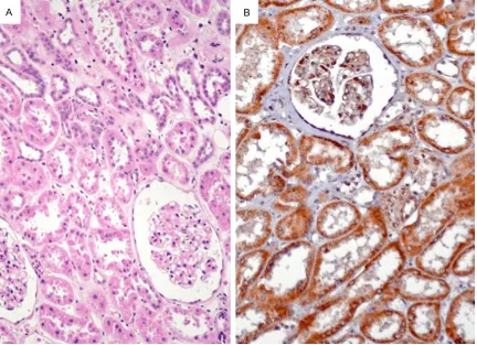
The EphA1 protein was positively expressed in normal renal tubes, but not expressed in renal glomerulus (Figure 1). The subcellular location of EphA1 protein was in cytoplasm.
The relationship between the expression of EphA1 protein in RCC and clinicopathologic parameters
Expression levels of EphA1 protein between cancer cells and adjacent normal cells were compared (Figures 2 and 3). The EphA1 protein was negatively or weakly expressed in 93 out of 144 ccRCC (64.6%) and positively expressed in 51 out of 144 ccRCC (35.4%). The high level
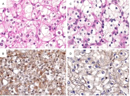
expression of the EphA1 protein was signifi -cantly associated with younger patients (P<0.001), sex (P=0.016) and lower nuclear grade (P<0.001) (Table 1). Fuhrman (nuclear)
grade criterion used in present study is as fol-lows. Grade I: Small, round, uniform nuclei (10 microns), inconspicuous nucleoli, look like lym-phocytes (very rare). Grade II: Slightly irregular nuclei, see nucleoli at 40× only, nuclear diam-eter 15 microns, open chromatin (40% of tumors). Grade III: See nucleoli at 10×, nuclei very irregular, diameter 20 microns, open chro-matin (30-40% of tumors). Grade IV: Mitoses; bizarre, multilobated, pleomorphic cells plus grade 3 features, macronucleoli (15% of tumors).
No significant relation between the expression
of EphA1 and tumor diameter was found (P=0.316) (Table 1).
Expression of EphA1 in chromophobe RCC and papillary RCC
[image:3.612.92.524.72.385.2]The EphA1 protein was positively expressed in all samples of chromophobe RCC and papillary RCC (Figure 4).
Expression of EphA1 in RCC
Discussion
Receptor tyrosine kinases of Eph family and the ligands of ephrin play important roles in the vascular development, tissue-border forma-tion, cell migraforma-tion, axon guidance and angio-genesis. Abnormal expression of Eph receptor tyrosine kinases in cancers is related to malig-nant transformation, tumor metastasis, tumor differentiation and prognosis. EphA1 is widely expressed in normal tissues including lung, small intestinal, kidney, bladder, thymus, and colon [8]. The expression level of EphA1 in human cancers is variable. Over-expression of EphA1 was observed in certain types of tumors including ovarian carcinoma [9], and head and neck squamous cell carcinoma [10]. Reduced expression of EphA1 was detected in prostate cancer cell lines [11], breast carcinoma cell lines [12], and basal cell carcinomas and squa-mous cell carcinoma specimens of the skin [13]. However, the role of EphA1 in the carcino-genesis of renal cell carcinoma is unknown. Wang et al previously reported that
down-regu-lation of EphA1 in colorectal carcinomas corre-lates with invasion and metastasis, and reduced EphA1 expression is associated with a poor overall survival [7]. Other group demon-strated that epigenetic silencing of EphA1 expression in colorectal cancer is correlated with poor survival [14]. Wang et al also found that increased expression of EphA1 protein in prostate cancers correlated with high Gleason score [15], and high expression of EphA1 in esophageal squamous cell carcinoma is asso-ciated with lymph node metastasis and advanced disease [16].
[image:4.612.90.525.72.392.2]Tcf pathway promotes EphB expression in
[image:5.612.91.522.72.383.2]colorectal cancer cells and the Ras-MAP kinae is the “Fuhrman Nuclear Grade” [19]. Fuhrman grade is on a scale of I-IV, where grade I carries Figure 3. The EphA1 protein was negatively expressed in grade III ccRCC (A: H&E, B: IHC, ×200) and in grade VI ccRCC (C: H&E, D: IHC, ×200).
Table 1. Expression of EphA1 protein in ccRCC and the association to clinicopathologic parameters
No. EphA1 protein P value 0, 1+ 2+ 3+
Sex
Male 102 18 54 30 0.016 Female 42 9 12 21 Age (years)
<50 39 3 9 27 <0.001 50-70 87 18 45 24
>70 18 3 12 3 Tumor diameter (cm)
<7 6 3 0 3 0.316 7-10 42 9 15 18
>10 96 15 51 30 Nuclear grade
I 60 6 12 42 <0.001 II 69 12 49 8
III+IV 15 9 5 1
pathway promotes EphA2 expression in breast cancer cells. Tumor suppressor activities have been reported for Eph sig-naling in colorectal, breast, prostate and skin cancer cells.
In this study, we evaluated the expres-sion of EphA1 in a set of renal cell carci-nomas and analyzed its association with clinicopathologic parameters. Our data showed that the high level expression of
[image:5.612.91.331.456.684.2]Expression of EphA1 in RCC
the best prognosis and grade IV the worst. Nuclear grade means that the system is based on just the appearance of the nuclei of the can-cer cells, rather than the appearance or struc-ture of the cells as a whole. Nuclear character-istics used in the Fuhrman Grade particularly indicate how actively the cells are making protein.
Down-regulation of EphA1 in colorectal cancer was reported and the mechanism of hyper-methylation of CpG island in promoter region has been demonstrated [7, 14]. In this study, we found that EphA1 protein was positively expressed in all normal renal tubes and only expressed in about 35% ccRCC (IHC score 3+). The EphA1 protein was negatively or weakly expressed in 65% of ccRCC (IHC score 0, 1+, and 2+). The mechanism for loss of EphA1
pro-tein in ccRCC should be confirmed in the next
study program.
In conclusion, the EphA1 protein was positively expressed in normal renal tubes and parts of ccRCC. Expression of EphA1 protein is
associ-ated with nuclear grade of ccRCC, which may be a new biomarker for the prognosis of ccRCC.
Acknowledgements
This work was supported in part by the Na- tional Natural Science Foundation of China (81371611).
Disclosure of conflict of interest
None.
Address correspondence to: Dr. Hanfeng Xu, De-
partment of Oncology, Second Affiliated Hospital,
Southeast University, Nanjing 210003, Jiangsu, China. E-mail: xuhanfeng1820@163.com; Dr. Jian- dong Wang, Department of Pathology, Jinling Hospital, Nanjing 210002, China. E-mail: jd_wang@ outlook.com
References
[image:6.612.91.526.73.387.2][1] Siegel R, Ma J, Zou Z and Jemal A. Cancer sta-tistics, 2014. CA Cancer J Clin 2014; 64: 9-29.
[2] Cairns P. Renal cell carcinoma. Cancer Biomark 2010; 9: 461-473.
[3] Lessi F, Mazzanti CM, Tomei S, Di Cristofano C, Minervini A, Menicagli M, Apollo A, Masieri L, Collecchi P, Minervini R, Carini M and Bevilacqua G. VHL and HIF-1alpha: gene varia-tions and prognosis in early-stage clear cell renal cell carcinoma. Med Oncol 2014; 31: 840.
[4] Singh RB and Amare Kadam PS. Investigation of tumor suppressor genes apart from VHL on 3p by deletion mapping in sporadic clear cell renal cell carcinoma (cRCC). Urol Oncol 2013; 31: 1333-1342.
[5] Choueiri TK, Fay AP, Gagnon R, Lin Y, Bahamon B, Brown V, Rosenberg JE, Hutson TE, Baker-Neblett KL, Carpenter C, Liu Y, Pandite L and Signoretti S. The role of aberrant VHL/HIF pathway elements in predicting clinical out-come to pazopanib therapy in patients with metastatic clear-cell renal cell carcinoma. Clin Cancer Res 2013; 19: 5218-5226.
[6] Wang J, Dong Y, Wang X, Ma H, Sheng Z, Li G, Lu G, Sugimura H and Zhou X. Expression of EphA1 in gastric carcinomas is associated with metastasis and survival. Oncol Rep 2010; 24: 1577-1584.
[7] Dong Y, Wang J, Sheng Z, Li G, Ma H, Wang X, Zhang R, Lu G, Hu Q, Sugimura H and Zhou X. Downregulation of EphA1 in colorectal carcino-mas correlates with invasion and metastasis. Mod Pathol 2009; 22: 151-160.
[8] Hafner C, Schmitz G, Meyer S, Bataille F, Hau P, Langmann T, Dietmaier W, Landthaler M and Vogt T. Differential gene expression of Eph re-ceptors and ephrins in benign human tissues and cancers. Clin Chem 2004; 50: 490-499. [9] Herath NI, Spanevello MD, Sabesan S, Newton
T, Cummings M, Duffy S, Lincoln D, Boyle G, Parsons PG and Boyd AW. Over-expression of Eph and ephrin genes in advanced ovarian cancer: ephrin gene expression correlates with shortened survival. BMC Cancer 2006; 6: 144. [10] Lin HS, Berry GJ, Fee WE Jr, Terris DJ and Sun
Z. Identification of tyrosine kinases overex -pressed in head and neck cancer. Arch Otolaryngol Head Neck Surg 2004; 130: 311-316.
[11] Fox BP, Tabone CJ and Kandpal RP. Potential clinical relevance of Eph receptors and ephrin ligands expressed in prostate carcinoma cell lines. Biochem Biophys Res Commun 2006; 342: 1263-1272.
[12] Fox BP and Kandpal RP. Invasiveness of breast
carcinoma cells and transcript profile: Eph
receptors and ephrin ligands as molecular markers of potential diagnostic and prognostic application. Biochem Biophys Res Commun 2004; 318: 882-892.
[13] Hafner C, Becker B, Landthaler M and Vogt T.
Expression profile of Eph receptors and ephrin
ligands in human skin and downregulation of EphA1 in nonmelanoma skin cancer. Mod Pathol 2006; 19: 1369-1377.
[14] Herath NI, Doecke J, Spanevello MD, Leggett BA and Boyd AW. Epigenetic silencing of EphA1 expression in colorectal cancer is correlated with poor survival. Br J Cancer 2009; 100: 1095-1102.
[15] Peng L, Wang H, Dong Y, Ma J, Wen J, Wu J, Wang X, Zhou X and Wang J. Increased expres-sion of EphA1 protein in prostate cancers cor-relates with high Gleason score. Int J Clin Exp Pathol 2013; 6: 1854-1860.
[16] Wang J, Ma J, Dong Y, Shen Z, Ma H, Wang X, Shi S, Wu J, Lu G, Peng L and Zhoud X. High expression of EphA1 in esophageal squamous cell carcinoma is associated with lymph node metastasis and advanced disease. APMIS 2013; 121: 30-37.
[17] Klein R. Bidirectional modulation of synaptic functions by Eph/ephrin signaling. Nat Neurosci 2009; 12: 15-20.
[18] Pasquale EB. Eph-ephrin bidirectional signal-ing in physiology and disease. Cell 2008; 133: 38-52.