Introduction
Liver cancer ranks the fifth among all cancers worldwide and it is the third most frequent etio-logical factor of cancer-caused deaths. Hepato- cellular carcinoma (HCC), the predominant form of adult liver malignancies, accounts for between 85% and 90% of liver cancers [1-3]. Several risk factors are related to the tumori-genesis of HCC, including chronic hepatitis virus infection and fatty liver diseases [4-7]. Previous studies have not proven adequate evi-dences for the prevention, diagnosis and treat-ment of HCC. Thus, understanding and insight into the mechanisms contributing to tumorigen-esis and aggressiveness in HCC is necessary and essential for more precise diagnosis and effective treatments.
Recent evidences have pointed to a relation-ship between several long non-coding RNAs (lncRNAs) and metastasis, drug resistance and other clinical outcome in several types of can-cers [8-13]. Essentially, lncRNAs are involved in the pathogenesis of HCC, such as HOX tran-script antisense RNA (HOTAIR), metastasis associated lung adenocarcinoma transcript 1 (MALAT1) and H19 [14-17]. NEAT1, a nuclear-restricted long non-coding RNA, encodes two isoforms: 3700-nucleotide (nt) NEAT1_1 and 23,000-nt NEAT1_2 [18]. This non-coding RNA has been recently revealed to localize specifi-cally to paraspeckles. And NEAT1 is involved in the process of gene expression regulation by retaining mRNAs for editing in the nucleus [19]. Cumulatively, studies suggest that
down-regu-Original Article
Clinical implication of long non-coding RNA
NEAT1 expression in hepatocellular carcinoma patients
Sien Guo1*, Wenjie Chen2*, Yihuan Luo2, Fanghui Ren2, Tengfei Zhong2, Minhua Rong3, Yiwu Dang2, Zhenbo Feng2, Gang Chen2
1Department of Hepatobiliary Surgery, First Affiliated Hospital of Guangxi Medical University, 6 Shuangyong Road,
Nanning, Guangxi Zhuang Autonomous Region 530021, P. R. China; 2Department of Pathology, First Affiliated Hospital of Guangxi Medical University, 6 Shuangyong Road, Nanning, Guangxi Zhuang Autonomous Region 530021, P. R. China; 3Department of Research, Affiliated Cancer Hospital, Guangxi Medical University, 71 Hedi Road, Nanning, Guangxi Zhuang Autonomous Region 530021, P. R. China. *Equal contributors.
Received February 11, 2015; Accepted April 12, 2015; Epub May 1, 2015; Published May 15, 2015
Abstract: Hepatocellular carcinoma (HCC), a primary malignancy of the liver, is associated with high mortality rate and poor prognosis. Emerging evidence showed that novel biomarkers are required toward a better understanding of the biological mechanisms of HCC. NEAT1 (nuclear paraspeckle assembly transcript 1, also known as MENε/β), a novel long non-coding RNA (lncRNA), serves as a crucial regulator in several cancers. However, the correlation between NEAT1 expression with tumorigenesis and metastasis in HCC tissues remains out of the question so far. In the current study, the aim was to evaluate the potential role of NEAT1 expression in HCC tissues and its relation
-ship with clinicopathological parameters. Method: The expression of NEAT1 was detected by qRT-PCR, in 95 cases of adjacent non-cancerous liver and their paired HCC tissues, respectively. The associations of NEAT1 with clinico
-pathological features and other biological factors were further analyzed. Result: Our results revealed that NEAT1 appeared to have higher expression in the HCC tissues, compared with the adjacent non-cancerous liver tissues. High levels of NEAT1 promoted the clinical features of HCC, including the number of tumor nodes, metastasis, clinical TNM stage, the status of portal vein tumor embolus, vaso-invasion and the infiltration of tumor cells. Ad
-ditionally, high NEAT1 expression levels were significantly associated with the expression level of MDTH, NM23 and MALAT1. Conclusion: Our study demonstrates that NEAT1 acts as a pivotal player in tumorigenesis and metastasis of hepatocellular carcinoma.
lated NEAT1 expression is involved in lung, liver, esophageal and retinal cancers [20]. Intere- stingly, evidence also argued that high NEAT1 expression is involved in the metastasis of can-cers like lung cancer [21]. To date, the emerg-ing potential role of NEAT1 in HCC is yet unclear.
In light of these findings and previous reports, we hypothesized an appealing association be- tween NEAT1 expression and HCC. To investi-gate this hypothesis, we evaluated the expres-sion of NEAT1 in adjacent non-cancerous liver and HCC tissues, in a set of 95 primary tumors, and investigated the relationship between NEAT1 expression and clinical, histological, pathological and other biological factors in HCC.
Materials and methods
Patients
Patients were selected from the First Affiliated Hospital of the Guangxi Medical University (Nanning, Guangxi, China) during March, 2010 to December, 2011. Seventy-five males and twenty female individuals were enrolled in the study, with ages between 29 and 82 years old. Additionally, the clinicopathological parameters were collected such as age, gender, differentia-tion, tumor size, tumor nodes, metastasis, clini-cal TNM stages, the presence or absence of portal vein tumor embolus, vaso-invasion, cap-sular infiltration and cirrhosis, and other bio-markers, for instance, serum AFP level detect-ed by ELISA; expression of metadherin (MTDH), NM23, P53, P21, vascular endothelial growth factor (VEGF), as well as microvessel density (MVD) stained by using CD34 with immunohis-tochemistry; and expression of a lncRNA, MALAT1 by using real time RT-qPCR. Table 1 showed all these features in detail. The Ethical Committee of First Affiliated Hospital, Guangxi Medical University, China approved the current study. We obtained informed consent from all participating patients. In accordance with the Helsinki Declaration, related research proce-dure was carried out. All samples were reviewed and diagnosed by two independent patho- logists.
RT-qPCR
RNA isolation and RNA normalization were per-formed by the method reported before [22, 23]. Extracted RNA was analyzed by Real-time qPCR
using Applied Biosystems PCR7900. The expre- ssion of NEAT1 was examined by Reverse tran-scription (RT) and qPCR kits according to the instructions described previously [22, 23]. Pri- mers were as below: NEAT1 Forward-5’-TGGC- TAGCTCAGGGCTTCAG-3’, NEAT1 Reverse-5’-TC- TCCTTGCCAAGCTTCCTTC-3’; GAPDH Forward-5’-TGAACGGGAAGCTCACTGG-3’, GAPDH Rever- se-5’-TCCACCACCCTGTTGCTGTA-3’. The NEAT1 expression was calculated with the formula 2-Δcq [22-24].
Statistics
All statistical analyses were performed using SPSS 20. For the analysis of the significance of difference between two groups, we performed Student’s t test. Accordingly, we also applied One-way analysis of variance (ANOVA) test to distinguish its significance of difference, as we divided differentiation into three groups. The strengths of the associations between NEAT1 expression and clinicopathological parameters were tested with the Spearman rank correla-tion. The effectiveness of NEAT1 for predicting the risk of the clinicopathological parameters was evaluated by ROC curve. We used Kaplan-Meier survival method and log-rank test to esti-mate the impact of NEAT1 on recurrence of HCC. All tests were 2-sided, and statistical sig-nificance was considered as P<0.05.
Results
Overexpression of NEAT1 in HCC
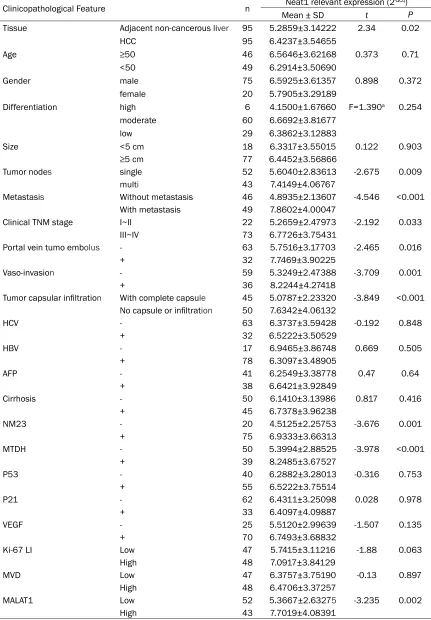
Analysis of 95 matched samples (95 adjacent non-cancerous liver tissues and 95 HCC tis-sues) by RT-qPCR detected significantly increased levels of NEAT1 in tumor samples compared to the non-tumor group (6.42±3.55 vs. 5.29±3.14, P=0.02, Figure 1A).
Associations of NEAT1 with clinicopathological
factors
Table 1. Association of NEAT1 expression with clinicopathological parameters in HCC
Clinicopathological Feature n Neat1 relevant expression (2-ΔCq)
Mean ± SD t P
Tissue Adjacent non-cancerous liver 95 5.2859±3.14222 2.34 0.02
HCC 95 6.4237±3.54655
Age ≥50 46 6.5646±3.62168 0.373 0.71
<50 49 6.2914±3.50690
Gender male 75 6.5925±3.61357 0.898 0.372
female 20 5.7905±3.29189
Differentiation high 6 4.1500±1.67660 F=1.390a 0.254
moderate 60 6.6692±3.81677
low 29 6.3862±3.12883
Size <5 cm 18 6.3317±3.55015 0.122 0.903
≥5 cm 77 6.4452±3.56866
Tumor nodes single 52 5.6040±2.83613 -2.675 0.009
multi 43 7.4149±4.06767
Metastasis Without metastasis 46 4.8935±2.13607 -4.546 <0.001
With metastasis 49 7.8602±4.00047
Clinical TNM stage I~II 22 5.2659±2.47973 -2.192 0.033
III~IV 73 6.7726±3.75431
Portal vein tumo embolus - 63 5.7516±3.17703 -2.465 0.016
+ 32 7.7469±3.90225
Vaso-invasion - 59 5.3249±2.47388 -3.709 0.001
+ 36 8.2244±4.27418
Tumor capsular infiltration With complete capsule 45 5.0787±2.23320 -3.849 <0.001 No capsule or infiltration 50 7.6342±4.06132
HCV - 63 6.3737±3.59428 -0.192 0.848
+ 32 6.5222±3.50529
HBV - 17 6.9465±3.86748 0.669 0.505
+ 78 6.3097±3.48905
AFP - 41 6.2549±3.38778 0.47 0.64
+ 38 6.6421±3.92849
Cirrhosis - 50 6.1410±3.13986 0.817 0.416
+ 45 6.7378±3.96238
NM23 - 20 4.5125±2.25753 -3.676 0.001
+ 75 6.9333±3.66313
MTDH - 50 5.3994±2.88525 -3.978 <0.001
+ 39 8.2485±3.67527
P53 - 40 6.2882±3.28013 -0.316 0.753
+ 55 6.5222±3.75514
P21 - 62 6.4311±3.25098 0.028 0.978
+ 33 6.4097±4.09887
VEGF - 25 5.5120±2.99639 -1.507 0.135
+ 70 6.7493±3.68832
Ki-67 LI Low 47 5.7415±3.11216 -1.88 0.063
High 48 7.0917±3.84129
MVD Low 47 6.3757±3.75190 -0.13 0.897
High 48 6.4706±3.37257
MALAT1 Low 52 5.3667±2.63275 -3.235 0.002
High 43 7.7019±4.08391
tumor embolus (r=0.255, P=0.013), vaso-inva-sion (r=0.341, P=0.001), tumor capsule infiltra-tion (r=0.324, P=0.001), MTDH level (r=0.278,
P<0.001), NM23 level (r=0.287, P=0.005) and MALAT1 level (r=0.180, P=0.013). Meanwhile, NEAT1 level in HCC with multi-tumor nodes was significantly higher (7.41±4.07) than that in the group with single tumor node (5.60±2.84,
P=0.009). NEAT1 level in HCC tissues with metastasis was remarkably upregulated (7.86±4.00) as compared to that without metastasis (4.89±2.14, P<0.001). Significantly increasing level of NEAT1 was found in clinical TNM stage III~IV (6.77±3.75), as compared to that in the stage I~II (5.27±2.48, P=0.033). NEAT1 appeared to have higher expression in the HCC tissues with the portal vein tumor embolus (7.75±3.90) than that without (5.75±3.18, P=0.016). Moreover, higher NEAT1 expression was detected in vaso-invasion (8.22±4.27) than that without (5.32±2.47,
[image:4.612.311.519.74.244.2] [image:4.612.92.298.79.566.2]P=0.001). Furthermore, NEAT1 levels were also significantly higher in the group of tumors with-out capsule or with tumor cell infiltration (7.63±4.06) than that in the corresponding group (5.08±2.23, P<0.001, Figure 1B). Additionally, the level of NEAT1 was significantly higher in high MTDH group (8.25±3.68) than in low MDTH group (5.40±2.89, P<0.001). The positive of NM23 also disclosed a higher expression of NEAT1 (6.93±3.66) than the neg-ative one (4.51±2.26, P=0.001). Additionally, the high MALAT1 expression group revealed a Figure 1.A. Overexpression of NEAT1 in HCC. Higher NEAT1 expression level was observed in HCC compared to that in the adjacent non-cancerous live (P=0.020). B. The relationships between NEAT1 clinicopathologi -cal features. Nodes: 1. multi-tumor nodes; 2. single tumor node; Metastasis: 1. with metastasis; 2. without metastasis; TNM: 1. TNM III-IV 2. TNM I-II; Embolus: 1. portal vein tumor embolus (+); 2. portal vein tumor embolus (-); Vaso-invasion: 1. (+); 2. (-); Capsular: 1. no capsule or infiltration; 2. With complete capsule; C. Associations of NEAT1 expression with biological fac -tors in HCC. NM23:1. (+); 2. (-); MDTH: 1. (+); 2. low level (-); MALAT1: 1. high expression 2. low expression; *P<0.05; **P<0.01; ***P<0.001.
significantly higher NEAT1 level (7.70±4.08) than the low expression group (5.37±2.63,
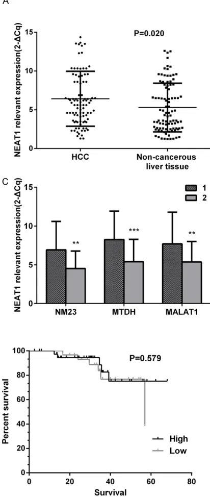
P=0.002, Figure 1C). We next divided the patients into two groups using the median NEAT1 expression as cutoff value
[image:5.612.90.522.72.561.2](medi-an=5.90). The mean recurrence time was 58.90 months in patients with higher NEAT1 expression and 50.60 months in patients with lower NEAT1 expression, but there is no statisti-cal significance (P=0.579, Figure 2).
Diagnostic value of NEAT1 for clinicopathologi-cal parameters by ROC curve analysis
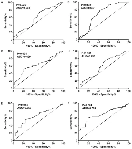
As the ROC curve indicated, NEAT1 of 3.7 repre-sented a significant diagnostic cut off value for HCC (AUC=0.594, P=0.025, Figure 3A). We fur-ther explored the predictive value of NEAT1 for other clinicopathological parameters in the 95 HCC samples by ROC curve. NEAT1 of 6.32 in the prediction of capsule or infiltration resulted in AUC of 0.687 (P=0.02, Figure 3B). As NEAT1 ≥6.45, it was also considered to be a valuable parameter in the prediction of tumor node (AUC=0.629, P=0.031), metastasis (AUC=0.73,
P<0.001), the portal vein tumor embolus (AUC=0.656, P=0.014) and vaso-invasion (AUC=0.703, P=0.001), respectively (Figure 3C-F).
Discussion
Accumulating evidence indicates that the involvement in functional lncRNAs is implicated in HCC pathogenesis [25]. For example, GAS5 expression was decreased in human HCC and was associated with advanced tumor progres-sion [26]. Moreover, MALAT1 expresprogres-sion level was proved to be an independent prognostic factor for HCC. Furthermore, the higher expres-sion of MALAT1 was also found to be related with shortened disease-free survival post liver transplantation [16]. Another lncRNA overex-pressed in HCC was HOTAIR. HCC patients with high expression of HOTAIR exhibited significant-ly poorer prognosis than those with low HOTAIR expression [15].
NEAT1 has been extensively demonstrated to be involved in several cancers. Kim et al. have demonstrated that NEAT1 was up-regulated in stage III serous ovarian carcinoma [27]. A recent study described that upregulation of NEAT1 was observed in acute promyelocytic leukemia samples and cell lines [28]. Dimple et al. conducted the study that NEAT1 could act as one of the differentially regulated lncRNAs in prostate cancer by activating prostate cancer genes [29]. The report by Choudhry et al. also showed that high tumor NEAT1 expression con-fers a poor outcome in breast cancer patients [30]. The objective of this study was to seek to probe the potentials of NEAT1, a long non-cod-ing RNA recently characterized by our group in HCC. To our knowledge, Gibb et al. was the first
one to indicate a direct association of NEAT1 expression with liver cancers via GEO acces-sioned human short SAGE libraries. Inter- estingly, NEAT1 presented a lower expression in liver cancers than in normal tissues in the previous study [20]. Contrary to the result of Gibb et al., our result revealed that high NEAT1 expression was detected in HCC. A possible explanation for this discrepancy is that NEAT1 was detected both in human and non-human origin in their study. In our current study, the significant higher expression of NEAT1 was detected in 95 cases of HCC tissues, compared with adjacent non-cancerous liver tissues. Taken together with the previous studies, we demonstrate that the regulation of NEAT1 con-tributes to tumorigenesis in HCC. Further large-scale studies might facilitate the discovery of NEAT1 molecular regulation in hepatocarcino-genesis of HCC.
It was interesting to discover the correlation between NEAT1 expression level and clinico-pathological parameters in HCC. The expres-sion level of NEAT1 was strongly correlated with the numbers of tumor nodes, metastasis and TNM stage in HCC patients. Moreover, we found significant correlations between high NEAT1 levels and portal vein tumor embolus, vaso-invasion and tumor capsular infiltration. The results also indicate that NEAT1 affects the infiltration of tumor cells, migration and inva-sion in HCC. More specifically, NEAT1 appears to be associated with MTDH level. The previous studies suggested that MTDH was regarded as a major event in metastasis and poor outcome in HCC patients [31, 32]. Of note, higher NEAT1 expression was detected in the cases of NM23 being positive. RUN et al carried out the study that overexpression of NM23 contributes to the hepatocarcinogenesis in HCC [33]. Given the strong association with NM23 level, the evi-dence of upregulated NEAT1 expression in HCC suggests a novel pro-oncogenic potential role in hepatocarcinogenesis. All together, our data indicate that the upregulation of NEAT1 increas-es tumorigenincreas-esis and metastasis in HCC.
reducing apoptosis, contributing to tumorigen-esis [30]. More recently, the cellular levels of NEAT1_1/2 were found to be down-regulated in a particular class of cells in MALAT1-knockout mice, which indicates that MALAT1 transcribed from the adjacent region has an effect on the expression of NEAT1_1/2 in specific cells [34]. In agreement with the previous report, high NEAT1 expression was found to be associated with MALAT1 upregulation in our study. MALAT1 has been associated with metastasis and poor prognosis of HCC according to histology find-ings. MALAT1 is well-known as an oncogene, because it regulates alternative splicing of endogenous target genes that are involved in cancer [16]. Moreover, we hypothesized that NEAT1 may play an essential role in the prog-ress and metastasis of HCC, by affecting the expression of MALAT1. Taken together, the results suggest NEAT1 may modulate in tumori-genesis and metastasis in HCC. The molecular mechanism driving this regulation is still unclear.
In conclusion, our results suggest that NEAT1 may represent a valuable predictive marker of tumorigenesis and metastasis of human HCC. Additional research of larger series into the molecular mechanism of NEAT1 in HCC is needed.
Acknowledgements
The study was supported partly by the Fund of Guangxi Natural Scientific Research (No. 2013GXNSFBA019191), Guangxi Provincial Health Bureau Scientific Research Project (Z2014054), Youth Science Foundation of Guangxi Medical University (GXMUYSF201311), Guangxi University Science and Technology Research Projects (LX2014075), and the Fund of National Natural Science Foundation of China (NSFC 81360327). The funders had no role in study design, data collection and analy-sis, decision to publish, or preparation of the manuscript.
Disclosure of conflict of interest
None.
Address correspondence to: Drs. Gang Chen and Zhenbo Feng, Department of Pathology, First Affi-liated Hospital of Guangxi Medical University, 6 Shuangyong Road, Nanning, Guangxi Zhuang
Auto-nomous Region 530021, P. R. China. Fax: +86 771 5358943; E-mail: [email protected] (GC); [email protected] (ZBF)
References
[1] Ma L, Chua MS, Andrisani O and So S. Epi
-genetics in hepatocellular carcinoma: an up
-date and future therapy perspectives. World J
Gastroenterol 2014; 20: 333-45.
[2] Lafaro KJ, Demirjian AN and Pawlik TM. Epide
-miology of Hepatocellular Carcinoma. Surg On
-col Clin N Am 2015; 24: 1-17.
[3] Taura N, Fukushima N, Yastuhashi H, Takami Y, Seike M, Watanabe H, Mizuta T, Sasaki Y, Na
-gata K, Tabara A, Komorizono Y, Taketomi A, Matsumoto S, Tamai T, Muro T, Nakao K, Fu
-kuizumi K, Maeshiro T, Inoue O and Sata M. The incidence of hepatocellular carcinoma as
-sociated with hepatitis C infection decreased in Kyushu area. Med Sci Monit 2011; 17:
Ph7-11.
[4] El-Serag HB and Rudolph KL. Hepatocellular carcinoma: epidemiology and molecular carci
-nogenesis. Gastroenterology 2007; 132:
2557-76.
[5] Hou W and Bonkovsky HL. Non-coding RNAs in hepatitis C-induced hepatocellular carcinoma: dysregulation and implications for early detec
-tion, diagnosis and therapy. World J Gastroen -terol 2013; 19: 7836-45.
[6] Kretzer IF, do Livramento A, da Cunha J, Gon
-calves S, Tosin I, Spada C and Treitinger A. Hepatitis C worldwide and in Brazil: silent epi
-demic--data on disease including incidence, transmission, prevention, and treatment. Sci
-entificWorldJournal 2014; 2014: 827849. [7] Amr S, Iarocci EA, Nasr GR, Saleh D, Blancato
J, Shetty K and Loffredo CA. Multiple pregnan
-cies, hepatitis C, and risk for hepatocellular carcinoma in Egyptian women. BMC Cancer
2014; 14: 893.
[8] Zhang H, Chen Z, Wang X, Huang Z, He Z and Chen Y. Long non-coding RNA: a new player in cancer. J Hematol Oncol 2013; 6: 37.
[9] Malek E, Jagannathan S and Driscoll JJ. Corre
-lation of long non-coding RNA expression with metastasis, drug resistance and clinical out
-come in cancer. Oncotarget 2014; 5: 8027-38. [10] Mizrahi I, Mazeh H, Grinbaum R, Beglaibter N,
Wilschanski M, Pavlov V, Adileh M, Stojadi
-novic A, Avital I, Gure AO, Halle D and Nissan A. Colon Cancer Associated Transcript-1 (CCAT1) Expression in Adenocarcinoma of the Stom -ach. J Cancer 2015; 6: 105-10.
[11] Ding C, Cheng S, Yang Z, Lv Z, Xiao H, Du C, Peng C, Xie H, Zhou L, Wu J and Zheng S. Long non-coding RNA HOTAIR promotes cell migra
[12] Gao W, Chan JY and Wong TS. Long non-coding RNA deregulation in tongue squamous cell car
-cinoma. Biomed Res Int 2014; 2014: 405860. [13] Weber DG, Johnen G, Casjens S, Bryk O, Pesch
B, Jockel KH, Kollmeier J and Bruning T. Evalu
-ation of long noncoding RNA MALAT1 as a can
-didate blood-based biomarker for the diagno
-sis of non-small cell lung cancer. BMC Res
Notes 2013; 6: 518.
[14] Pan YF, Qin T, Feng L and Yu ZJ. Expression pro
-file of altered long non-coding RNAs in patients with HBV-associated hepatocellular carcino
-ma. J Huazhong Univ Sci Technolog Med Sci
2013; 33: 96-101.
[15] Ishibashi M, Kogo R, Shibata K, Sawada G, Takahashi Y, Kurashige J, Akiyoshi S, Sasaki S, Iwaya T, Sudo T, Sugimachi K, Mimori K, Waka
-bayashi G and Mori M. Clinical significance of the expression of long non-coding RNA HOTAIR in primary hepatocellular carcinoma. Oncol
Rep 2013; 29: 946-50.
[16] Lai MC, Yang Z, Zhou L, Zhu QQ, Xie HY, Zhang F, Wu LM, Chen LM and Zheng SS. Long non-coding RNA MALAT-1 overexpression predicts tumor recurrence of hepatocellular carcinoma after liver transplantation. Med Oncol 2012;
29: 1810-6.
[17] Zhang L, Yang F, Yuan JH, Yuan SX, Zhou WP, Huo XS, Xu D, Bi HS, Wang F and Sun SH. Epi
-genetic activation of the MiR-200 family con
-tributes to H19-mediated metastasis suppres
-sion in hepatocellular carcinoma. Carcino-genesis 2013; 34: 577-86.
[18] Naganuma T and Hirose T. Paraspeckle forma
-tion during the biogenesis of long non-coding RNAs. RNA Biol 2013; 10: 456-61.
[19] Clemson CM, Hutchinson JN, Sara SA, Ens
-minger AW, Fox AH, Chess A and Lawrence JB. An architectural role for a nuclear noncoding RNA: NEAT1 RNA is essential for the structure of paraspeckles. Mol Cell 2009; 33: 717-26. [20] Gibb EA, Vucic EA, Enfield KS, Stewart GL, Lo
-nergan KM, Kennett JY, Becker-Santos DD, Ma
-cAulay CE, Lam S, Brown CJ and Lam WL. Hu
-man cancer long non-coding RNA transcrip-tomes. PLoS One 2011; 6: e25915.
[21] Zhao W, An Y, Liang Y and Xie XW. Role of HO
-TAIR long noncoding RNA in metastatic pro
-gression of lung cancer. Eur Rev Med Pharma -col Sci 2014; 18: 1930-6.
[22] Rong M, He R, Dang Y and Chen G. Expression and clinicopathological significance of miR-146a in hepatocellular carcinoma tissues. Ups
J Med Sci 2014; 119: 19-24.
[23] Chen G, Umelo IA, Lv S, Teugels E, Fostier K, Kronenberger P, Dewaele A, Sadones J, Geers C and De Greve J. miR-146a inhibits cell growth, cell migration and induces apoptosis in non-small cell lung cancer cells. PLoS One
2013; 8: e60317.
[24] Dang YW, Zeng J, He RQ, Rong MH, Luo DZ and
bition, motility suppression and apoptosis in
-duction in hepatocellular carcinoma cells. Asian Pac J Cancer Prev 2014; 15: 4969-76. [25] Fang TT, Sun XJ, Chen J, Zhao Y, Sun RX, Ren N
and Liu BB. Long Non-coding RNAs are differ
-entially expressed in hepatocellular carcinoma cell lines with differing metastatic potential. Asian Pac J Cancer Prev 2014; 15: 10513-124. [26] Tu ZQ, Li RJ, Mei JZ and Li XH. Down-regulation
of long non-coding RNA GAS5 is associated with the prognosis of hepatocellular carcino -ma. Int J Clin Exp Pathol 2014; 7: 4303-9.
[27] Kim YS, Hwan JD, Bae S, Bae DH and Shick WA. Identification of differentially expressed genes using an annealing control primer sys
-tem in stage III serous ovarian carcinoma. BMC
Cancer 2010; 10: 576.
[28] Zeng C, Xu Y, Xu L, Yu X, Cheng J, Yang L, Chen S and Li Y. Inhibition of long non-coding RNA NEAT1 impairs myeloid differentiation in acute promyelocytic leukemia cells. BMC Cancer
2014; 14: 693.
[29] Chakravarty D, Sboner A, Nair SS, Giannopou
-lou E, Li R, Hennig S, Mosquera JM, Pauwels J, Park K, Kossai M, MacDonald TY, Fontugne J, Erho N, Vergara IA, Ghadessi M, Davicioni E, Jenkins RB, Palanisamy N, Chen Z, Nakagawa S, Hirose T, Bander NH, Beltran H, Fox AH, Ele
-mento O and Rubin MA. The oestrogen recep
-tor alpha-regulated lncRNA NEAT1 is a critical modulator of prostate cancer. Nat Commun
2014; 5: 5383.
[30] Choudhry H, Albukhari A, Morotti M, Hider S, Moralli D, Smythies J, Schodel J, Green CM, Camps C, Buffa F, Ratcliffe P, Ragoussis J, Har
-ris AL and Mole DR. Tumor hypoxia induces nuclear paraspeckle formation through HIF-2alpha dependent transcriptional activation of NEAT1 leading to cancer cell survival. Onco
-gene 2014; [Epub ahead of print].
[31] Ahn S, Hyeon J and Park CK. Metadherin is a prognostic predictor of hepatocellular carcino
-ma after curative hepatectomy. Gut Liver
2013; 7: 206-12.
[32] Zhu K, Dai Z, Pan Q, Wang Z, Yang GH, Yu L, Ding ZB, Shi GM, Ke AW, Yang XR, Tao ZH, Zhao YM, Qin Y, Zeng HY, Tang ZY, Fan J and Zhou J. Metadherin promotes hepatocellular carcino
-ma metastasis through induction of epithelial-mesenchymal transition. Clin Cancer Res
2011; 17: 7294-302.
[33] Lee MJ, Xu DY, Li H, Yu GR, Leem SH, Chu IS, Kim IH and Kim DG. Pro-oncogenic potential of NM23-H2 in hepatocellular carcinoma. Exp
Mol Med 2012; 44: 214-24.
[34] Nakagawa S, Ip JY, Shioi G, Tripathi V, Zong X, Hirose T and Prasanth KV. Malat1 is not an es