Original Article
Promoter methylation inhibits SCCA1 expression in
nasopharyngeal carcinoma cells
Shuang-Xiang Tan1, Rui-Cheng Hu1,2, Jing Li1, Gui-Xiang Gan2, Li-Le Wang2, Qian Peng1, Qi Zuo1, Cheng-Xu Wang1
1Hunan Province Institute of Gerontology, Hunan Province People Hospital, Changsha, China; 2Department of Respiratory Medicine, Hunan Province People Hospital, Changsha, China
Received August 12, 2016; Accepted August 24, 2016; Epub November 1, 2016; Published November 15, 2016
Abstract: Squamous cell carcinoma antigen 1 (SCCA1) is implicated in the growth, differentiation and metastasis of squamous cell carcinoma. Previously we found that SCCA1 protein level was lower in nasopharyngeal carcinoma (NPC) than in nasopharyngeal epithelial tissue. This study aimed to investigate the mechanism by which SCCA1 expression is downregulated in NPC. Four NPC cell lines including CNE1, CNE2, 5-8F, 6-10B, and immortalized non-neoplastic human nasopharyngeal epithelial cell line NP69 were cultured in vitro. Methylation of SCCA1 promoter
was detected by methylation specific polymerase chain reaction, and mRNA expression level was detected by re -verse transcriptional polymerase chain reaction. Subsequently, NPC cell lines were treated with different
concentra-tion (0, 0.1, 1, 5 and 10 μmol/L) of 5-aza-2’-deoxycytidine (5-aza-2dC) for 72 h. Methylaconcentra-tion status and mRNA and
protein expression of SCCA1 gene were detected. The results showed that SCCA1 promoter was methylated in all four NPC cell lines but not in NP69 cell line, and methylation status was correlated with SCCA1 expression level as well as the differentiation and metastasis potential of the cells. 5-aza-2dC reversed SCCA1 promoter methylation and increased mRNA and protein expression levels of SCCA1 in all four NPC cell lines. In conclusion, SCCA1 pro-moter is methylated in NPC cells and contributes to the downregulation of SCCA1 in NPC.
Keywords: Nasopharyngeal carcinoma, methylation, SCCA1, deoxycytidine
Introduction
Nasopharyngeal carcinoma (NPC) has a high incidence in southern China and Southeast Asia, with significant racial and geographic dis-tribution characteristics [1]. Although the pa- thogenesis of NPC has not yet been fully eluci-dated, it is generally believed that the incidence of NPC is related to genetic factors, viral infec-tions and environmental factors. In recent years, a growing number of studies have shown that DNA methylation is involved in the occur-rence and development of NPC. More than 30 genes have been detected to be methylated in NPC, and they regulate apoptosis, cell cycle regulation, cell adhesion, stress response and DNA mismatch repair [2-6].
SCCA1 gene is located on human chromosome 18q21.3 region. SCCA1 protein was isolated as a 45 kDa antigen from cervical squamous cell carcinoma. SCCA1 inhibits the activity of
prote-ases L, S and K [7]. SCCA1 is mainly expressed in normal squamous epithelium and cervical cancer, vaginal cancer, lung cancer, esophageal cancer and other squamous cell carcinoma [8]. SCCA1 expression level is low in a variety of tumors and negatively correlated with tumor differentiation.
Materials and methods
Cell culture and treatment
Nasopharyngeal carcinoma cell line CNE1 (well differentiated), CNE2 (poorly differentiated), 5-8F (poorly differentiated, tumor, metastasis), 6-10B (poorly differentiated, tumor, no trans-fer) and non-tumor immortalized human nose pharyngeal epithelial cell line NP69-SV40T were provided by the Cancer proteomics labo-ratory of the Ministry of Health, Xiangya Hospital, Central South University. The cells were cultured in RPMI 1640 medium supple-mented with 10% fetal calf serum at 37°C in a
5% CO2 incubator. Cells were treated with dif-ferent concentrations of 5-aza-2dC (Sigma, 0 μmol/L, 0.1 μmol/L, 1 μmol/L, 5 μmol/L, 10 μmol/L) for 72 h, and then the medium was replaced with fresh medium without 5-aza-2dC and cultured for 48 h, cells were collected for further experiments.
Methylation specific polymerase chain reac-tion (MSP)
Genomic DNA was extracted from cells using Genomic DNA Purification kit (Sigma) and DNA integrity was confirmed by 1.4% agarose gel electrophoresis. Genomic DNA (1 μg) was modified with EZ-DNA methylation kit (Zymo, Orange, CA, USA). The primers were designed using MethyPrimer software (http://www.μro- gene.org/meth primer/index1.html) as follows: methylation primers for SCCA1 5’-TATAAAAG- GATTTATTGTATGATTCGA-3’ and 5’-ATTATACTAT- CACCATCATAATCTCGTT-3’, product 180 bp; un-methylation primers for SCCA1 5’-TATAAAAGG- ATTTATTGTATGATTTGA-3’ and 5’-AAAATTATAC- TATCACCATCATAATCTCAT-3’, product 183 bp. PCR reaction conditions were as follows: 95°C denaturation for 5 min, then 95°C denaturation 30 s, 54°C annealing 30 s, 72°C extension 30 s for 35 cycles, and finally 72°C for 6 min. PCR products were electrophoresed on 2% agarose gel and stained with ethidium bromide, the images were scanned using UV gel imaging sys-tem. The percentage of methylation = methyl primers amplified bands A value/(methylated primer a value of the amplified bands + unmeth-ylated primers a band value) × 100%. The experiment was repeated three times.
RT-PCR
[image:2.612.88.286.71.376.2]Total RNA was extracted from cells using Trizol (Invitrogen) and dissolved in diethylpyrocarbon-ate-treated deionized water. Reverse transcrip-tion was performed using Reverse transcriptranscrip-tion kit (Takara, Japan). The primers were designed using Primer3 v 0.4.0 software (http://frodo wi.mit.edu) as follows: SCCA1 5’-TCCTGGGTG- GAAAGTCAAAC-3’ and 5’-AGCGAGGCAAAATG- AAAAGA-3’, product 227 bp. GAPDH 5’-GTCAG- TGGTGGACCTGACCT-3’ and 5’-TGAGGAGGGG- AGATTCAGTG-3’, product 400 bp. PCR products were electrophoresed on 1.5% agarose gel and stained with ethidium bromide, the images were scanned using UV gel imaging system. The experiment was repeated three times.
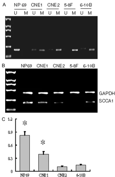
Figure 1. SCCA1 promoter methylation and SCCA1m-RNA expression in NPC cell lines. A. Typical MSP re-sults. The left lane was loaded with DL2000 marker,
“U” lanes were loaded with PCR products amplified by primer specific for unmethylated SCCA1 promoter,
and “M” lanes were loaded with PCR products
Western blot analysis
Cells were collected and lysed in modified RIPA lysis buffer (50 mmol/LTris-HCl pH8.0, 150 mmol/L NaCl, 0.5% sodium deoxycholate, 1 mmol/LEDTA, 1% NP-40, 0.1 mg/L PMSF, 2 mg/L bright endostatin) on ice for 30 min. The supernatants were collected after centrifuga-tion at 12,000 r/min for 30 min at 4°C. The pro-tein concentration of the supernatants was measured, and then total cell proteins were separated using 10% SDS-polyacrylamide gel electrophoresis and transferred to PVDF
mem-6-10B) and immortalized human cells NP69 was detected by MSP. The results showed that SCCA1 promoter was fully methylated in 5-8F cells, partly methylated in other three NPC cells, but not methylated in NP69 cells (Figure 1A).
[image:3.612.97.374.74.293.2]Next, SCCA1 mRNA expression levels in these cells were detected by RT-PCR. The results showed that SCCA1 mRNA expression level was un-detected in 5-8F cells, and was signifi-cantly lower in three NPC cell lines (CNEl, CNE2 and 6-10B) than in NP69 cells (Figure 1B, 1C).
Figure 2. SCCA1 promoter methylation and SCCA1mRNA expression in NPC cell lines after 5-aza-2dC treatment. A. Typical MSP results in each cell line. The left lane was loaded with DL2000 marker, “U” lanes were loaded with
PCR products amplified by primer specific for unmethylated SCCA1 promoter, and “M” lanes were loaded with PCR products amplified by primer specific
for methylated SCCA1 promoter. With increased concentration of 5-aza-2dC, less SCCA1 promoter methylation was observed. B. Typical RT-PCR results. The left lane was loaded with DL2000 marker. With increased concentration of 5-aza-2dC, higher SCCA1 mRNA expression was observed.
branes (Millipore). The mem-branes were blocked with 5% nonfat milk at room tempera-ture for 2 h, incubated with primary antibody for SCCA1 or β-actin (Abcam) at 4°C over-night, and then incubated with horseradish peroxidase-labeled goat anti-rabbit IgG antibody (Santa Cruz Biotech) at room temperature for 1 h. The membranes were washed and incubated with sub-strates using enhanced che-miluminescence kit (Amers- han Biosciences), then expo- sed to X-ray film and the imag-es were analyzed by plus 5.1 software.
Statistical analysis
The data were presented as mean ± standard deviation (_x
± sd). Data from two samples were compared using t test and data from multiple sam-ples were compared using single factor analysis of vari-ance. P<0.05 was considered statistically significant.
Results
SCCA1 promoter was meth-ylated and SCCA1 mRNA expression was decreased in NPC cells
Promoter methylation status of SCCA1 gene in four NPC cell lines (CNEl, CNE2, 5-8F, Table 1. 5-aza-2dC reversed SCCA1 promoter methylation in NPC
cells
Cell line n 5-aza-2dC concentration
0 µmol/L 0.1 µmol/L 1 µmol/L 5 µmol/L 10 µmol/L
CNE1 3 81±7 67±6* 52±7*,∆ 10±3*,∆,▲ 0±0
CNE2 3 92±4 86±6 53±9*,∆ 42±5*,∆,▲ 7±2*,∆,▲
5-8 F 3 100±0 95±2* 51±8*,∆ 31±4*,∆,▲ 6±2*,∆,▲,□
6-10B 3 90±5 76±7* 21±7*,∆ 18±5*,∆ 8±3*,∆,▲,□
*Compared with 0 µmol/L 5-aza-2dC group, P<0.05; ∆compared with 0.1 µmol/L
[image:3.612.91.378.440.520.2]5-aza-2dC reversed SCCA1 promoter methyla-tion and increased SCCA1 mRNA expression level in NPC cells
5-aza-2dC is a known DNA demethylation agent. Thus we treated NPC cells with differe- nt concentrations (0.1 μmol/L, 1 μmol/L, 5 μmol/L, 10 μmol/L) of 5-aza-2dC. MSP analysis showed that 5-aza-2dC reversed SCCA1 pro-moter methylation in four NPC cell lines in a dose-dependent manner (Figure 2A; Table 1). RT-PCR analysis showed that 5-aza-2dC increased SCCA1 mRNA expression level in four NPC cell lines in a dose-dependent man-ner (Figure 2B; Table 2).
5-aza-2dC upregulated SCCA1 protein expres-sion in NPC cells
To confirm that 5-aza-2dC could upregulate SCCA1 expression in NPC cells, we detected
SCCA1 protein levels in four NPC cell lines treat-ed with 5 μmol/L 5-aza-2dC or untreattreat-ed. Western blot analysis showed that after 5-aza 2dC treatment, SCCA1 protein expression lev-els were significantly higher in all four NPC cell lines (Figure 3).
Discussion
SCCA1 is mainly expressed in squamous epithelium, and its expression is decreased in a variety of squamous cell carcinoma [8]. In addition, SCCA1 expression level was correlat-ed with the differentiation of squamous cell carcinoma [10, 11]. SCCA1 has been proposed to be useful for assessing the prognosis of patients and predicting tumor recurrence or tumor progression [12].
In our previous study we found that SCCA1 expression level was lower in NPC tissue than in non-neoplastic nasopharyngeal epithelial membrane [9]. To investigate the mechanism underlying SCCA1 downregulation in NPC, we focused on methylation status of SCCA1 pro-moter and analyzed the relationship between SCCA1 promoter methylation and SCCA1 ex- pression levels. Our results showed that SCCA1 promoter was methylated in NPC cells and this was correlated with decreased SCCA1 expres-sion levels. To further analyze the causal rela-tionship between SCCA1 promoter methylation and SCCA1 expression levels in NPC cells. We treated NPC cells with 5-aza-2dC and found that demethylation of SCCA1 promoter by 5-aza-2dC led to increased SCCA1 mRNA and protein expression levels. These results con-firm that low expression of SCCA1 in NPC cells is caused by promoter methylation of SCCA1. A recent study reported that SCCA1 methyla-tion was correlated with lymph node metasta-sis of hepatocellular carcinoma [13]. In this study we found that SCCA1 promoter methyla-Table 2. 5-aza-2-dC increased SCCA1 mRNA expression in NPC cells
Cell line n 5-aza-2dC concentration
0 µmol/L 0.1 µmol/L 1 µmol/L 5 µmol/L 10 µmol/L
CNE1 3 0.35±0.08 0.41±0.10 0.59±0.12*,∆ 0.83±0.14*,∆,▲ 0.98±0.13*,∆,▲
CNE2 3 0.24±0.06 0.26±0.10 0.38±0.13* 0.48±0.15*,∆ 0.81±0.13*,∆,▲,□
5-8 F 3 0±0 0.13±0.04* 0.24±0.04*,∆ 0.31±0.05*,∆,▲ 0.76±0.11*,∆,▲,□
6-10B 3 0.12±0.04 0.16±0.07 0.31±0.09*,∆ 0.56±0.10*,∆,▲ 0.61±0.12*,∆,▲
*Compared with 0 µmol/L 5-aza-2dC group, P<0.05; ∆compared with 0.1 µmol/L 5-aza-2dC group, P<0.05; ▲compared with 1
[image:4.612.90.524.88.165.2]µmol/L 5-aza-2dC group, P<0.05; □compared with 5 µmol/L 5-aza-2dC group, P<0.05.
Figure 3. 5-aza-2dC treatment increased SCCA1 protein expression in NPC cell lines. A. Typical West-ern blot analysis in each NPC cell line treated with 5 µmol/L 5-aza-2dC or untreated. β-actin was loading
[image:4.612.92.287.208.387.2]tion was the highest in highly metastatic cell line 5-8F among all NPC cell lines. Our data are consistent with previous study and suggest that SCCA1 may inhibit NPC lymph node metas-tasis while SCCA1 promoter methylation may lead to the downregulation of SCCA1 and con-sequent lymph node metastasis.
In conclusion, our results suggest that promot-er methylation is one important cause for the downregulation of SCCA1 in NPC. Demethylation agents may help reverse SCCA1 promoter methylation and inhibit NPC progression and metastasis.
Acknowledgements
This study was supported by Hunan Province Natural Science Foundation (No. 11JJ3110). Disclosure of conflict of interest
None.
Address correspondence to: Rui-Cheng Hu, Hunan Province Institute of Gerontology, Hunan Province People Hospital, Changsha 410016, China; De- partment of Respiratory Medicine, Hunan Province People Hospital, Changsha 410016, China. Tel: 86-13723886638; E-mail: 770107828@qq.com
References
[1] Wei WI, Sham JS. Nasopharyngeal carcinoma. Lancet 2005; 365: 2041-2054.
[2] Cheung lW, Ching YP, Nicholls JM, Ling MT, Wong YC, Hui N, Cheung A, Tsao SW, Wang Q, Yeun PW, Lo KW, Jin DY, Wang X. Epigenetic inactivation of CHFR in nasopharyngeal carci-noma through promoter methylation. Mol Car-cinog 2005; 3: 237-245.
[3] Tsao SW, Liu Y, Wang X, Yuen PW, Leung SY, Yuen ST, Pan J, Nicholls JM, Cheung AL, Wong YC. The association of E-cadherin expression and the methylation status of the E-eadherin gene in nasopharyngeal carcinoma cells. Eur J Cancer 2003; 39: 524-531.
[4] Tsai CN, Tsai CL, Tse KP, Chang HY, Chang YS. The Epstein-Barr virus oncogene product la-tent membrane protein l induces the down-
regμlation of E-cadherin gene expression via
activation of DNA methyltransferases. Proc Natl Acad Sci U S A 2002; 6199: 10084-10089.
[5] Sun D, Zhang Z, Van DN, Huang G, Ernberg I, Hu L. Aberrant methylation of CDHl 3 gene in nasopharyngeal carcinoma could serve as a potential diagnostic biomarker. Oral Cancer 2006; 43: 82-87.
[6] Ying J, Li H, Seng TJ, Langford C, Srivastava G, Tsao SW, Putti T, Murray P, Chan AT, Tao Q.
Functional epigenetics identifies a protocad -herin PCDHl0 as a candidate tumor suppres-sor for nasopharyngeal, esophageal and mul-tiple other carcinomas with frequent meth- ylation. Oncogene 2006; 25: 1070-1080. [7] Vidalino L, Doria A, Quarta S, Zen M, Gatta A,
Pontisso P. SERPINB3, apoptosis and autoim-munity. Autoimmun Rev 2009; 9: 108-112. [8] Kugimoto T, Morita K, Omura K. Development
of oral cancer screening test by detection of squamous cell carcinoma among exfoliated oral mucosal cells. Oral Oncol 2012; 48: 794-798.
[9] Cheng AL, Huang WG, Chen ZC, Peng F, Zhang PF, Li MY, Li F, Li JL, Li C, Yi H, Yi B, Xiao ZQ.
Identification of novel nasopharyngeal carci -noma biomarkers by laser capture microdis-section and proteomic analysis. Clin Cancer Res 2008; 14: 435-45.
[10] Xu Y, Cao LQ, Jin LY, Chen ZC, Zeng GQ, Tang CE, Li GQ, Duan CJ, Peng F, Xiao ZQ, Li C. Quan-titative proteomic study of human lung squa-mous carcinoma and normal bronchial epithe-lial acquired by laser capture microdissection. J Biomed Biotechnol 2012; 2012: 510418. [11] Qi Y, Chiu JF, Wang L, Kwong DL, He QY.
Com-parative proteomic analysis of esophageal squamous cell carcinoma. Proteomics 2005; 5: 2960-2971.
[12] Roijer E, de Brμijn HW, Dahlen U, ten Hoor K,
Lundin M, Nilsson K, Soderstrom K, Nilsson O. Squamous cell carcinoma antigen is forms in serum from cervical cancer patients. Tumor Biol 2006; 27: 142-152.