Original Article
Tumor suppressor BLU exerts growth inhibition by
blocking ERK signaling and disrupting cell cycle
progression through RAS pathway interference
Xiangning Zhang1*, Song-Jun Shao3*, Jia-Hui Zhou1,5, Xiao-Wu Li4, Biying Zheng2, Zunnan Huang1, Zhiwei He1
1Department of Pathophysiology, Chinese American Collaborative Cancer Research Institute, Guangdong Provincial Key Laboratory of Molecular Diagnostics, Guangdong Medical University, Dongguan, Guangdong, People’s Republic of China; 2Department of Microbiology, Guangdong Medical University, Dongguan, Guangdong, People’s Republic of China; 3Departments of Respiratory and Critical Medicine, Guizhou Provincial People’s Hospital and Guizhou Medical University, Guiyang, Guizhou, People’s Republic of China; 4Department of General Surgery, Guangdong Provincial People’s Hospital, Southern Medical University, Guangzhou, Guangdong, People’s Republic of China; 5Department of Pathology, Lishui Manicipal Central Hospital, Lishui, Zhejiang, People’s Republic of China. *Equal contributors.
Received November 30, 2017; Accepted December 22, 2017; Epub January 1, 2018; Published January 15, 2018
Abstract: We have previously reported that the 3p21 tumor suppressor BLU regulates cell cycle by blocking JNK/ MAPK signaling. Another member of the MAPK family, extracellular signal response kinase (ERK), is induced by the RAS-RAF-MEK-ERK pathway and is targeted in anticancer therapy. The effects of BLU on tumor growth were evaluated by measuring the size of nasopharyngeal carcinoma (NPC) xenografted tumors intra-tumorally injected with BLU adenovirus 5 (BLU Ad5) and the viability of NPC cells transferred with BLU. Tumor size was correlated with downregulation of the ERK pathway by BLU. Phosphorylation of ERK and Elk reporter activities were assayed. The regulated cyclins D1 and B1 were measured by CCND1 and CCNB1 gene promoter activity by co-transfection of BLU, RAS V12G, together with BLU+RAS V12G, pCD316+RAS V12G. The cell cycle phase distribution was determined by FACS-based DNA content assay. The data showed that growth of the xenografted tumor was inhibited and viability of HONE-1 cells was reduced by recombinant BLU. BLU down-regulated ERK signaling by reducing protein substrate phosphorylation, inhibiting Elk reporter activity, and blocking promoter activities of the CCND1 gene and reduced cyclins D1 expression to arrest the cell cycle at the G1 phase. The population of G2/M cells was also remarkably decreased. HRAS V12G activated ERK and cyclin D1 and B1 promoters, and the effects were antagonized by BLU. Taken together, our results suggested that BLU inhibited ERK signaling, downregulated cyclins D1 and B1, and pre-vented cell cycle progression through interfering with HRAS V12G signaling to exert tumor suppression.
Keywords:BLU/ZMYND10, H-RAS, MAPK/ERK, cyclin D1, cyclin B1, cell cycle
Introduction
Carcinogenesis is a multistep, multifactorial process involving anomalies at the cytogenetic and molecular levels, including a loss of homo-zygosity (LOH) on chromosomal regions, gene amplification, and hypermethylation at the pro -moters of tumor suppressor genes (TSGs) [1]. BLU is a candidate TSG mapped to a frequently lost chromosomal region, 3p21. Its inactivation has been seen in a variety of tumors of epithe-lial origin, including breast, lung, cervical, and nasopharyngeal cancers [2, 3]. In nasopharyn-geal carcinoma (NPC), BLU expression is
N-terminal kinase (JNK), which is a member of the mitogen-activated protein kinase (MAPK) family that integrates signals of growth, prolif-eration, and apoptosis, and inactivates the pro-moter of the cyclin D1 coding gene CCND1, and dampens expression of cyclin D1 to arrest the cell cycle at the G1 phase [11].
MAPK family proteins are activated by epider-mal growth factor receptor (EGFR), the most frequently employed target of anticancer thera-py, through activation of RAS proteins which transmit signals of receptor occupied growth factors, cytokines, and other stimuli [12]. Mutation-activated RAS proteins are implicated in malignant transformation [13, 14] and are modulated by interacting with a group of pro-teins that display a conserved domain to bind the small molecule guanidine nucleotide asso-ciating protein. Members of this family com-prise RASSF1 and RASSF5/NORE1, which neg-atively regulate the activity of RAS proteins [15, 16]. Activated RAS proteins induce a kinase cascade involving kinases RAF and an MAPK family protein, extracellular signal-regulated kinase (ERK) [17, 18].
In the present study, we tested whether tumor suppression by BLU is exerted by targeting the RAF-MEK-ERK pathway, through interfering with the RAS oncogene with a high frequency of mutational activation in human cancers.
Materials and methods
Cells, plasmids and packaging of viral vector of BLU
HONE1 cells derived from a Chinese patient with undifferentiated NPC [19, 20] were main-tained in RPMI-1640 that was supplemented with 10% fetal calf serum (FCS) (Gibco Biotech nology, Guangzhou, China) and 1% penicillin+ streptomycin (PEST) antibiotics. SKOV3 cells were derived from a serious ovarian cancer patient, and purchased from Cell Bank, Institute of Life Science, Chinese Academy of Science (CAS) Shanghai Branch. BLU pCD316 was constructed by inserting the cDNA of BLU/ ZMYND10 into the cytomegalovirus-derived shuttle plasmid pCD316 as described previ-ously [8, 11]. The recombinant shuttle plasmid was co-transfected to packaging 293 cells with at least two viral structural protein coding genes to generate recombinant BLU
adenovi-rus type 5 (BLU Ad5) which express BLU on infection of target cells. The purified cell lysate was used in infection to transfer BLU. The expression vector/plasmid of H-RAS V12G carrying a point mutation on codon 12 of H-RAS and the empty vector pBABE were a kind gift from Professor Bob Weinberg (Massachusetts Institute of Technology, Boston, MA, USA) [21] and Dr. Elena Kashuba (Karolinska Institutet, Stockholm, Sweden) [22]. The ERK reporter plasmids pFA+pFR with Elk were purchased from Strategene (Guangzhou, China) [23]. Reporter plasmids for the promoters of the CCND1 and CCNB1 genes were kind gifts from Professors Richard Pestell (Temple University, Philadelphia, PA, USA) [24] and Kurt England (University of Leipzig, Leipzig, Germany) [25, 26].
Antibodies
The murine anti-human cyclin B1 mAb, GNS3 (8A5D12; Millipore, Guangzhou, China); rabbit anti-human cyclin D1 polyclonal antibody (Cell Signaling Technology, Guangzhou, China); goat anti-human BLU/ZYND10 polyclonal (Abcam, Cambridge, U.K.); Anti-p44/42 MAPK/ERK1/2 Rabbit mAb (#4695), and anti-Phospho-p44/42 ERK1/2 (Thr202/Tyr204) Rabbit mAb (#4370) purchased from Cell Signaling Technology were used in probing the proteins transferred to nitrocellulose filter. The reaction of primary antibodies binding was developed with ultra-red labelled Odyssey antibodies against the immunoglobulin (Ig) of the primary antibodies (Li-COR Biosciences).
Cell viability assay
Up to 1,000 cells were seeded into each well of a 96 well tissue culture plate. After overnight (ON) incubation cells were challenged with BLU Ad5 at doses of 0, 5, 10, 20, and 40 PFU/cell in triplicate for 24 h. The cells were then washed twice with sterile PBS and incubated with the Cell Counting Kit-8 (CCK-8) reagent (Dojindo, Guangzhou, China). Absorbance values were read on a BioTek ELISA plate reader.
Western blotting
min, and stored at -20°C until use. Total pro-teins were separated by 10% SDS-polyacryla- mide gel electrophoresis (PAGE), and electrob-lotted. After blocking with 5% skim milk in PBS for 1 h at RT or overnight at 4°C, the filters were
[image:3.612.91.526.75.518.2]probed with specific primary antibodies. Mem-branes were washed with 0.1% Tween 20 in PBS. The reaction was developed with ultra-red labelled Odyssey antibodies against the Ig of primary antibodies.
Figure 1. Re-expression of BLU inhibited in vitro and in vivo growth of HONE1 cells. HONE-1 cells were infected with
BLU Ad5 and viewed under a fluorescence microscope to screen for tagged EGFP (A); Total proteins of the infected
cells (left panel) and from the xenografted tumors (right panel) were tested with immunoblotting, probing with
specific antibodies for the presence of BLU (B). The cells were challenged with BLU Ad5 at the indicated doses, an -dviability was evaluated. Data are presented as the mean ± SD derived from at least three independent analyses; and the asterisks (*) denoted P<0.05 (C). After injected intra-tumorally with BLU Ad5, the xenografted tumor grew for two weeks before harvest, and BLU Ad5 injected (Upper) and control tumors (Lower) were compared (D). The tumors of control (E, left panel) and injected with BLU Ad5 (E, right panel) were incised and sectioned, stained with
Transfection
Nearly confluent HONE-1 and SKOV3 cells were seeded in 6- or 12-well plates. After ON incuba-tion, plasmids BLU or HRAS V12G and their corresponding empty vectors (pCD316 and pBABE) were co-transfected with the plasmids pFR+pFA2, the Elk1 reporter, or the reporters of the CCNB1, CCNB2 and CCND1 promoters with the internal control by mixing with FuGene HD (Roche, Shanghai, China).
Flow cytometry
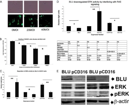
[image:4.612.94.518.74.418.2]After synchronizing, HONE-1 cells were trans-fected with BLU pCD316, the pCD316 vector, RAS V12G pBABE, and pBABE for 24 or 48 h, as described above. Harvested cells were fixed in 80% ethanol overnight at -20°C. Then resus-pended in 100 μg/ml propidium iodide (PI; Sigma Aldrich). Flow cytometry was carried out on a BD FACScan (San Jose, CA, USA), with data analyzed using Cycle 32 software (Phoenix Figure 2. Expression of BLU inhibited ERK signaling in HONE-1, and the ovarian cell line SKOV3 cells. The presence of BLU in the infected SKOV3 cells was confirmed by tagged EGFP signals (A). The viability of SKOV3 cells infected
with recombinant BLU Ad5 virus at indicated doses was assayed with CCK8 kit. Data are represented as the mean ± SD derived from at least three independent tests, and the asterisks(*) denoted P<0.05 (B). The BLU, mock trans-fected HONE-1 and SKOV3 cells were co-transtrans-fected with the reporter plasmids pFA2+pFR. Also with pCD316+RAS V12G, BLU+RAS V12G, and pFA2+pFR to test the activity of ERK reporter (C). Data are presented as the mean ± SD that were derived from at least three independent tests, and the asterisks(*) denoted P<0.05. Total ERK (middle, upper panel) and phosphorylated ERK (middle, lower panel) of BLU Ad5 and mock infected HONE-1 and SKOV3
cells were tested with specific antibodies (D). Presence of BLU was confirmed simultaneously by probing with spe
-cific anti-BLU antibody (Upper panel). The expression of BLU by HONE1 (left panel) and SKOV3 (right panel) cells on adenoviral infection was confirmed by immunoblotting, probing with specific anti-BLU antibody the samples of
were indicated; the proteins obtained from BLU Ad5 infected and mock cells as indicated were also tested for phos-phorylation of ERK by probing with antibodies against total and phosphorylated ERK. The loading was calibrated by
Flow Systems, Inc., San Diego, CA, USA). Cell cycle distribution was then assessed.
Xenografted tumors in nude mice
The protocols of animal experiments were app- roved by the Ethics Committee of Guangdong
Re-expression of the tumor suppressor BLU inhibits in vitro and in vivo growth of HONE1 cells and viability of SKOV3 cells
BLU was re-expressed by transfer with vector BLU Ad5 in two lines negative for BLU: NPC-derived HONE-1 cells, and ovarian cancer-Figure 3. The activities of CCND1 and CCNB1 genes were inhibited and
in-duced by BLU and RAS V12G. HONE-1 cells were co-transfected with BLU,
pCD316, RAS V12G, pBABE, together with CCND1 (A) and CCNB1 (hB1) (B and C) promoter reporter plasmids. pCD316+RAS and BLU+RAS V12G were also included. The data in relative light units is presented as the mean ± SD that is derived from at least three independent experiments. The symbols *indicated P<0.05, and **indicated P<0.01 on when comparing values from two adjacent columns. Levels of cyclins D1 and B1 were measured
with Western blotting (D and E). The blots were probed with specific
anti-cyclins D1 and B1 antibodies (upper panel; left: BLU, right: pCD316) and anti-b-actin mAb (lower panel).
Medical University Affiliated Hospital (Permit no. 2015-051KT). BALB/c nude mice (4-6 weeks old) of both gen-ders were used. Up to 2×10⁶ HONE-1 cells were re-sus-pended in 0.2 ml serum-free RPMI, and injected subcuta-neously into mice. Palpable tumors were identified two weeks thereafter. For treat-ment, up to 3.5×10⁶ PFU BLU Ad5 was intra-tumorally inject-ed. Tumors were allowed to grow for an additional two weeks before harvesting. Be- fore the injection and during this period, the longest and shortest tumor diameters we- re measured with a Vernier caliper. Tumor volume was derived as v = ab2/2 (where a and b are the longest and shortest diameters, respec-tively). At the time of harvest, the tumors were weighed. The means ± standard deviation (SD) of tumor sizes was calcu-lated to establish growth cur- ves in the treatment and con-trol groups.
Statistical analysis
Statistical analysis was per-formed using the SSSP soft-ware package. Quantitative data were presented as mean ± SD from at least three in- dependent experiments. The Student’s T-test was used for group comparisons and a val- ue of P<0.05 was considered statistically significant.
[image:5.612.90.374.69.505.2]derived SKOV3 cells. The expression was evi-denced by the fluorescence of the tagged EGFP signal viewed microscopically (Figures 1A and
2A) and immunoblotting, probed with specific antibody (Figures 1D and 2B). Transferred BLU
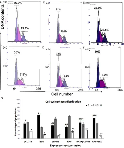
[image:6.612.95.522.71.580.2]Ad5 by viral infection reduced viability of HONE-1 cells in a dose-dependent manner (Figure 1B); the finding was also noted in ovarian can -cer-derived SKOV3 cells (Figure 2C). When HONE-1 cells were xenografted in nude mice by Figure 4.Alteration of cell cycle phase distribution by BLU. Cells were transfected with 1 μg of the plasmids pCD316
(A), BLU (B), pBABE (C), RAS V12G (D), PCD316+RAS V12G (E) and BLU+RAS V12G (F) and DNA content was flow
subcutaneously injection, palpable lesions of tumor were identified, and the radius of the tumors were recorded and size was calculated according to an established formula. The BLU Ad5 suspension and medium control were then intra-tumorally injected to the tumors. During the period of observation, the size of the xeno-grafted tumors was calculated as above. After two weeks of growth, the tumors were harvest-ed by excision, The incorporation of the human NPC cells and expression of BLU were con-firmed hisopathologcally and immunoblotting respectively (Figure 1C and 1D); the size of the BLU transferred and control tumors was com-pared (Figure 1E) and the growth curve was plotted according to the recorded data (Figure 1F). The inhibition of growth by BLU was suggested.
BLU down-regulated ERK signaling when com-pared to RAS V12G, and phosphorylation of ERK in HONE1 and SKOV3 cells
The RAS-RAF-MEK-ERK pathway is activated in a cascade manner when integrating extracellu-lar stimuli, including ligation of growth factors with cognate receptors, to stimulate cell growth
gest that BLU inhibited ERK signaling through interfering with RAS activity.
BLU inhibited the promoter activity of CCND1 and CCNB1 and reduced expression levels of cyclins D and B1 in HONE1 cells
[image:7.612.91.372.75.284.2]Cyclins are activated in a chain of phosphoryla-tion and dephosphorylaphosphoryla-tion and the phosphory-lation of several amino residues is catalyzed by ERK and other MAPK family kinases. Activated cyclins serve as regulatory subunits of the cyclin-cyclin dependent kinase (CDK) complex [27, for a review see 28]; groups D and B cyclins function to promote transition of G1/S and G2/M phases in cell cycle respectively. The reporter plasmid for the promoters of cyclin D1 and cyclin B1 coding genes CCND1, and CCNB1 were co-transfected with BLU, HRAS V12G vec-tors, and their corresponding empty vectors. Since cyclin B1 is also induced by activation of the RAF-MEK-ERK pathway, CCNB1 promoter activity and expression of cyclin B1 were mea-sured by the luciferase assay and Western blot-ting. The data showed that activity of CCNB1 and CCND1 promoters was inhibited by BLU, and elevated by HRAS V12G (Figure 3A-C). The Figure 5. Scheme of engagement of ERK pathway by BLU. The tumor
sup-pressor BLU was re-expressed by viral transduction or gene transfection. The expression of BLU inhibited the phosphorylation of ERK that was catalyzed by an upstream kinase(s), which opposed the effect of RAS proteins. Next, the activity of the CCND1 and CCNB1 gene promoters was down-regulated, leading to reduced expression of cyclins D1 and B1, and induction of G1 cell cycle arrest. It remains to be elucidated whether reduced proportions of cells in the G2/M phase of the cell cycle was related to the change of cyclin B1 level.
and proliferation. The signal-ing of ERK is regulated by oncoproteins and coding prod-ucts of TSGs. In SKOV3 and HONE-1 cells, ERK signaling was downregulated by BLU, but induced by mutant RAS as demonstrated by co-transfec-tion with BLU pCD316 and RAS V12G plasmids, their cor-responding empty vectors, together with reporter plas-mids pFA2 and pFR (Figure 2B
sug-level of cyclins D1 and B1 was reduced in BLU expressing cells, correlating with ERK inhibition by BLU (Figure 3D and 3E). These findings sug -gested that BLU exerts proliferation inhibition and tumor suppression by inhibition of cyclin D1 and B1 gene promoter, i.e. transcriptional blocking of cyclin B1 and cyclin D1 expression and thus prevented cell cycle progression. The assay of reporter activity of CCND1 sug-gests that BLU was antagonistic with the induc-tion of CCND1 promoter by HRAS through RAS-RAF-MEK-ERK.
The expression arrested cell cycle of HONE1 at G1 phase and correlated with its effect on cyclin D1 expression
The progression of cell cycle is regulated by complexes of cyclin and CDK. BLU has been shown to downregulate cyclin D1 and cyclin B1. The arrest of cell cycle at the G1 phase was well-correlated with alterations of cyclin D1 caused by BLU (Figure 4A and 4B). In line with the published finding that HRAS V12G induced G2/M arrest, we found that the G2/M popula-tion of HONE-1 cells that had been transfected with H-RAS was remarkably increased in com-parison with the mock (Figure 4C and 4D) con-trol. In BLU expressing cells, the G2/M popula-tion was decreased as revealed by DNA con-tent. It might be reasoned that the change in DNA content might result from an accumulation of cells in the G1 phase of the cell cycle due in part to cell cycle arrest, as previously reported [29]. There is, however, the possibility that entry of cells from the S to the G2/M phase of the cell cycle was prevented because cyclin B1 expression was blocked by BLU (Figure 3B). In a co-transfection system with BLU+HRAS V12G, G1 arrest was registered, and the G2/M cell population was reduced by BLU when compared with cells expressing pCD316+RAS V12G (Figure 4E and 4F). The data further sug-gested that BLU could interfere with the signal-ing of the mutant HRAS (Figure 4G).
Our results suggest that the tumor suppressor BLU inhibits tumor growth by blocking ERK sig-naling and downstream expression of effectors like cyclin D1. The changes then led to arrest of the cell cycle in the G1 phase. Moreover, down-regulation of the ERK pathway also reduced cyclin B1 expression. BLU also reduced the population of cells in the G2/M phase of the cycle as compared with induction of G2/M
arrest by HRAS V12G. ERK targeting growth and tumor suppression by BLU is summarized in Figure 5.
Discussion
BLU was mapped to the minimal region of dif-ferentially deleted fragments of varying sizes on chromosome 3p21. It is frequently lost in a variety of human tumors, notably of epithelial origin [2, 3, 30]. Its coding product is a protein with a zinc finger myeloid Nervy DEAF-1 (ZMYND) motif on its carboxylterminus, shared by members of a protein family. The activities of modified histone reader, transcription repres -sion have been described in other members [9, 10].
BLU has been reported to inhibit cell cycle pro-gression by engaging the RAF-MEK-JNK signal-ing pathway, similar to the tumor suppressor and the RAS effector RASSF1A [15] and inhibits angiogenesis by reducing VEGF levels and other cytokines [31], and enhances chemother-apeutic agent-induced apoptosis [32, 33]. Our observation suggested that BLU inhibited NF- κB signaling, down-regulated NF-κB dependent anti-apoptotic factors like cFLIP and cIAP2 and potentiated death receptor-induced apoptosis [8]. Its effect on NF-κB signaling could be explained by BLU-mediated post-translational modifications (PTMs) of IKKα, a kinase that phosphorylates IkappaB whose dissociation from NF-κB leading to its activation.
targeted to the RAF-MEK1/2-JNK pathway [11]. We reasoned whether BLU also targeted path-ways that involved other members of the MAPK family to exert tumor suppression. We thus compared regulation of ERK activated by, pro-to-oncogenes RAS and its downstream effec-tors cyclins D1 and B1 by HRAS V12G and BLU, and tested their interaction in a co-transfection system.
In the present study, it was shown that in HONE1 and SKOV3 cells, BLU inhibited signal-ing of ERK. In the context, BLU behaved simi-larly to a family of proteins that include RASSF. RASSF1A, the founding member of the family and RASSF5/NORE which inhibit tumor growth and proliferation by targeting signaling mole-cules of the MAPK family [9, 10]. MAPK pro- teins promote G1/S cell cycle phase transition by inducing cyclin D1, whose over-expression contributes to neoplastic transformation [36, 37]. RASSF5/NORE also targets cyclin B1 which promotes G2/M cell cycle progression by complexing with the CDK1 complex [38, 39]. Cyclin B1 plays an indispensable role in regulat-ing cell entry into mitosis [40]. Growth inhibition by suppressing the cyclin B1/CDK1 complex and upstream signaling pathway(s) involves networks include the EGFR/ERK pathway [41]. Similar with that BLU arrested cell cycle at the G1 phase in CNE-2 cells [11]; it also induced G1 arrest in HONE-1 cells (Figure 4A and 4B). The data supported that G1 arrest is the frequent target of tumor suppressors. However, we found that the G2/M phase in BLU expressing cells decreased. In line with previous reports, HRAS V12G induced G2/M arrest by activating the RAF-MEK-ERK pathway [42, 43]. While a decrease in the G2/M population might be explained by an increase in the G1 population, we observed that cyclin B1, which regulates entry into the G/M phase,was also reduced by BLU, and that G2/M arrest that is induced by RAS V12G was reversed by BLU (Figure 4E and 4F). We reasoned that BLU could prevent entry into G2/M by affecting cyclin B1 activity, a finding that warrants future in depth investi-gations.
Effects of BLU on the ERK-cyclin D1 axis may account for its in vitro and in vivo growth and proliferation inhibition. In the present study, BLU inhibited the human NPC xenograft in nude mice upon intra-tumoral injection, and prevent-ed tumor cell growth in vitro by inducing G1 cell cycle arrest. These effects correlated with
down-regulation of ERK signaling, which were activated by HRAS V12G. Recently, it was reported that silencing of cyclin B by microRNA 15a/16-1, inhibits tumor growth [44, 43]. Toge- ther with the finding of inhibition of cell cycle progression by cyclin B1 by BLU, it is proposed that cyclin B1 might serve as an anti-cancer therapeutic target.
Our data suggested that xenografted tumor in- hibition by BLU was incomplete, prompting fur-ther efforts to test whefur-ther concurrent adminis-tration of an additional tumor suppressive factor would completely eliminatethe tumors. Indeed it was reported that adenoviral transfer of IL-24 and TRAIL, leads to complete eradica-tion of xenografted human hepatoma in mice [46]. Our recent publication suggested that BLU enhanced TRAIL-induced apoptosis [8]. The efficacy of concurrent administration of BLU and a pro-apoptotic factor in anti-tumor biotherapy could be evaluated in the in vivo sys-tem described above.
In conclusion, a tumor suppressor BLU, down-regulated ERK signaling and its downstream effector cyclins D1 and B1, which disrupted cell cycle progression and subsequent suppression of tumor growth. The process might be achieved by interfering with RAS-mediated signaling.
Acknowledgements
The work is supported by research grants from the National Natural Science Foundation of China (Grant Numbers: 81372137 to ZWH and 31770774 to ZNH) and the Provincial Medical Research Fund of Guangdong (2014A267 and 2017113104128629 to XZ). We would like to thank Drs. Qian Tao, Chinese University of Hong Kong, Hong Kong; Bob Weinberg, MIT, Boston, Massachusetts; Elena Kashuba, Karolinska Institutet, Stockholm, Sweden; Kurt Engeland, University of Leipzig, Leipzig, Germany and Richard G, Pestell, Thomas Jefferson University, Philadelphia, Pennsylvania for their kind gifts of the precious reagents. We would also like to thank Dr. Michael Lerman, Stamford, Conecticut for his critical reading of our manuscript.
Disclosure of conflict of interest
None.
Key Laboratory of Molecular Diagnostics, Guang- dong Medical University, 1 Xincheng Avenue,
Songshan Lake Scientific and Industrial Park,
Dongguan 523808, Guangdong, China. Tel: +86 769 2289 6405; Fax: +86769 2289 6100; E-mail: zhangxn_2006@126.com (XNZ); zn_huang@yahoo. com (ZNH); zhiweihe688@yahoo.com (ZWH)
References
[1] Hanahan D, Weinberg RA. Hallmarks of can-cer: the next generation. Cell 2011; 144: 646-74.
[2] Zabarovsky ER, Lerman MI, Minna JD. Tumor suppressor genes on chromosome 3p involved in the pathogenesis of lung and other cancers. Oncogene 2002; 21: 6915-35.
[3] Hesson LB, Cooper WN, Latif F. Evaluation of the 3p21.3 tumour-suppressor gene cluster. Oncogene 2007; 26: 7283-301.
[4] Qiu GH, Tan LK, Loh KS, Lim CY, Srivastava G, Tsai ST, Tsao SW, Tao Q. The candidate tumor suppressor gene BLU, located at the common-ly deleted region 3p21.3, is an E2F-regulated, stress-responsive gene and inactivated by both epigenetic and genetic mechanisms in nasopharyngeal carcinoma. Oncogene 2004; 23: 4793-806.
[5] Liu XQ, Chen HK, Zhang XS, Pan ZG, Li A, Feng QS, Long QX, Wang XZ, Zeng YX. Alterations of BLU, a candidate tumor suppressor gene on chromosome 3p21.3, in human nasopharyn-geal carcinoma. Int J Cancer 2003; 106: 60-5. [6] Guo R, Zheng L, Park JW, Lv R, Chen H, Jiao F,
Xu W, Mu S, Wen H, Qiu J, Wang Z, Yang P, Wu F, Hui J, Fu X, Shi X, Shi YG, Xing Y, Lan F, Shi Y. BS69/ZMYND11 reads and connects histone H3.3 lysine 36 trimethylation-decorated chro-matin to regulated pre-mRNA processing. Mol Cell 2014; 56: 298-310.
[7] Adhikary S, Sanyal S, Basu M, Sengupta I, Sen S, Srivastava DK, Roy S, Das C. Selective recognition of H3.1K36 dimethylation/H4K16 acetylation facilitates the regulation of all-trans-retinoic acid (ATRA)-responsive genes by putative chromatin reader ZMYND8. J Biol Chem 2016; 291: 2664-81.
[8] Zhou J, Huang Z, Wang Z, Liu S, Grandien A, Ernberg I, He Z, Zhang X. Tumor suppressor BLU promotes TRAIL-induced apoptosis by
downregulating NF-κB signaling in nasopha -ryngeal carcinoma. Oncotarget 2017; 8: 43853-43865.
[9] Whang YM, Kim YH, Kim JS and Yoo YD. RASSF1A suppresses the c-Jun-NH2-kinase pathway and inhibits cell cycle progression. Cancer Res 2005; 65: 3682-3690.
[10] Yoo YA, Na AR, Lee MS, Yoon S, Kim JS and Yoo YD. RASSF1A suppresses oncogenic
H-Ras-induced c-Jun N-terminal kinase activation. Int J Oncol 2006;29: 1541-7.
[11] Zhang X, Liu H, Li B, Huang P, Shao J, He Z. Tumor suppressor BLU inhibits proliferation of nasopharyngeal carcinoma cells by regulation of cell cycle, c-Jun N-terminal kinase and the cyclin D1 promoter. BMC Cancer 2012; 12: 267.
[12] Sahu RP, Zhang R, Batra S, Shi Y, Srivastava SK. Benzylisothiocyanate-mediated generation of reactive oxygen species causes cell cycle ar-rest and induces apoptosis via activation of MAPK in human pancreatic cancer cells. Carcinogenesis 2009; 30: 1744-1753.
[13] Macara IG, Lounsbury KM, Richards SA, McKiernan C, Bar-Sagi D. The Ras superfamily of GTPases. FASEB J 1996; 10: 625-630. [14] Karnoub AE, Weinberg RA. Ras oncogenes:
split personalities. Nat Rev Mol Cell Biol 2008; 9: 517-31.
[15] Agathanggelou A, Cooper WN, Latif F. Role of the Ras-association domain family 1 tumor suppressor gene in human cancers. Cancer Res 2005; 65: 3497-3508.
[16] Moshnikova A, Frye J, Shay JW, Minna JD, Khokhlatchev AV. The growth and tumor sup-pressor NORE1A is a cytoskeletal protein that suppresses growth by inhibition of the ERK pathway. J Biol Chem 2006; 281: 8143-52. [17] McCubrey JA, Steelman LS, Chappell WH,
Abrams SL, Wong EW, Chang F, Lehmann B, Terrian DM, Milella M, Tafuri A, Stivala F, Libra M, Basecke J, Evangelisti C, Martelli AM, Franklin RA. Roles of the Raf/MEK/ERK path-way in cell growth, malignant transformation and drug resistance. Biochim Biophys Acta 2007; 1773: 1263-84.
[18] Friday BB, Adjei AA. Advances in targeting the Ras/Raf/MEK/Erk mitogen-activated protein kinase cascade with MEK inhibitors for cancer therapy. Clin Cancer Res 2008; 14: 342-6. [19] Dittmer DP, Hilscher CJ, Gulley ML, Yang EV,
Chen M, Glaser R. Multiple pathways for Epstein-Barr virus episome loss from nasopha-ryngeal carcinoma. Int J Cancer 2008; 123: 2105-12.
[20] Li L, Zhang Y, Fan Y, Sun K, Su X, Du Z, Tsao SW, Loh TK, Sun H, Chan AT, Zeng YX, Chan WY, Chan FK, Tao Q. Characterization of the
naso-pharyngeal carcinoma methylome identifies
aberrant disruption of key signaling pathways and methylated tumor suppressor genes. Epigenomics 2015; 7: 155-73.
[21] Chipperfield RG, Jones SS, Lo KM, Weinberg
RA. Activation of Ha-ras p21 by substitution, deletion, and insertion mutations. Mol Cell Biol 1985; 5: 1809-13.
complementary helper-free packaging cell-line. Nucleic Acids Res 1990; 18: 3587-3596. [23] Park KS, Jeon SH, Kim SE, Bahk YY, Holmen
SL, Williams BO, Chung KC, Surh YJ, Choi KY. APC inhibits ERK pathway activation and cel-lular proliferation induced by RAS. J Cell Sci 2006; 119: 819-27.
[24] Albanese C, Johnson J, Watanabe G, Eklund N, Vu D, Arnold A, Pestell RG. Transforming p21ras mutants and c-Ets-2 activate the cyclin D1 promoter throughdistinguishable regions. J Biol Chem 1995; 270: 23589-23597.
[25] Müller GA, Quaas M, Schümann M, Krause E, Padi M, Fischer M, Litovchick L, DeCaprio JA, Engeland K. The CHR promoter element con-trols cell cycle-dependent gene transcription and binds the DREAM and MMB complexes. Nucleic Acids Res 2004; 20: 1561-1578. [26] Quaas M, Müller GA, Engeland K. p53 can
re-press transcription of cell cycle genes through a p21(WAF1/CIP1)-dependent switch from MMB to DREAM protein complex binding at CHR promoter elements. Cell Cycle 2012; 11: 4661-4672.
[27] Obaya AJ and Sedivy JM. Regulation of cyclin-Cdk activity in mammalian cells. Cell Mol Life Sci 2002; 59: 126-42.
[28] Vervoorts J and Lüscher B. Post-translational regulation of the tumor suppressor p27 KIP1. Cell Mol Life Sci 2008; 65: 3255-3264. [29] Tao CL, Lin H, Chen SQ. The regulation of ERK
and p-ERK expression by cisplatin and soraf- enib in gastric cancer cells. Gene 2014; 552: 106-115.
[30] Lerman MI, Minna JD. The 630-kb lung cancer homozygous deletion region on human
chro-mosome 3p21.3: identification and evaluation
of the resident candidate tumor suppressor genes. The international lung cancer chromo-some 3p21.3 tumor suppressor gene consor-tium. Cancer Res 2000; 60: 6116-33.
[31] Cheng Y, Ho RL, Chan KC, Kan R, Tung E, Lung HL, Yau WL, Cheung AK, Ko JM, Zhang ZF, Luo DZ, Feng ZB, Chen S, Guan XY, Kwong D, Stanbridge EJ, Lung ML. Anti-angiogenic path-way in association of the 3p21 mapped BLU gene in nasopharyngeal carcinoma. Oncogene 2015; 34: 4219-28.
[32] Park ST, Byun HJ, Kim BR, Dong SM, Park SH, Jang PR, Rho SB. Tumor suppressor BLU pro-motes paclitaxel antitumor activity by inducing apoptosis through the down-regulation of Bcl-2 expression in tumorigenesis. Biochem Biophys Res Commun 2013; 435: 153-159.
[33] Yoo HJ, Kim BR, Byun HJ, Park SY, Rho SB. BLU enhances the effects of anti-angiogenic activi-ty in combination with gemcitabine-based che-motherapeutic agents. Int J Biochem Cell Biol 2013; 45: 1236-45.
[34] Cho KR, Vogelstein B. Genetic alterations in the adenoma--carcinoma sequence. Cancer 1992; 70: 1727-31.
[35] Tam IY, Chung LP, Suen WS, Wang E, Wong MC, Ho KK, Lam WK, Chiu SW, Girard L, Minna JD, Gazdar AF, Wong MP. Distinct epidermal growth factor receptor and KRAS mutation patterns in non-small cell lung cancer patients with differ-ent tobacco exposure and clinicopathologic features. Clin Cancer Res 2006; 12: 1647-53. [36] Resnitzky D and Reed SI. Different roles for cy-clins D1 and E in regulation of the G1-to-S tran-sition. Mol Cell Biol 1995; 15: 3463-3469. [37] Casimiro MC, Crosariol M, Loro E, Li Z, Pestell
RG. Cyclins and cell cycle control in cancer and disease. Genes Cancer 2012; 3: 649-57. [38] MargolisSS, Perry JA, Weitzel DH, Freel CD,
Yoshida M, Haystead TA, Kornbluth S. A role for PP1 in the Cdc2/Cyclin B-mediated positive feedback activation of Cdc25. Mol Biol Cell 2006; 17: 1779-1789.
[39] Moon DO, Kim MO, Kang SH, Lee KJ, Heo MS, Choi KS, Choi YH, Kim GY. Induction of G2/M arrest, endoreduplication, and apoptosis by actin depolymerization agent pextenotoxin-2 in human leukemia cells, involving activation of ERK and JNK. Biochem Pharmacol 2008; 76: 312-321.
[40] Lindqvist A, van Zon W, Karlsson Rosenthal C, Wolthuis RM. Cyclin B1-Cdk1 activation contin-ues after centrosome separation to control mi-totic progression. PLoS Biol 2007; 5: e123. [41] Lim YC, Cha YY. Epigallocatech3-gallate
in-duces growth inhibition and apoptosis of hum- an anaplastic thyroid carcinoma cells through suppression of EGFR/ERK pathway and cyclin B1/CDK1 complex. J Surg Oncol 2011; 104: 776-80.
[42] Lowe SW, Cepero E and Evan G. Intrinsic tu-mour suppression. Nature 2004; 432: 307-315.
[43] Zhang Z, Miao L, Lv C, Sun H, Wei S, Wang B, Huang C and Jiao B. Wentilactone B induces G2/M phase arrest and apoptosis via the Ras/ Raf/MAPK signaling pathway in human hepa-toma SMMC-7721 cells. Cell Death Dis 2013; 4: e657.
[44] Crombez L, Morris MC, Dufort S, Aldrian-Herra- da G, Nguyen Q, McMaster G, Coll JL, Heitz F, Divita G. Targeting cyclin B1 through peptide-based delivery of siRNA prevents tumour growth. Nucleic Acids Res 2009; 37: 4559-69. [45] Dai L, Wang W, Zhang S, Jiang Q, Wang R, Dai
L, Cheng L, Yang Y, Wei YQ, Deng HX. Vector-based miR-15a/16-1 plasmid inhibits colon cancer growth in vivo. Cell Biol Int 2012; 36: 765-70.