Original Article
Sensitivities of periodic acid-Schiff staining, Grocott’s
silver staining and calcofluor white staining in
the diagnosis of human sporotrichosis
Sha Lv1, Han-Fei Wu2, Bing Wang1, Ming-Rui Zhang1, Lian-Lian Song3, Fu-Qiu Li1
1Department of Dermatology, The Second Hospital of Jilin University, Jilin, China; Departments of 2General
Surgery, 3Pathology, The First Clinical Hospital of Academy of Science of TCM in Jilin Province, Jilin, China
Received July 5, 2019; Accepted August 26, 2019; Epub September 1, 2019; Published September 15, 2019
Abstract: Objective: This study aimed to investigate the sensitivity of periodic acid-Schiff (PAS) staining, Grocott’s silver staining (GSS) and calcofluor white (CFW) staining in the diagnosis of sporotrichosis. Methods: Paraffin em-bedded tissues (n = 100) which were diagnosed with sporotrichosis by fungal culture were subjected to PAS, GSS, and CFW staining, and the detection rate of sporotrichosis was determined. Results: The sensitivity of PAS, GSS, and CFW staining was 31%, 40% and 74%, respectively, in the diagnosis of sporotrichosis. Conclusion: CFW staining has a high sensitivity in the diagnosis of sporotrichosis, and sections are easily observed and can be repeatedly stained after CFW staining. For patients suspected to have sporotrichosis, CFW staining may be employed for early diagnosis before a fungal culture.
Keywords: Sporotrichosis, special histopathological staining, calcofluor white
Introduction
Sporotrichosis is a subacute or chronic infec-tion caused by thermodimorphic fungi of the genus Sporothrix [1], and in recent years, the prevalence of sporotrichosis is increasing wo- rldwide [2]. Humans usually acquire the infec-tion through traumatic inoculainfec-tion of the fun-gus during outdoor activities (such as farming, gardening, animal husbandry) and similar occu-pations [3]. However, zoonotic sporotrichosis seems to be occupation-independent and any contact with an infected animal can predispose to the infection [2]. Some parts of China, espe-cially Northeast areas (including Jilin, Heilong- jiang and Liaoning), have a relatively high preva-lence of sporotrichosis [4]. Jilin is an important agricultural province in northeast China and Sporothrix isolates have been noted cornstalks, dead branches, rotten wood, sludge, soil, and tree bark in this region with a rate from 8.6% to 15% [5, 6]. This may be one of the reasons for the high prevalence of sporotrichosis in Jilin. Based on its clinical manifestations,
sporotri-chosis can be classified into fixed cutaneous,
lymphocutaneous, disseminated cutaneous, and extracutaneous forms [7]. Recently, a new
classification was proposed as new clinical pre
-sentations were identified, to better describe
the clinical features of sporotrichosis [8]. The lesions of sporotrichosis may be plaque-like, nodular, verrucous, or ulcerated papules. Spe- cial clinical presentations were also observed in our department, such as acne-like, verruca-like, and cutaneous tuberculosis-like lesions [4]. The diagnosis of atypical sporotrichosis is still challenging because of its similarities to many other dermatologic diseases in clinical presentations [9].
The primary screening of sporotrichosis is based on clinical experience. Histopathological examination and fungal culture are usually employed once sporotrichosis is suspected. The most important use that is given to the tis-sue obtained from a biopsy is for a fungal
cul-ture because it is rarely identified in the histo -pathology study with haematoxylin and eosin
eks, which may delay the antifungal treatment in some cases [11]. The PAS and GSS staining
are mandatory due to the difficult observation
of fungal structures in the tissues [12]. Special histochemical stains, such as GSS, has been proposed for the diagnosis of human and ani-mal sporotrichosis and are usually employed to enhance the visualization of yeast-like cells in tissues [13, 14].
Calcofluor white (CFW) that can bind to chitin and cellulose and fluorescein that can emit flu -orescence after exposure to UV can be used for the detection of fungal components [15]. CFW staining has been used in the auxiliary diagnosis of candida, onychomycosis, and fun-gal pathogen of corneal ulcer [16-18]. In China, CFW was also reported to be used in the diag-nosis of sporotrichosis with favorable perfor-mance. In our previous study, results showed it was easier to observe yeast-like forms by CFW. Convenient tools that can enable rapid and reli-able results are highly desirreli-able for the diagno-sis of sporotrichodiagno-sis, which is crucial for early treatment [19]. The present study investigated the sensitivity of PAS, GSS, and CFW staining in the diagnosis of sporotrichosis.
Materials and methods
Samples
100 paraffin embedded tissues that were
dia-gnosed with sporotrichosis by fungal culture were employed for independent PAS, GSS and CFW staining in three sections from the same tissue, respectively.
PAS
After routine deparaffinization, sections were
washed with distilled water for 1-2 min, and then treated with periodic acid solution for 10 min. After washing in distilled water, sections were subjected to Schiff staining for 10-15 min,
followed by washing in flowing water. Nuclear
staining with hematoxylin was done for 2-5 min,
followed by washing in flowing water. After dehy -dration and transparentization, mounting was done with neutral gum. The mold, glycogen and neutral mucus material are red and the nucleus is blue on PAS staining. Materials were pur-chased from Zhuhai Beisuo Biotech Co., Ltd.
GSS
After routine deparaffinization, sections were
treated with periodic acid solution for 15 min,
followed by washing in flowing water. Then, sec -tions were incubated with hexamethylamine silver at 62°C for 30-60 min until black was present on the yellow-brown background. Fo-
llowing washing in flowing water, sections were
treated with sodium thiosulfate for 3 min,
fol-lowed by washing in flowing water.
Counter-staining was performed with eosin for 30-60 s, followed by routine dehydration and transpar-entization. Mounting was done with neutral
gum. Mold, elastic fibers and some reticular fibers are black on GSS. Materials were pur -chased from Zhuhai Beisuo Biotech Co., Ltd. Fluorescence staining
After deparaffinization and hydration, sections
were stained for 2 min. Following mounting,
sections were observed under a fluorescence microscope. Positive result was defined once identifiable strong or weak green fluorescence was observed; very weak fluorescence or no
fl-uorescence was considered negative; repeated examination was needed for suspected
posi-tive tissues. Fungal fluorescence staining solu -tion was purchased from Jiangsu Laifu Shidai Technology Co., Ltd.
Statistical analysis
Sections were assessed by an experienced pathologist and a skin pathologist. The pres-ence of yeast-like forms consistent with Spo- rothrix schenkii was considered positive. Sta- tistical analysis was performed with SPSS ver-sion 21.0, and quantitative data were com-pared by Chi square test. A value of P < 0.05
was considered significant.
Results
100 tissues were diagnosed with sporotricho-sis by fungal culture before this study. Among these tissues, the detection rate of sporotri-chosis was 31% (n = 31) for PAS, 40% (n = 40) for GSS and 74% (n = 74) for CFW. Statistical analysis showed the detection rate of CFW was
significantly higher than that of PAS and GSS,
but there was no marked difference between PAS and GSS (Table 1). In 100 tissues,
-sue on silver staining, but negative result was
found on fluorescence staining. Moreover, the
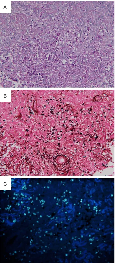
detection of PAS+GSS+CFW was only 77%, which was comparable to that of CFW (P > 0.05) (Table 2). On PAS, round or oval spores could be observed, the sporular wall was red, and the nucleus was blue (Figures1A and 2A). On GSS, black round spores were observed (Figures 1B and 2B). On CFW, round spores
with strong or weak green fluorescence were
noted (Figures 1C and 2C). As shown in Figur- es, the spores were observable by the three stains when the amount of spores was relative-ly large; when the amount of spores was small,
spores with strong or weak fluorescence were
observable on CFW (Figure 3). Discussion
Unlike tumors with relatively high homogeneity, fungal infections possess heterogenicity, and therefore the diagnosis of fungal infection is dependent on the sections used.
In the diagnosis of cat sporotrichosis, Silva et al found the sensitivity of GSS was as high as 91.3% [20]. Miranda investigated the histopa-thology of cat sporotrichosis and they found the sensitivity was 94% for GSS in the diagno-sis of Cat sporotrichodiagno-sis, but the sensitivity of PAS in the diagnosis of cat sporotrichosis is sti- ll unclear. In addition, they also proposed that the skin lesions had a large amount of spores in cats with sporotrichosis, and more a precise method is needed when the amount of fungi is small [13, 21]. In the diagnosis of dog
[image:3.612.324.523.70.525.2]sporotri-chosis, Miranda found the sensitivity of PAS and GSS was 19.5% and 43.7%, respectively [22]. In the present study, the sensitivity of PAS was 31% and that of GSS was 40% in the dia- gnosis of human sporotrichosis. In 100 tissu- es, a large amount of spores were observed in 10 tissues (> 10 spores per section), and no more than 5 spores were observable in the
Table 1. Detection rate of CFW, PAS, and GSS in the diagnosis of Sporotrichosis
Method Negative Positive Detection rate PAS 69 31 31%a,c
GSS 60 40 40%b
CFW 26 74 74%
[image:3.612.91.287.96.150.2]Notes: aP < 0.05 vs CFW; bP < 0.05 vs CFW; cP > 0.05 vs GSS.
Table 2. Detection rate of PAS+GSS+CFW and CFW alone
Methods Detection rate
CFW 74%a
PAS+GSS+CFW 77%
Notes: aP > 0.05 vs PAS+GSS+CFW.
[image:3.612.90.287.218.256.2]majority of tissues. This indicates that the am- ount of spores in human sporotrichosis lesions is small.
In our study, only 3 tissues were positive on PAS and 1 was positive on silver staining am- ong 100 tissues, but they were negative on CFW; in the remaining tissues positive on PAS
and GSS, positivity was seen on CFW. Among the three methods, the detection rate of CFW
was significantly higher than that of PAS and
GSS, but there was no marked difference be- tween PAS and GSS. Moreover, the detection rate of CFW+PAS+CFW (77%) was comparable to that of CFW alone. This suggests that com-bined staining may not achieve a better posi-tive rate as compared to CFW alone. In addi-tion, sections can be observed within 2-min
CFW fluorescence staining. Observation may
be repeated within 10 min and counterstaining is feasible after washing in water, which facili-tates the rapid assessment of sections. When the amount of spores is large, the spores sh-
ow round or oval fluorescence, and strong fluo -rescence is suggestive of positive staining. When the amount of spores is small, false po-
sitive staining is suggested if fluorescence
movement is observed on moving the covers-
[image:4.612.323.523.70.375.2]lip; true positive staining is indicated if fluores -cence location remains stable on moving the coverslip. If the amount of spores is small, it is recommended to repeat examination three Figure 2. PAS, GSS, and CFW staining of the same
tissue (No 52). A: 400×; on PAS, the sporular wall was red and the nucleus in addition to the wall was red in round or oval spores; B: 400×; on GSS, round or oval blackbrown spores were observed. C: 200×; spores with strong or weak green fluorescence were observed.
[image:4.612.89.289.71.519.2]times; true positive staining is confirmed if the fluorescence location remains stable.
Our results showed, when the amount of fungi
was large, the spores were easily identified
after different stains. In the sections of No 50 and No 52 (Figures 1A-C, 2A-C), a large amount of spores was observed under a microscope, which indicates that an alternative staining
may be employed if a stains fails to confirm
the presence of fungi.
When the amount of fungi was small, CFW staining achieved a higher detection rate. In
CFW, the fluorescein binds to the wall of fungi to form complexes which emit fluorescence at the specific excitation wavelength. Our results
showed observation and assessment of sec-tions was relatively convenient and easy after
CFW: round or oval fluorescence was sugges
-tive of posi-tive staining; the elastic fibers with blue fluorescence could be discriminated
bas-ed on the morphology.
Of note, the detection rate was comparable between GSS and PAS. The fungal morphology after PAS is superior to that after GSS [23]. GSS has complex procedures and is time-consum-ing, and over-staining is common with GSS. In
the present study, the findings after PAS and
GSS were interfered with by glycogen, neutral
mucus, and inflammatory cells, and therefore
the assessment of PAS and GSS should be done by an experienced pathologist and/or skin pathologist.
Conclusion
In conclusion, our study indicates the amount of fungi in human sporotrichosis tissues is re- latively small; among three staining methods, CFW is easy to perform, observation of sec-tions is simple and convenient, and the sensi-tivity is higher. We recommend CFW for fungal staining if possible before a fungal culture be- cause CFW is simple, observation is easy and results can be rapidly obtained. Of note, the
fluorescence will quench, which makes the
long-term storage of sections impossible. Disclosure of conflict of interest
None.
Address correspondence to: Fu-Qiu Li, Department of Dermatology, The Second Hospital of Jilin Univer-
sity, 218 Ziqiang Street, Changchun 130041, Jilin, China. Tel: +86-431-81105019; E-mail: Lifuqiu1234 @126.com
References
[1] Orofino-Costa R, Macedo PM, Rodrigues AM and Bernardes-Engemann AR. Sporotrichosis: an update on epidemiology, etiopathogenesis, laboratory and clinical therapeutics. An Bras Dermatol 2017; 92: 606-620.
[2] Moussa TAA, Kadasa NMS, Al Zahrani HS, Ahmed SA, Feng P, Gerrits van den Ende AHG, Zhang Y, Kano R, Li F, Li S, Song Y, Dong B, Ros-sato L, Dolatabadi S and Hoog S. Origin and distribution of Sporothrix globosa causing sap-ronoses in Asia. J Med Microbiol 2017; 66: 560-569.
[3] Chakrabarti A, Bonifaz A, Gutierrez-Galhardo MC, Mochizuki T and Li S. Global epidemiology of sporotrichosis. Med Mycol 2015; 53: 3-14. [4] Zhang YQ, Xu XG, Zhang M, Jiang P, Zhou XY, Li
ZZ and Zhang MF. Sporotrichosis: clinical and histopathological manifestations. Am J Derma-topathol 2011; 33: 296-302.
[5] Tian YP, Zhong SX, Zhang CY and Jin XZ. Isola-tion of Sporothrix schenckii and dematiacious fungi. J Jilin Univ (Med Ed) 2004; 30: 360-365. [6] Jin XZ, Li FQ and Zhu MJ. Isolation of Sporothrix
from Reed and Soil. Chin J Dermatol 1998; 298-299.
[7] Zhao L, Cui Y, Zhen Y, Yao L, Shi Y, Song Y, Chen R and Li S. Genetic variation of Sporothrix glo-bosa isolates from diverse geographic and clinical origins in China. Emerg Microbes Infect 2017; 6: e88.
[8] Lopes-Bezerra LM, Schubach A and Costa RO. Sporothrix schenckii and sporotrichosis. An Acad Bras Cienc 2006; 78: 293-308.
[9] Fischman Gompertz O, Rodrigues AM, Fer-nandes GF, Bentubo HD, de Camargo ZP and Petri V. Atypical clinical presentation of sporo-trichosis caused by sporothrix globosa resis-tant to itraconazole. Am J Trop Med Hyg 2016; 94: 1218-1222.
[10] Conti Diaz IA. Epidemiology of sporotrichosis in Latin America. Mycopathologia 1989; 108: 113-116.
[11] Jessica N, Sonia RL, Rodrigo C, Isabella DF, Tania MP, Jeferson C, Anna BF and Sandro A. Diagnostic accuracy assessment of cytopatho-logical examination of feline sporotrichosis. Med Mycol 2015; 53: 880-884.
[12] Mayayo Artal E. Histopathological diagnosis of mycoses. Rev Iberoam Micol 2004; 21: 1-9. [13] Miranda LH, Conceicao-Silva F, Quintella LP,
clinical presentation. Comp Immunol Microbiol Infect Dis 2013; 36: 425-432.
[14] Quintella LP, Passos SR, do Vale AC, Galhardo MC, Barros MB, Cuzzi T, Reis Rdos S, de Carv-alho MH, Zappa MB and Schubach Ade O. His-topathology of cutaneous sporotrichosis in Rio de Janeiro: a series of 119 consecutive cases. J Cutan Pathol 2011; 38: 25-32.
[15] Hamer EC, Moore CB and Denning DW. Com-parison of two fluorescent whiteners, Calcoflu-or and BlankophCalcoflu-or, fCalcoflu-or the detection of fungal elements in clinical specimens in the diagnos-tic laboratory. Clin Microbiol Infect 2006; 12: 181-184.
[16] Bonifaz A, Rios-Yuil JM, Arenas R, Araiza J, Fer-nandez R, Mercadillo-Perez P and Ponce-Olive-ra RM. Comparison of direct microscopy, cul-ture and calcofluor white for the diagnosis of onychomycosis. Rev Iberoam Micol 2013; 30: 109-111.
[17] Chander J, Chakrabarti A, Sharma A, Saini JS and Panigarhi D. Evaluation of Calcofluor stain-ing in the diagnosis of fungal corneal ulcer. My-coses 1993; 36: 243-245.
[18] Sanketh DS, Patil S and Rao RS. Estimating the frequency of Candida in oral squamous cell carcinoma using Calcofluor White fluorescent stain. J Investig Clin Dent 2016; 7: 304-307.
[19] Pereira SA, Menezes RC, Gremiao ID, Silva JN, Honse Cde O, Figueiredo FB, da Silva DT, Kita-da AA, dos Reis EG and Schubach TM. Sensi-tivity of cytopathological examination in the di-agnosis of feline sporotrichosis. J Feline Med Surg 2011; 13: 220-223.
[20] Silva JN, Miranda LHM, Menezes RC, Gremiao IDF, Oliveira RVC, Vieira SMM, Conceicao-Silva F, Ferreiro L and Pereira SA. Comparison of the sensitivity of three methods for the early diag-nosis of sporotrichosis in cats. J Comp Pathol 2018; 160: 72-78.
[21] Gremiao ID, Menezes RC, Schubach TM, Figueiredo AB, Cavalcanti MC and Pereira SA. Feline sporotrichosis: epidemiological and clin-ical aspects. Med Mycol 2015; 53: 15-21. [22] Miranda LH, Quintella LP, Menezes RC, dos
Santos IB, Oliveira RV, Figueiredo FB, Lopes-Bezerra LM and Schubach TM. Evaluation of immunohistochemistry for the diagnosis of sporotrichosis in dogs. Vet J 2011; 190: 408-411.