Original Article
TERT
promoter mutated WHO grades II and III
gliomas are located preferentially in the
frontal lobe and avoid the midline
Ze-Lin Sun1,3,5*, Aden Ka-Yin Chan2*, Ling-Chao Chen1,5, Chao Tang1,5, Zhen-Yu Zhang1, Xiao-Jie Ding1, Yang Wang1, Chong-Ran Sun4, Ho-Keung Ng2, Yu Yao1,5, Liang-Fu Zhou1
1Department of Neurosurgery, Huashan Hospital, Fudan University, Shanghai 200040, China; 2Department of
Anatomical and Cellular Pathology, Prince of Wales Hospital, The Chinese University of Hong Kong, Hong Kong, China; 3Department of Neurosurgery, North China University of Science and Technology Affiliated Hospital,
Tangshan 063000, Hebei Province, P. R. China; 4Department of Neurosurgery, The Second Affiliated Hospital of
Zhejiang University College of Medicine, Hangzhou 310000, China; 5Neurosurgical Immunology Laboratory of
Huashan Hospital, Fudan University, Shanghai, Chian. *Equal contributors.
Received July 29, 2015; Accepted August 28, 2015; Epub September 1, 2015; Published September 15, 2015
Abstract: The promoter region of telomerase reverse transcriptase (TERTp) and isocitrate dehydrogenase (IDH) have been regarded as biomarkers with distinct clinical and phenotypic features. Investigated the possible cor-relations between tumor location and genetic alterations would enhance our understanding of gliomagenesis and heterogeneity of glioma. We examined mutations of TERTp and IDH by direct sequencing and fluorescence in-situ
hybridization in a cohort of 225 grades II and III diffuse gliomas. Correlation analysis between molecular markers and tumor locations was performed by Chi-square tests/Fisher’s exact test and multivariate logistic regression analysis. We found gliomas in frontal lobe showed higher frequency of TERTp mutation (P=0.0337) and simul-taneously mutations of IDH and TERTp (IDHmut-TERTpmut) (P=0.0281) than frequency of biomarkers mutation of tumors in no-Frontal lobes, while lower frequency of TERTp mutation (P<0.0001) and simultaneously wild type of
IDH and TERTp(IDHwt-TERTpwt) (P<0.0001) in midline than no-midline lobes. Logistic regression analysis indicated that locations of tumors associated with TERTp mutation (OR=0.540, 95% CI 0.324-0.900, P=0.018) and status of combinations of IDH and TERTp (IDHmut-TERTpmut vs. IDHwt-TERTp wt OR=0.162, 95% CI 0.075-0.350, P<0.001). In conclusion, grades II and III gliomas harboring TERTp mutation were located preferentially in the frontal lobe and rarely in midline. Association of IDH-TERTp status and tumor location suggests their potential values in molecular classification of grades II and III gliomas.
Keywords: Gliomas, heterogeneity, IDH mutation, TERT promoter mutation, tumor location
Introduction
Gliomas are the most common primary brain malignancies. They were classified by World Health Organization (WHO) into four grades according to the morphological resemblance of the neoplastic cells to normal glial tissues [1, 2]. While glioblastomas (grade IV) had the most dismal prognosis [3-5], grades II and III gliomas showed a relatively favorable but highly vari-able survival [2]. Although the great improve had been made on the diagnosis and treatment of grades II and III gliomas in the past decades, patients with grades II and III gliomas had an inevitable recurrence and mortal result.
It had been increasingly valued by researchers that gliomas with diverse genetic abnormalities might arise from distinct cell types of origin and might be an important cause of tumor hetero-geneity [6, 7]. The association between genetic signature and tumor location is important for understanding the spatial origin of tumorigene-sis, sub-classifying this kind of aggressive can-cers and developing more intensive treatment measures to prolong the life of patients with grades II and III gliomas.
In the recent years, isocitrate dehydrogenase
ximately 70% to 80% of grades II and III diffuse gliomas and secondary glioblastomas, exclud-ing ependymoma and pilocytic astrocytomas, harbor mutations at codon 132 (R132) of the
isocitrate dehydrogenase 1 gene (IDH1) or at
codon 172 (R172) of the isocitrate
dehydroge-nase 2 gene (IDH2) [5, 8-11]. Many studies
reported the preferential localization of glio-blastomas [12-14] and lower-grade diffuse astrocytic gliomas [5, 6, 9, 12, 15-19] with IDH
mutation in frontal lobe, suggesting this kind of tumors may arise from distinct cell types of origin.
Concurrently, it is well known thatthe promoter region of telomerase reverse transcriptase
(TERTp) is a driver event in cancer development [19]. Shortening of telomere repeats cap at the ends of eukaryotic chromosomes with each cell division trigger cell death or senescence even-tually [20]. TERTp encodes catalytic subunits of telomerase which maintain telomere length, and delay cellular senescence [20, 21]. Tumors with TERTp mutation present distinct clinical and phenotypic features [19]. Analysis of the spatial distribution of TERTp mutation in grade II and III gliomas and combined analysis with the spatial distribution of IDH mutation may enhance the understanding of gliomagenesis. It would be a new perspective for neurosur-geons to consider grade II and III gliomas because of the association between genetic alterations and distinct prognosis of gliomas [22-25]. To our knowledge, there is no report about the regional distribution of TERTp muta-tion in WHO grade II and III gliomas. In this study, we examined the TERTp and IDH muta-tion of 225 WHO grade II and III gliomas, con-firmed the brain lobes their located and analy-sis the associations of TERTp mutation, IDH
mutation and tumor location.
Patients and methods
Patients and tissue samples
A total of 225 grades II and III gliomas with for-malin-fixed paraffin-embedded tissues avail-able and imaging studies (MRI) at the time of the diagnosis or in the preoperative period available were selected from the Department of Neurosurgery, Huashan hospital (Shanghai, China) between 2001 and 2011 and from the Department of Anatomical and Cellular Patho- logy, Prince of Wales Hospital (Hong Kong)
between 1990 and 2012 [26, 27]. To confirm the tumors location perfectly, detailed radio-logical reports, operative reports and profiles of postoperative MRI were studied also. According to the 2007 WHO classification [2], there were 96 diffuse astrocytomas (WHO grade II; AII), 20 oligodendrogliomas (WHO grade II; OII), 47 oli-goastrocytomas (WHO grade II; OAII), 54 ana-plastic astrocytomas (WHO grade III; AAIII), 5 anaplastic oligodendrogliomas (WHO grade III; AOIII), 3 anaplastic oligoastrocytomas (WHO grade III; AOAIII). The cohort overlapped partly with previous studies [26, 27]. This study was approved by the Ethics Committee of Shanghai Huashan Hospital and the New Territories East Cluster-Chinese University of Hong Kong Ethics Committee.
Tumor location
To consider the tumor locations, imaging pro-files (MRI) and the clinical pro-files were retrospec-tively reviewed by two neurosurgeons that had no idea of the molecular status of the patient. If there were disagree with the tumor categori- zation between them, a senior neurosurgeon would have the right to judge the tumor involve-ment. To simplify the analysis, the tumors were primarily assigned into three kinds of locations: frontal, midline and others [28]. Frontal gliomas included only the tumors located entirely in frontal lobe. Midline location included corpus callosum, thalamencephalon, periventricular location, brainstem and thoracic spinal cord [7], where tumors entirely located in. Others lobes included insular lobe (including insular, frontotemporal-insular, temporal-insular and frontal-insular lobe), temporal lobe (including temporal, frontotemporal, tempoparietal, tem-poral-occipital lobe), parietal lobe (including parietal, frontoparietal, parietal-occipital lobe) and Occipital lobe & Cerebellum (including occipital lobe and cerebellum).
Mutational analysis of TERTp and IDH
Among the 225 grade II and III gliomas that had been detected IDH mutation, 213 grade II and III gliomas were examined for mutations of
tumor area with tumor content greater than 70% was used for subsequent polymerase chain reaction (PCR) analysis. Primers sequenc-es (the forward primer IDH1F: 5’-CGGTCTT- CAGAGAAGCCATT-3’ and reverse primer IDH1-R: 5’-CACATTATTGCCAACATGAC-3’; forward primer IDH2-F: 5’-AGCCCATCATCTGCAAAAAC-3’ and reverse primer IDH2-R 5’-CTAGGCGAGGAGCT- CCAGT-3’) were used to amplify fragments of PCR, while a 163 bp fragment spanning the two mutational hotspots (C228T and C250T) in pro-moter region of TERTp were amplified with TERT-F (5’-GTCCTGCCCCTTCACCTT-3’) and TE- RT-R (5’-CAGCGCTGCCTGAAACTC-3’). Sequen- cing was performed using Big Dye Terminator Cycle Sequencing kit v1.1. The products were resolved in Genetic Analyzer 3130xl and ana-lyzed by Sequencing Analysis software.
one IDH1-R132S mutation, one IDH2-R172M mutation, three IDH2-R172K mutation and 144 IDH1-R132H mutations. IDH mutations were found in 65% (91/140) male patients and 68.24% (58/85) female patients. There were 117 patients younger than or equal to 40 years and 108 patients older than 40 years, with 72.12% (75/108) and 63.25% (74/117) harbor-ing IDH mutations, respectively. Among the cohort, 68.75% (66/96) of AII, 90% (18/20) of OII and 82.98% (39/47) of OAII harbored IDH
mutations. Grade III gliomas, including AAIII, AOIII and AOAIII showed IDH mutations in 41.94% (26/62) of cases (Table 1 and Table S1).
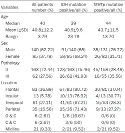
[image:3.612.91.341.95.360.2]Mutation in TERTp was found in 61 of 213 (28.64%) grade II and III gliomas examined. The
Table 1. Clinical and molecular data of 225 WHO grade II and III gliomas
Variables number (%)All patients positive/all (%)IDH mutation TERTppositive/all (%) mutation Age
Median 40 39 44
Mean (±SD) 40.8±12.2 40.9±9.6 43.7±11.5
Range 3-79 23-79 13-70
Sex
Male 140 (62.22) 91/140 (65) 35/131 (26.72)
Female 85 (37.78) 58/85 (68.24) 26/82 (31.71)
Pathology
II 163 (72.44) 123/163 (75.46) 45/158 (28.48)
III 62 (27.56) 26/62 (41.93) 16/55 (35.56)
Location
Frontal 83 (36.89) 67/83 (80.72) 30/81 (37.04)
Insular 13 (5.78) 10/13 (76.92) 4/13 (30.77)
Temporal 61 (27.11) 41/61 (67.21) 15/53 (28.3)
Parietal 35 (15.56) 25/35 (71.43) 9/33 (27.27)
O & C 6 (2.67) 1/6 (16.67) 0/6 (0)
C & C 6 (2.67) 3/6 (50) 0/6 (0)
Midline 21 (9.33) 2/21 (9.52) 2/21 (9.52)
Abbreviations: O & C, Occipital & Cerebellum; C & C, Corpus callosum & Cingulate gyrus. Frontal including tumors located entirely in frontal lobe; Midline including tumors located entirely in corpus callosum, thalamen-cephalon, periventricular location, brainstem and thoracic spinal cord; Insular including tumors involved in insular, frontotemporal-insular, temporal-insular and frontal-insular lobe; Temporal including tumors involved in temporal, frontotemporal, tempoparietal, temporal-occipital lobe; Parietal including tumors involved in parietal, frontoparietal, parietal-occipital lobe; O & C including tumors located entirely in occipital lobe and cerebellum; C & C including tumors located entirely in corpus callosum and cingulate gyrus.
Statistical analysis
Fisher’s exact test (or Chi-square tests when n>10) were performed to assess the genotype distribution of IDH and
TERTp mutation in different tumor
locations. Main effects multivariate logistic regression analysis was used to identify the factors associated with status of biomarker of this cohort of grade II and III gliomas. Gender (val-ues: 1= female, 0= male), age (val(val-ues: 1= group of patients ≤40 years old, 2≥40 years old), pathology (values: 1= GradeII, 2= Grade III) and locations of tumors (values: 1= Frontal, 2= Others lobe, 3= Midline) were selected as independent variables for the analysis of each biomarker status. Value of β, odds ratio (ORs), 95% confidence interval (95% CIs) and p-values of fac-tors with status of each biomarkers status were calculated respectively. All statistical tests were two-sided, and the threshold for statistical signifi-cance was P<0.05. Analyses were conducted with SPSS for Windows ver-sion 20.0 (SPSS Inc, Chicago, IL, USA).
Results
Cohort characteristics and molecular data
mutations were found in 26.72% (35/131) of male patients and 31.7% (26/82) of female patients. There were110 patients younger than or equal to 40 years and 103 patients older than 40 years, with 19.42% (20/103) and 36.36% (40/110) showing TERTp mutation, respectively. Among the cohort, 14.89% (14/94) of AII, 73.68% (14/19) of OII, 37.78% (17/45) of OAII, 22.45% (11/49) of AAIII, 100% (3/3) of AOIII and 66.67% (2/3) of AOAIII har-bored TERTp mutation respectively (Table 1).
Correlation between biomarker status and tu-mor location
To discover the tendency of the distributions of biomarker status on brain lobes, we calculated the approximate mutation rate of each bio-marker across brain lobes. Figure 1A showed
[image:4.612.92.523.71.312.2] [image:4.612.326.522.477.544.2]IDH mutation was identified in 67 of 83 (80.72%) frontal tumors, 10 of 13 (76.92%) insular tumors, 41 of 61 (67.21%) temporal tumors, 25 of 35 (71.43%) parietal tumors, 3 of Figure 1.Correlations between locations distribution and molecular status of WHO grade II and III gliomas. The rate of IDH mutation (A) and TERT promoter mutation (C) of WHO grade II and III gliomas decreased gradually from frontal lobe to Midline. WHO grade II and III gliomas with IDH mutation (B) and TERT promoter mutation (D) is more preferentially located in Frontal and repulsively in midline. Footnotes: Frontal including tumors located entirely in frontal lobe; Midline including tumors located entirely in corpus callosum, thalamencephalon, periventricular loca-tion, brainstem and thoracic spinal cord; Insular lobe including tumors involved in insular, frontotemporal-insular, temporal-insular and frontal-insular lobe; Temporal lobe including tumors involved in temporal, frontotemporal, tempoparietal, temporal-occipital lobe; Parietal lobe including tumors involved in parietal, frontoparietal, parietal-occipital lobe; Occipital lobe & Cerebellum including tumors located entirely in parietal-occipital lobe and cerebellum.
Table 2. ORs, 95% CIs and p-values of classes of gliomas location according to status of IDH
Variables β P OR 95% CI
Gender -0.286 0.396 0.751 0.388-1.455
Age -0.387 0.237 0.679 0.358-1.289
Pathology -1.483 0.000 0.227 0.114-0.453
Location -1.456 0.000 0.233 0.132-0.413
Abbreviations: CI, confidence interval; OR, odds ratio.
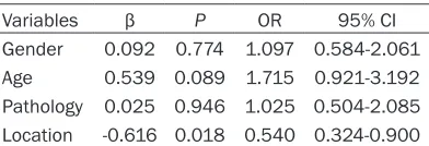
Table 3. ORs, 95% CIs and p-values of class-es of gliomas location according to status of
TERTp
Variables β P OR 95% CI
Gender 0.092 0.774 1.097 0.584-2.061
Age 0.539 0.089 1.715 0.921-3.192
Pathology 0.025 0.946 1.025 0.504-2.085
Location -0.616 0.018 0.540 0.324-0.900
[image:4.612.92.288.478.544.2]6 (50%) corpus callosum & cingulate gyrus tumors, 1 of 6 (16.67%) occipital & cerebellar tumors and 2 of 21 (9.52%) midline tumors. We found from Figure 1A that there was a degres-sive tendency of mutation rate of IDH from Frontal to Midline. Chi-square test or Fisher’s exact test had been used to find out that the rate of IDH mutation in the cohort of grade II and grade III gliomas were higher in the frontal lobe than non-frontal region (P=0.0004, Chi-square test) and lower in the midline than non-midline location (P<0.0001, Fisher’s exact test) (Figure 1B). Results of binary logistic regress confirmed this discover. As shown in Table 2, Locations of tumors after adjustment for gen-der, age and pathology (β=-1.456, OR=0.233, 95% CI 0.132-0.413, P<0.001) was found to be independently associated with status of
IDH1/2. With the values of β is negative, the rate of IDH mutation has a degressive tendency from Frontal to midline.
TERTp mutation was found in 37.04% (30/81)
of frontal tumors, 30.77% (4/13) of insular tumors, 28.30% (15/53) temporal tumors, 9 of 33 (27.27%) parietal tumors, 0 of 6 (0%) corpus
pathology (β=-0.616, OR=0.540, 95% CI 0.324-0.900, P=0.018) was found to be independent-ly associated with status of TERTp. With the values of β is negative, the rate of TERTp muta-tion has a degressive tendency from frontal lobe to midline also (Table 3).
Distribution of grade II and III gliomas with combined status of IDH and TERTp
As the results shown above, IDH mutation and
TERTp mutation in grades II and III gliomas
shared similar spatial distributions across brain lobes. Therefore, we analyzed the regional dis-tribution of the tumors according to combined
status of IDH and TERTp (IDH-TERTp). Results
of Chi-square test or Fisher’s exact test identi-fied that simultaneously mutations of IDH and
TERTp (IDHmut-TERTpmut) subgroup was prefer-entially located in frontal lobe (P=0.0281) and simultaneously wild type of IDH and TERTp
[image:5.612.89.526.85.153.2](IDHwt-TERTpwt) subgroup was preferentially lo- cated in midline regions (P<0.0001). We identi-0.0001). We identi-fied the rate of IDHmut-TERTpwt were lower in midline tumors (P=0.0004), but did not identify any other association between IDHmut-TERTpwt
Table 4. Regional distribution of the tumors according to combined status of IDH and TERTp
Number Frontal P/N no Frontal P/N P (F vs. NF) Midline P/N no Midline P/N P (M vs. NM)
IDHwt-TERTpwt 57 10/71 47/85 0.0002 17/4 40/152 <0.0001
IDHwt-TERTpmut 13 5/76 8/124 1.0000 2/19 7/185 0.6309
IDHmut-TERTpwt 95 41/40 54/78 0.2015 2/19 93/99 0.0004
IDHmut-TERTpmut 48 25/56 23/109 0.0281 0/21 48/144 0.0050
Abbreviations: P/N, numbers of gliomas with positive/negative biomarker mutation; P (F vs. NF), P values between Frontal and no-Frontal; P (M vs. NM), P values between Midline and no-Midline.
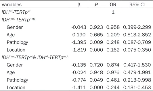
Table 5. ORs, 95% CIs and p-values of classes of gliomas location according to combined status of IDH and TERTp
Variables β P OR 95% CI
IDHwt-TERTpwt 1
IDHmut-TERTpmut
Gender -0.043 0.923 0.958 0.399-2.299
Age 0.190 0.665 1.209 0.513-2.852
Pathology -1.395 0.009 0.248 0.087-0.709
Location -1.819 0.000 0.162 0.075-0.350
IDHmut-TERTpwt& IDHwt-TERTpmut
Gender -0.135 0.720 0.874 0.417-1.830
Age -0.024 0.948 0.976 0.479-1.991
Pathology -0.774 0.049 0.461 0.213-0.998
Location -1.411 0.000 0.244 0.131-0.453
Abbreviations: CI, confidence interval; OR, odds ratio.
callosum & cingulate gyrus tumors, 0 of 6 (0%) occipital & cerebellar tumors and 2 of 21 (9.52%) midline tumors (Figure 1C). We found also that there was a degressive ten-dency of mutation rate of TERTp
from Frontal to Midline. The rat of
TERTp mutation in the cohort of
[image:5.612.90.356.221.380.2]subgroup and IDHwt-TERTpmut subgroup with location (Table 4). To confirm the result, we used a main effects multivariate logistic regres-sion analysis to identify if the IDH-TERTp status correlated with tumor locations. For dependent variables, the IDHmut-TERTpmut subgroup was set as 1, while subgroup of IDHmut-TERTpwt and
[image:6.612.93.520.97.349.2]IDHwt-TERTpmut were set as 2, IDHwt-TERTpwt subgroup was set as 3 (Table S1). As shown in
Table 6, Locations of tumors after adjustment for gender, age and pathology (IDHmut-TERTpmut vs. IDHwt-TERTpwt β=-1.819, OR=0.162, 95% CI 0.075-0.350, P<0.001; IDHmut-TERTpwt & IDHwt
-TERTpmut vs. IDHwt-TERTpwt β=-1.411, OR=0.244, 95% CI 0.131-0.453, P<0.001) was found to be independently associated with status of IDH
-TERTp. The result of the regression analysis
shown there was a depressive of rate of IDH
-TERTp mutation from Frontal to Midline (Table
5).
Biomarker distribution in subgroups by age, gender and histology
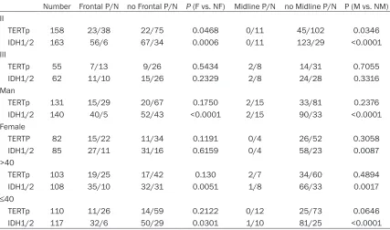
We further evaluated the effects of age, gender and histology on the spatial distribution of molecular markers as shown in Table 6. In both
age subgroups of patients above 40 years and patients at or under 40 years, IDH mutation frequency was significantly higher in frontal tumors and lower in midline tumors. For gender, the frequencies of IDH mutation were higher in frontal tumors and lower in midline tumors of male patients. Among the female patients, there was no statistical difference in IDH muta-tion rate between different tumor locamuta-tions. There were not any associations between
TERTp mutation and tumor locations in
sub-groups of gliomas classified by age and gender. Evaluating the cohort according to histological grade, biomarker-location associations were mainly identified in grade II tumors, with frontal tumors demonstrating higher rate of IDH muta-tion and TERTp mutation, and midline tumors showing lower rate of IDH mutation and TERTp
mutation. Results of regression analysis shown pathology independently associated with IDH-
1/2 mutation (β=-1.483, OR=0.227, 95% CI 0.114-0.453, P<0.001) (Table 2) and status of
IDH-TERTp (IDHmut-TERTpmut vs. IDHwt-TERTpwt β=-1.395, OR=0.248, 95% CI 0.087-0.709,
P=0.009; IDHmut-TERTpwt & IDHwt-TERTpmut vs.
IDHwt-TERTpwt β=-0.774, OR=0.049, 95% CI
0.213-0.998, P=0.049) (Table 5).
Table 6. Correlations between locations distribution and molecular status of subgroups of WHO grade II and III gliomas
Number Frontal P/N no Frontal P/N P (F vs. NF) Midline P/N no Midline P/N P (M vs. NM)
II
TERTp 158 23/38 22/75 0.0468 0/11 45/102 0.0346
IDH1/2 163 56/6 67/34 0.0006 0/11 123/29 <0.0001
III
TERTp 55 7/13 9/26 0.5434 2/8 14/31 0.7055
IDH1/2 62 11/10 15/26 0.2329 2/8 24/28 0.3316
Man
TERTp 131 15/29 20/67 0.1750 2/15 33/81 0.2376
IDH1/2 140 40/5 52/43 <0.0001 2/15 90/33 <0.0001
Female
TERTP 82 15/22 11/34 0.1191 0/4 26/52 0.3058
IDH1/2 85 27/11 31/16 0.6159 0/4 58/23 0.0087
>40
TERTp 103 19/25 17/42 0.130 2/7 34/60 0.4894
IDH1/2 108 35/10 32/31 0.0051 1/8 66/33 0.0017
≤40
TERTp 110 11/26 14/59 0.2122 0/12 25/73 0.0646
IDH1/2 117 32/6 50/29 0.0301 1/10 81/25 <0.0001
Discussion
In this study, we found that WHO grade II and III gliomas located in frontal lobe were preferen-tially associated with IDH, TERTp and IDHmut
-TERTpmut, while gliomas in midline were
prefer-entially associated with IDH, TERTp and IDHwt
-TERTpwt, evenly after adjustment for gender,
age and pathology. It is in concordance with previous data [6, 15, 18] that frontal lobe was a preferential location for gliomas harboring
IDH mutation as compared to other cerebral regions. Chen et al. recently reported that the glutaminergic neurotransmitter specialization of human neocortex, especially frontal lobe, created a metabolic niche favorable for the development of IDH1 mutant tumors [30]. Their findings may explain the biological mechanisms for the preferential distribution of IDH mutated grades II and III gliomas in frontal lobes. Midline location in this study included corpus callosum, thalamencephalon, periventricular location, br- ainstem and thoracic spinal cord. The mencha-nism of the special distribution of gliomas in midline without IDH mutation remained undis-covered. In the subset analysis, association between IDH mutation and frontal localization was observed in male patients but not female patients. Given that the case numbers in the individual gender subsets of the cohort were small, further study evaluating the effect of gender on the regional distribution of IDH muta-tion in a larger cohort should be conducted. We found that grades II and III gliomas in fron-tal lobe show high TERTp mutation rate, on the counterparts, low TERTp mutation rate in mid-line. To the best of our knowledge, this is the first study reported it. Dominik et al. had classi-fied glioblastomas into six subgroups based on their global DNA methylation patterns [7]. They suggested there were a specific anatomically-defined subset of gliomas with H3F3A-K27M mutation almost exclusively arose from midline locations, which was consistent with results of several other researchers’ studies [31, 32]. Our results of this report suggested that there might be an anatomically-defined subset of gli-omas with biological character of IDHwt-TERTpwt in grades II and III diffuse gliomas located in midline. Although the biological mechanism of the special distribution of gliomas with TERTp
mutation requires further elucidation, our results shed light on potential anatomical
cel-lular origins of grades II and III gliomas and improved further research in this field.
As our previously study showed, TERTp muta-tion had been recognized as a dismal molecu-lar marker in prognostic classification of diffuse gliomas [26]. Killela et al. had identified that gliomas exhibit IDHmut-TERTpmut had a best prognosis for exhibiting a median over survival (OS) of 125 months, while gliomas exhibit
TERTp mutation alone with a poorest OS (11.5
months) in their cohort of gliomas [19]. In keep-ing with this study, Eckel-Passow et al. recently found that patients with IDHwt-TERTpwt and with-out co-deletion of 1p19q had poor OS than
IDHmut-TERTpmut or IDH mutation alone, but bet-ter OS than TERTp mutation alone, after adjust-ment for age and grade [22]. Data of this study may imply neurosurgeons that grades II and III gliomas located in frontal lobe where is more accessible to surgery would be associated with different prognostic biomarkers. Moreover, our results suggest a gloomier prognosis of pa- tients with grades II and III gliomas located in midline for being barely inaccessible to surgery and associated with poor prognostic biomark-ers. So, there would be a more crucial need to design new therapeutic agents for grades II and III gliomas located in midline.
In conclusion, we have investigated the spatial distribution of grades II and III gliomas with specially status of IDH, TERTp and IDH-TERTp, suggested that there are some anatomically-defined subset of gliomas with special biomark-ers, enhanced neurosurgeons’ understanding of grades II and III gliomas in specific area of brain, and would stimulated more researchers to study in this field.
Acknowledgements
This work was supported by grant 13JC14080- 00 from the Science and Technology Com-mission of Shanghai Municipality, the Research Special Fund for Public Welfare Industry of Health (No. 201402008), and the National key clinical specialist construction Programs of China (Oncology). We thank Dr. Xiao-Qin Wang for helpful instructions about statistics treat- ment.
Disclosure of conflict of interest
Address correspondence to: Drs. Liang-Fu Zhou and Yu Yao, Department of Neurosurgery, Huashan Hospital, Fudan University, 12 Wulumuqi Zhong Road, Shanghai 200040, China. Tel: 086-21-528- 88305; 086-021-52887034; E-mail: lfzhouc@126. com (LFZ); [email protected] (YY)
References
[1] Levine AJ and Puzio-Kuter AM. The control of the metabolic switch in cancers by oncogenes and tumor suppressor genes. Science 2010; 330: 1340-1344.
[2] Louis DN, Ohgaki H, Wiestler OD, Cavenee WK, Burger PC, Jouvet A, Scheithauer BW and Kleihues P. The 2007 WHO classification of tu-mours of the central nervous system. Acta Neuropathol 2007; 114: 97-109.
[3] Stupp R, Mason WP, van den Bent MJ, Weller M, Fisher B, Taphoorn MJ, Belanger K, Brandes AA, Marosi C, Bogdahn U, Curschmann J, Janzer RC, Ludwin SK, Gorlia T, Allgeier A, Lacombe D, Cairncross JG, Eisenhauer E, Mirimanoff RO, European Organisation for R, Treatment of Cancer Brain T, Radiotherapy G and National Cancer Institute of Canada Clinical Trials G. Radiotherapy plus concomi-tant and adjuvant temozolomide for glioblas-toma. N Engl J Med 2005; 352: 987-996. [4] Wen PY and Kesari S. Malignant gliomas in
adults. N Engl J Med 2008; 359: 492-507. [5] Yan H, Parsons DW, Jin G, McLendon R,
Rasheed BA, Yuan W, Kos I, Batinic-Haberle I, Jones S, Riggins GJ, Friedman H, Friedman A, Reardon D, Herndon J, Kinzler KW, Velculescu VE, Vogelstein B and Bigner DD. IDH1 and IDH2 mutations in gliomas. N Engl J Med 2009; 360: 765-773.
[6] Lai A, Kharbanda S, Pope WB, Tran A, Solis OE, Peale F, Forrest WF, Pujara K, Carrillo JA, Pandita A, Ellingson BM, Bowers CW, Soriano RH, Schmidt NO, Mohan S, Yong WH, Seshagiri S, Modrusan Z, Jiang Z, Aldape KD, Mischel PS, Liau LM, Escovedo CJ, Chen W, Nghiemphu PL, James CD, Prados MD, Westphal M, Lamszus K, Cloughesy T and Phillips HS. Evidence for sequenced molecular evolution of IDH1 mutant glioblastoma from a distinct cell of origin. J Clin Oncol 2011; 29: 4482-4490. [7] Sturm D, Witt H, Hovestadt V, Khuong-Quang
DA, Jones DT, Konermann C, Pfaff E, Tonjes M, Sill M, Bender S, Kool M, Zapatka M, Becker N, Zucknick M, Hielscher T, Liu XY, Fontebasso AM, Ryzhova M, Albrecht S, Jacob K, Wolter M, Ebinger M, Schuhmann MU, van Meter T, Fruhwald MC, Hauch H, Pekrun A, Radlwimmer B, Niehues T, von Komorowski G, Durken M, Kulozik AE, Madden J, Donson A, Foreman NK,
Drissi R, Fouladi M, Scheurlen W, von Deimling A, Monoranu C, Roggendorf W, Herold-Mende C, Unterberg A, Kramm CM, Felsberg J, Hartmann C, Wiestler B, Wick W, Milde T, Witt O, Lindroth AM, Schwartzentruber J, Faury D, Fleming A, Zakrzewska M, Liberski PP, Zakrzewski K, Hauser P, Garami M, Klekner A, Bognar L, Morrissy S, Cavalli F, Taylor MD, van Sluis P, Koster J, Versteeg R, Volckmann R, Mikkelsen T, Aldape K, Reifenberger G, Collins VP, Majewski J, Korshunov A, Lichter P, Plass C, Jabado N and Pfister SM. Hotspot mutations in H3F3A and IDH1 define distinct epigenetic and biological subgroups of glioblastoma. Cancer Cell 2012; 22: 425-437.
[8] Liu XY, Gerges N, Korshunov A, Sabha N, Khuong-Quang DA, Fontebasso AM, Fleming A, Hadjadj D, Schwartzentruber J, Majewski J, Dong Z, Siegel P, Albrecht S, Croul S, Jones DT, Kool M, Tonjes M, Reifenberger G, Faury D, Zadeh G, Pfister S and Jabado N. Frequent ATRX mutations and loss of expression in adult diffuse astrocytic tumors carrying IDH1/IDH2 and TP53 mutations. Acta Neuropathol 2012; 124: 615-625.
[9] Ohgaki H and Kleihues P. Genetic profile of as-trocytic and oligodendroglial gliomas. Brain Tumor Pathol 2011; 28: 177-183.
[10] Parsons DW, Jones S, Zhang X, Lin JC, Leary RJ, Angenendt P, Mankoo P, Carter H, Siu IM, Gallia GL, Olivi A, McLendon R, Rasheed BA, Keir S, Nikolskaya T, Nikolsky Y, Busam DA, Tekleab H, Diaz LA Jr, Hartigan J, Smith DR, Strausberg RL, Marie SK, Shinjo SM, Yan H, Riggins GJ, Bigner DD, Karchin R, Papadopoulos N, Parmigiani G, Vogelstein B, Velculescu VE and Kinzler KW. An integrated genomic analy-sis of human glioblastoma multiforme. Science 2008; 321: 1807-1812.
[11] Watanabe T, Nobusawa S, Kleihues P and Ohgaki H. IDH1 mutations are early events in the development of astrocytomas and oligo-dendrogliomas. Am J Pathol 2009; 174: 1149-1153.
[12] Gupta R, Webb-Myers R, Flanagan S and Buckland ME. Isocitrate dehydrogenase muta-tions in diffuse gliomas: clinical and aetiologi-cal implications. J Clin Pathol 2011; 64: 835-844.
[13] Ikota H, Nobusawa S, Tanaka Y, Yokoo H and Nakazato Y. High-throughput immunohisto-chemical profiling of primary brain tumors and non-neoplastic systemic organs with a specific antibody against the mutant isocitrate dehy-drogenase 1 R132H protein. Brain Tumor Pathol 2011; 28: 107-114.
gliomas: first series from a tertiary care centre in India with comprehensive review of litera-ture. Exp Mol Pathol 2011; 91: 385-393. [15] Gorovets D, Kannan K, Shen R, Kastenhuber
ER, Islamdoust N, Campos C, Pentsova E, Heguy A, Jhanwar SC, Mellinghoff IK, Chan TA and Huse JT. IDH mutation and neuroglial de-velopmental features define clinically distinct subclasses of lower grade diffuse astrocytic glioma. Clin Cancer Res 2012; 18: 2490-2501. [16] Qi S, Yu L, Li H, Ou Y, Qiu X, Ding Y, Han H and
Zhang X. Isocitrate dehydrogenase mutation is associated with tumor location and magnetic resonance imaging characteristics in astrocyt-ic neoplasms. Oncol Lett 2014; 7: 1895-1902. [17] Bourne TD and Schiff D. Update on molecular
findings, management and outcome in low-grade gliomas. Nat Rev Neurol 2010; 6: 695-701.
[18] Wang Y, Zhang T, Li S, Fan X, Ma J, Wang L and Jiang T. Anatomical localization of isocitrate dehydrogenase 1 mutation: a voxel-based ra-diographic study of 146 low-grade gliomas. Eur J Neurol 2015; 22: 348-354.
[19] Killela PJ, Pirozzi CJ, Healy P, Reitman ZJ, Lipp E, Rasheed BA, Yang R, Diplas BH, Wang Z, Greer PK, Zhu H, Wang CY, Carpenter AB, Friedman H, Friedman AH, Keir ST, He J, He Y, McLendon RE, Herndon JE 2nd, Yan H and Bigner DD. Mutations in IDH1, IDH2, and in the TERT promoter define clinically distinct sub-groups of adult malignant gliomas. Oncotarget 2014; 5: 1515-1525.
[20] Aubert G and Lansdorp PM. Telomeres and ag-ing. Physiol Rev 2008; 88: 557-579.
[21] Xu L, Li S and Stohr BA. The role of telomere biology in cancer. Annu Rev Pathol 2013; 8: 49-78.
[22] Eckel-Passow JE, Lachance DH, Molinaro AM, Walsh KM, Decker PA, Sicotte H, Pekmezci M, Rice T, Kosel ML, Smirnov IV, Sarkar G, Caron AA, Kollmeyer TM, Praska CE, Chada AR, Halder C, Hansen HM, McCoy LS, Bracci PM, Marshall R, Zheng S, Reis GF, Pico AR, O’Neill BP, Buckner JC, Giannini C, Huse JT, Perry A, Tihan T, Berger MS, Chang SM, Prados MD, Wiemels J, Wiencke JK, Wrensch MR and Jenkins RB. Glioma Groups Based on 1p/19q, IDH, and TERT Promoter Mutations in Tumors. N Engl J Med 2015; 372: 2499-2508.
[23] SongTao Q, Lei Y, Si G, YanQing D, HuiXia H, XueLin Z, LanXiao W and Fei Y. IDH mutations predict longer survival and response to temo-zolomide in secondary glioblastoma. Cancer Sci 2012; 103: 269-273.
[24] Houillier C, Wang X, Kaloshi G, Mokhtari K, Guillevin R, Laffaire J, Paris S, Boisselier B, Idbaih A, Laigle-Donadey F, Hoang-Xuan K, Sanson M and Delattre JY. IDH1 or IDH2
muta-tions predict longer survival and response to temozolomide in low-grade gliomas. Neurology 2010; 75: 1560-1566.
[25] Metellus P, Coulibaly B, Colin C, de Paula AM, Vasiljevic A, Taieb D, Barlier A, Boisselier B, Mokhtari K, Wang XW, Loundou A, Chapon F, Pineau S, Ouafik L, Chinot O and Figarella-Branger D. Absence of IDH mutation identifies a novel radiologic and molecular subtype of WHO grade II gliomas with dismal prognosis. Acta Neuropathol 2010; 120: 719-729. [26] Chan AK, Yao Y, Zhang Z, Chung NY, Liu JS, Li
KK, Shi Z, Chan DT, Poon WS, Zhou L and Ng HK. TERT promoter mutations contribute to subset prognostication of lower-grade gliomas. Mod Pathol 2015; 28: 177-186.
[27] Yao Y, Chan AK, Qin ZY, Chen LC, Zhang X, Pang JC, Li HM, Wang Y, Mao Y, Ng HK and Zhou LF. Mutation analysis of IDH1 in paired gliomas revealed IDH1 mutation was not associated with malignant progression but predicted lon-ger survival. PLoS One 2013; 8: e67421. [28] Laigle-Donadey F, Martin-Duverneuil N,
Lejeune J, Criniere E, Capelle L, Duffau H, Cornu P, Broet P, Kujas M, Mokhtari K, Carpentier A, Sanson M, Hoang-Xuan K, Thillet J and Delattre JY. Correlations between molec-ular profile and radiologic pattern in oligoden-droglial tumors. Neurology 2004; 63: 2360-2362.
[29] Chan AK, Yao Y, Zhang Z, Chung NY, Liu JS, Li KK, Shi Z, Chan DT, Poon WS, Zhou L and Ng HK. TERT promoter mutations contribute to subset prognostication of lower-grade gliomas. Mod Pathol 2015; 28: 177-86.
[30] Chen R, Nishimura MC, Kharbanda S, Peale F, Deng Y, Daemen A, Forrest WF, Kwong M, Hedehus M, Hatzivassiliou G, Friedman LS and Phillips HS. Hominoid-specific enzyme GLUD2 promotes growth of IDH1R132H glioma. Proc Natl Acad Sci U S A 2014; 111: 14217-14222. [31] Bechet D, Gielen GG, Korshunov A, Pfister SM,
Rousso C, Faury D, Fiset PO, Benlimane N, Lewis PW, Lu C, David Allis C, Kieran MW, Ligon KL, Pietsch T, Ellezam B, Albrecht S and Jabado N. Specific detection of methionine 27 muta-tion in histone 3 variants (H3K27M) in fixed tissue from high-grade astrocytomas. Acta Neuropathol 2014; 128: 733-741.
Zakrzewski K, Liberski PP, Dong Z, Siegel P, Kulozik AE, Zapatka M, Guha A, Malkin D, Felsberg J, Reifenberger G, von Deimling A, Ichimura K, Collins VP, Witt H, Milde T, Witt O, Zhang C, Castelo-Branco P, Lichter P, Faury D,
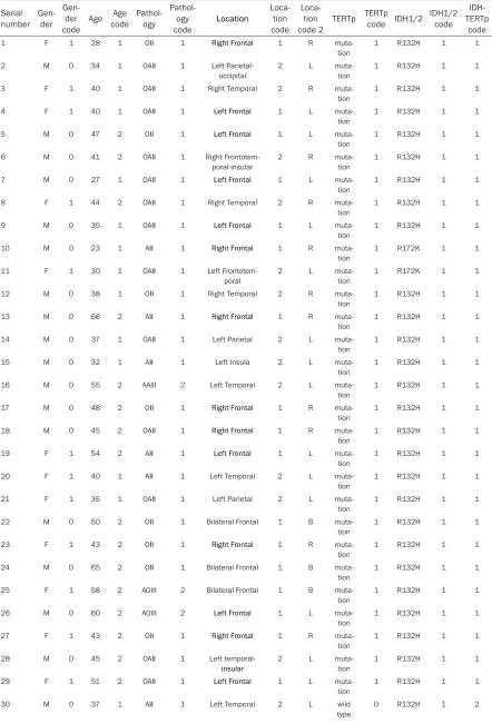
Table S1. Details about clinical and molecular data of 225 WHO grade II and III gliomas Serial
number Gen-der
Gen-der
code Age Age code
Pathol-ogy
Pathol-ogy
code Location
Loca-tion code
Loca-tion
code 2 TERTp TERTp code IDH1/2 IDH1/2
code IDH-TERTp
code
1 F 1 28 1 OII 1 Right Frontal 1 R
muta-tion 1 R132H 1 1
2 M 0 34 1 OAII 1 Left
Parietal-occipital 2 L muta-tion 1 R132H 1 1
3 F 1 40 1 OAII 1 Right Temporal 2 R
muta-tion
1 R132H 1 1
4 F 1 40 1 OAII 1 Left Frontal 1 L
muta-tion
1 R132H 1 1
5 M 0 47 2 OII 1 Left Frontal 1 L
muta-tion 1 R132H 1 1
6 M 0 41 2 OAII 1 Right
Frontotem-poral-insular 2 R muta-tion 1 R132H 1 1
7 M 0 27 1 OAII 1 Left Frontal 1 L
muta-tion
1 R132H 1 1
8 F 1 44 2 OAII 1 Right Temporal 2 R
muta-tion
1 R132H 1 1
9 M 0 35 1 OAII 1 Left Frontal 1 L
muta-tion 1 R132H 1 1
10 M 0 23 1 AII 1 Right Frontal 1 R
muta-tion 1 R172K 1 1
11 F 1 30 1 OAII 1 Left
Frontotem-poral
2 L
muta-tion
1 R172K 1 1
12 M 0 38 1 OII 1 Right Temporal 2 R
muta-tion
1 R132H 1 1
13 M 0 66 2 AII 1 Right Frontal 1 R
muta-tion 1 R132H 1 1
14 M 0 37 1 OAII 1 Left Parietal 2 L
muta-tion 1 R132H 1 1
15 M 0 32 1 AII 1 Left Insula 2 L
muta-tion
1 R132H 1 1
16 M 0 55 2 AAIII 2 Left Temporal 2 L
muta-tion
1 R132H 1 1
17 M 0 48 2 OII 1 Right Frontal 1 R
muta-tion 1 R132H 1 1
18 M 0 45 2 OAII 1 Right Frontal 1 R
muta-tion 1 R132H 1 1
19 F 1 54 2 AII 1 Left Frontal 1 L
muta-tion
1 R132H 1 1
20 F 1 40 1 AII 1 Left Temporal 2 L
muta-tion
1 R132H 1 1
21 F 1 35 1 OAII 1 Left Parietal 2 L
muta-tion 1 R132H 1 1
22 M 0 50 2 OII 1 Bilateral Frontal 1 B
muta-tion 1 R132H 1 1
23 F 1 43 2 OII 1 Right Frontal 1 R
muta-tion
1 R132H 1 1
24 M 0 65 2 OII 1 Bilateral Frontal 1 B
muta-tion
1 R132H 1 1
25 F 1 58 2 AOIII 2 Bilateral Frontal 1 B
muta-tion 1 R132H 1 1
26 M 0 60 2 AOIII 2 Left Frontal 1 L
muta-tion 1 R132H 1 1
27 F 1 43 2 OII 1 Right Frontal 1 R
muta-tion
1 R132H 1 1
28 M 0 45 2 OAII 1 Left
temporal-insular
2 L
muta-tion
1 R132H 1 1
29 F 1 51 2 OAII 1 Left Frontal 1 L
muta-tion 1 R132H 1 1
31 F 1 42 2 OAII 1 Left Parietal 2 L wild
type 0 R132H 1 2
32 F 1 42 2 OII 1 Right Frontal 1 R wild
type 0 R132H 1 2
33 M 0 26 1 OAII 1 Right Frontal 1 R wild type
0 R132H 1 2
34 M 0 57 2 OAII 1 Left Frontal 1 L wild
type
0 R132H 1 2
35 F 1 37 1 AII 1 Left Frontal 1 L wild
type
0 R132H 1 2
36 M 0 37 1 OAII 1 Right
Temporal-occipital and Lateral ventricle
2 R wild
type 0 R132H 1 2
37 M 0 49 2 OII 1 Left Frontal 1 L wild
type
0 R132H 1 2
38 F 1 41 2 OAII 1 Bilateral Frontal 1 B wild
type 0 R132H 1 2
39 M 0 50 2 OAII 1 Right Frontal 1 R ND ND R132H 1 ND
40 F 1 40 1 AII 1 Left Frontal 1 L
muta-tion 1 R132H 1 1
41 F 1 36 1 OII 1 Left Frontal 1 L
muta-tion 1 R132H 1 1
42 F 1 32 1 AII 1 Left Temporal 2 L
muta-tion
1 R132H 1 1
43 M 0 47 2 AII 1 Right Frontal 1 R
muta-tion
1 R132H 1 1
44 M 0 41 2 AAIII 2 Left Frontal 1 L
muta-tion 1 R132H 1 1
45 M 0 53 2 OII 1 Right Parietal 2 R
muta-tion 1 R132H 1 1
46 M 0 48 2 AII 1 Right Temporal 2 R
muta-tion
1 R132H 1 1
47 F 1 31 1 AII 1 Left Temporal 2 L
muta-tion
1 R132H 1 1
48 F 1 32 1 OII 1 Right Frontal 1 R
muta-tion 1 R132H 1 1
49 M 0 32 1 AOAIII 2 Right Frontal 1 R
muta-tion 1 R132H 1 1
50 F 1 34 1 AOIII 2 Left Frontoparietal 2 L
muta-tion
1 R132H 1 1
51 M 0 45 2 OAII 1 Right Frontal 1 R muta-tion
1 R132H 1 1
52 F 1 49 2 OII 1 Bilateral
Fronto-parietal 2 B muta-tion 1 R132H 1 1
53 F 1 42 2 OII 1 Left Temporal 2 L
muta-tion 1 R132H 1 1
54 M 0 36 1 AOAIII 2 Bilateral
Fronto-parietal
2 B
muta-tion
1 R132H 1 1
55 F 1 33 1 OAII 1 Left Frontal 1 L
muta-tion
1 R132H 1 1
56 M 0 49 2 OAII 1 Right
Frontotem-poral-insular 2 R muta-tion 1 R132H 1 1
57 F 1 58 2 AII 1 Right Frontal 1 R
muta-tion 1 wild type 0 2
58 F 1 36 1 AAIII 2 Right Frontal 1 R
muta-tion
1 wild
type
0 2
59 M 0 50 2 AAIII 2 Left Temporal 2 L
muta-tion
1 wild
type
0 2
60 F 1 46 2 AII 1 Cingulum 2 X
muta-tion 1 wild type 0 2
61 M 0 48 2 AAIII 2 Intraventricular 3 X
muta-tion 1 wild type 0 2
62 M 0 65 2 AAIII 2 Intraventricular 3 X
muta-tion
1 wild
type
63 F 1 57 2 AII 1 Right Frontal 1 R
muta-tion 1 wild type 0 2
64 M 0 60 2 AAIII 2 Right Temporal 2 R
muta-tion 1 wild type 0 2
65 M 0 70 2 AAIII 2 Right Parietal 2 R
muta-tion
1 wild
type
0 2
66 M 0 13 1 AAIII 2 Bilateral Parietal 2 B
muta-tion
1 wild
type
0 2
67 F 1 54 2 AAIII 2 Right Frontal 1 R
muta-tion
1 wild
type
0 2
68 M 0 48 2 AAIII 2 Right Frontal 1 R
muta-tion 1 wild type 0 2
69 M 0 46 2 AII 1 Right Temporal 2 R
muta-tion 1 wild type 0 2
70 F 1 45 2 AII 1 Left Parietal 2 L wild
type
0 R132H 1 2
71 M 0 38 1 AII 1 Right Frontal 1 R wild
type
0 R132H 1 2
72 F 1 46 2 AII 1 Left Temporal 2 L wild
type 0 R132H 1 2
73 M 0 39 1 AII 1 Right Frontal 1 R wild
type 0 R132H 1 2
74 M 0 58 2 AII 1 Right Temporal 2 R wild
type
0 R132H 1 2
75 M 0 32 1 AII 1 Left Parietal 2 L wild
type
0 R132H 1 2
76 M 0 34 1 AII 1 Left
Frontotempo-ral-insular 2 L wild type 0 R132H 1 2
77 F 1 52 2 OAII 1 Left Frontoparietal 2 L wild
type 0 R172K 1 2
78 F 1 42 2 OAII 1 Right Frontal 1 R wild type
0 R132H 1 2
79 F 1 35 1 OAII 1 Bilateral
Cerebel-lum
2 B wild
type
0 R132S 1 2
80 F 1 47 2 AII 1 Right
Frontotem-poral 2 R wild type 0 R132H 1 2
81 M 0 26 1 AII 1 Right Frontal 1 R wild
type 0 R132H 1 2
82 M 0 41 2 AII 1 Left Temporal 2 L wild
type
0 R132H 1 2
83 M 0 49 2 AII 1 Right Frontal 1 R wild
type
0 R132H 1 2
84 M 0 38 1 OAII 1 Right Frontal 1 R wild
type 0 R132H 1 2
85 F 1 55 2 OII 1 Right
Frontotem-poral 2 R wild type 0 R132H 1 2
86 M 0 79 2 AII 1 Left Frontal 1 L wild
type
0 R132H 1 2
87 M 0 28 1 AII 1 Left Temporal 2 L wild
type
0 R132H 1 2
88 F 1 46 2 OAII 1 Left
Frontotem-poral 2 L wild type 0 R132H 1 2
89 M 0 54 2 AII 1 Right
Frontotem-poral 2 R wild type 0 R132H 1 2
90 M 0 32 1 AII 1 Left Frontal 1 L wild
type
0 R132H 1 2
91 M 0 36 1 AII 1 Left Temporal 2 L wild
type
0 R132H 1 2
92 M 0 36 1 AII 1 Left Frontal 1 L wild
type 0 R132H 1 2
93 M 0 32 1 AII 1 Right Temporal 2 R wild
type 0 R132H 1 2
94 F 1 43 2 AII 1 Corpus callosum 2 X wild
type
95 M 0 31 1 AII 1 Left Temporal 2 L wild
type 0 R132H 1 2
96 M 0 29 1 AII 1 Left Insula 2 L wild
type 0 R132H 1 2
97 F 1 60 2 AII 1 Left Frontal 1 L wild
type
0 R132H 1 2
98 M 0 42 2 AII 1 Right Parietal 2 R wild
type
0 R132H 1 2
99 M 0 46 2 AII 1 Left
Frontotem-poral
2 L wild
type
0 R132H 1 2
100 M 0 31 1 AII 1 Right Temporal 2 R wild
type 0 R132H 1 2
101 M 0 46 2 AII 1 Left Frontal 1 L wild
type 0 R132H 1 2
102 F 1 34 1 OAII 1 Left Frontal 1 L wild type
0 R132H 1 2
103 M 0 68 2 AII 1 Left Frontal 1 L wild
type
0 R132H 1 2
104 F 1 45 2 AAIII 2 Left Temporal 2 L wild
type 0 R132H 1 2
105 M 0 49 2 AAIII 2 Right lateral
ven-tricle trigonal 3 X wild type 0 R132H 1 2
106 F 1 48 2 AAIII 2 Right
Frontopa-rietal
2 R wild
type
0 R132H 1 2
107 M 0 51 2 AAIII 2 Left Frontal 1 L wild
type
0 R132H 1 2
108 M 0 41 2 AAIII 2 Left Frontal 1 L wild
type 0 R132H 1 2
109 M 0 39 1 AAIII 2 Right Frontal 1 R wild
type 0 R132H 1 2
110 M 0 31 1 AAIII 2 Left
Frontotempo-ral-insular
2 L wild
type
0 R132H 1 2
111 F 1 42 2 OAII 1 Left Frontal 1 L wild type
0 R132H 1 2
112 F 1 31 1 AII 1 Left Frontal 1 L wild
type 0 R132H 1 2
113 M 0 40 1 AAIII 2 Bilateral Frontal 1 B wild
type 0 R132H 1 2
114 M 0 43 2 OAII 1 Right Frontal 1 R wild type
0 R132H 1 2
115 M 0 39 1 OAII 1 Left Temporal 2 L wild
type
0 R132H 1 2
116 M 0 38 1 AII 1 Left Parietal 2 L wild
type 0 R132H 1 2
117 M 0 28 1 AII 1 Left Frontal 1 L wild
type 0 R132H 1 2
118 F 1 50 2 AII 1 Right Parietal 2 R wild
type
0 R132H 1 2
119 F 1 49 2 AII 1 Left Temporal 2 L wild
type
0 R132H 1 2
120 F 1 33 1 AII 1 Right Insular 2 R wild
type 0 R132H 1 2
121 M 0 38 1 AII 1 Left Temporal 2 L wild
type 0 R132H 1 2
122 F 1 31 1 AII 1 Left Frontal 1 L wild
type
0 R132H 1 2
123 M 0 36 1 AII 1 Right Frontal 1 R wild type
0 R132H 1 2
124 F 1 34 1 AAIII 2 Frontal and
Cor-pus collosum 2 X wild type 0 R132H 1 2
125 M 0 31 1 AAIII 2 Brainstem 3 X wild
type 0 R132H 1 2
126 F 1 46 2 AAIII 2 Left Frontal 1 L wild
type
127 F 1 46 2 AII 1 Left Parietal 2 L wild
type 0 R132H 1 2
128 F 1 35 1 AAIII 2 Left
Frontotem-poral 2 L wild type 0 R132H 1 2
129 M 0 37 1 AAIII 2 Left Parietal 2 L wild
type
0 R132H 1 2
130 M 0 38 1 AII 1 Right Temporal 2 R wild
type
0 R132H 1 2
131 M 0 60 2 AAIII 2 Corpus callosum 2 X wild
type
0 R132H 1 2
132 M 0 38 1 AII 1 Right Frontal 1 R wild
type 0 R132H 1 2
133 M 0 35 1 AII 1 Left Temporal 2 L wild
type 0 R132H 1 2
134 M 0 32 1 AII 1 Left
Frontotempo-ral-insular
2 L wild
type
0 R132H 1 2
135 M 0 39 1 AII 1 Right Frontal 1 R wild type
0 R132H 1 2
136 M 0 42 2 OAII 1 Left Parietal and
Cental sulcus 2 L wild type 0 R132H 1 2
137 M 0 29 1 AII 1 Left Temporal 2 L wild
type 0 R132H 1 2
138 F 1 24 1 AII 1 Bilateral Frontal 1 B wild
type
0 R132H 1 2
139 M 0 46 2 AII 1 Bilateral Frontal 1 B wild
type
0 R132H 1 2
140 F 1 39 1 AII 1 Left Temporal 2 L wild
type 0 R132H 1 2
141 M 0 27 1 AAIII 2 Right Frontal 1 R wild
type 0 R132H 1 2
142 F 1 36 1 AII 1 Right Parietal 2 R wild
type
0 R132H 1 2
143 M 0 39 1 AII 1 Right Temporal 2 R wild
type
0 R132H 1 2
144 F 1 34 1 AII 1 Left Frontal 1 L wild
type 0 R132H 1 2
145 F 1 33 1 OAII 1 Right
Parietal-occipital 2 R wild type 0 R132H 1 2
146 F 1 30 1 OAII 1 Left Frontoparietal 2 L wild
type
0 R132H 1 2
147 M 0 35 1 AII 1 Left Temporal 2 L wild
type
0 R132H 1 2
148 M 0 33 1 OII 1 Left Parietal 2 L wild
type 0 R132H 1 2
149 F 1 46 2 AOAIII 2 Right Frontal 1 R wild
type 0 R132H 1 2
150 M 0 36 1 OAII 1 Left
Frontal-insular
2 L wild
type
0 R132H 1 2
151 M 0 32 1 OAII 1 Left Temporal 2 L wild
type
0 R132H 1 2
152 F 1 61 2 OAII 1 Right Frontal 1 R wild
type 0 R132H 1 2
153 F 1 47 2 AII 1 Left Frontal 1 L wild
type 0 R132H 1 2
154 F 1 25 1 OAII 1 Left Frontal 1 L wild type
0 wild
type
0 3
155 M 0 24 1 AII 1 Left Parietal 2 L wild
type
0 wild
type
0 3
156 F 1 40 1 OAII 1 Right Occipital 2 R wild
type 0 wild type 0 3
157 F 1 28 1 AII 1 Left Cerebellum 2 L wild
type 0 wild type 0 3
158 F 1 21 1 AII 1 Right Thalamus 3 X wild
type
0 wild
type
159 F 1 25 1 OAII 1 Right lateral
ven-tricle trigonal 3 X wild type 0 wild type 0 3
160 M 0 22 1 AII 1 Right Temporal 2 R wild
type 0 wild type 0 3
161 M 0 23 1 AII 1 Right Temporal 2 R wild
type
0 wild
type
0 3
162 M 0 35 1 AII 1 Left Parietal 2 L wild
type
0 wild
type
0 3
163 F 1 41 2 AII 1 Left Parietal 2 L wild
type
0 wild
type
0 3
164 F 1 29 1 AII 1 Left Parietal 2 L wild
type 0 wild type 0 3
165 M 0 52 2 AAIII 2 Right Temporal 2 R wild
type 0 wild type 0 3
166 M 0 26 1 AAIII 2 Bilateral Frontal
and Corpus Cal-losum
2 B wild
type
0 wild
type
0 3
167 M 0 38 1 AAIII 2 Right lateral
ven-tricle trigonal 3 X wild type 0 wild type 0 3
168 M 0 53 2 AII 1 Left
Frontotem-poral
2 L wild
type
0 wild
type
0 3
169 F 1 63 2 AAIII 2 Right
Tempopa-rietal
2 R wild
type
0 wild
type
0 3
170 M 0 65 2 AAIII 2 Left Parietal 2 L wild
type 0 wild type 0 3
171 M 0 45 2 AAIII 2 Left Frontal 1 L wild
type 0 wild type 0 3
172 M 0 34 1 AAIII 2 Left Frontal 1 L wild
type
0 wild
type
0 3
173 F 1 44 2 AAIII 2 Left Frontal 1 L wild
type
0 wild
type
0 3
174 M 0 50 2 AAIII 2 Left
Frontotem-poral 2 L wild type 0 wild type 0 3
175 M 0 58 2 AAIII 2 Right
Frontotem-poral-insular 2 R wild type 0 wild type 0 3
176 M 0 38 1 AAIII 2 Thoracic code
T6-8
3 X wild
type
0 wild
type
0 3
177 F 1 43 2 AAIII 2 Right Occipital 2 R wild
type
0 wild
type
0 3
178 M 0 34 1 AII 1 Right Occipital 2 R wild
type 0 wild type 0 3
179 F 1 51 2 AII 1 Corpus callosum 2 X wild
type 0 wild type 0 3
180 M 0 48 2 AAIII 2 Right Cerebellum 2 R wild
type
0 wild
type
0 3
181 M 0 51 2 AAIII 2 Right Basal
ganglion
3 X wild
type
0 wild
type
0 3
182 F 1 34 1 AAIII 2 Left Frontal 1 L wild
type 0 wild type 0 3
183 M 0 27 1 AAIII 2 Brainstem 3 X wild
type 0 wild type 0 3
184 M 0 33 1 AII 1 Intraventricular 3 X wild
type
0 wild
type
0 3
185 M 0 40 1 AII 1 Right Temporal 2 R wild
type
0 wild
type
0 3
186 M 0 14 1 AII 1 Right Temporal 2 R wild
type 0 wild type 0 3
187 M 0 45 2 AII 1 Right Temporal 2 R wild
type 0 wild type 0 3
188 M 0 48 2 AII 1 Left Thalamus,
Basal ganglion and midbrain
3 X wild
type
0 wild
type
0 3
189 F 1 46 2 AII 1 Left Parietal 2 L wild
190 M 0 58 2 AII 1 Right Insular 2 R wild
type 0 wild type 0 3
191 M 0 18 1 AAIII 2 Brainstem 3 X wild
type 0 wild type 0 3
192 M 0 59 2 AAIII 2 Left Parietal 2 L wild
type
0 wild
type
0 3
193 M 0 34 1 AAIII 2 Left Thalamus 3 X wild
type
0 wild
type
0 3
194 M 0 47 2 AII 1 Left Thalamus 3 X wild
type
0 wild
type
0 3
195 M 0 36 1 AII 1 Right Thalamus 3 X wild
type 0 wild type 0 3
196 M 0 46 2 AII 1 Brainstem 3 X wild
type 0 wild type 0 3
197 M 0 47 2 AII 1 Left Thalamus 3 X wild
type
0 wild
type
0 3
198 M 0 42 2 AAIII 2 Right Frontal 1 R wild
type
0 wild
type
0 3
199 M 0 59 2 AAIII 2 Left
temporal-insular 2 L wild type 0 wild type 0 3 200 F 1 56 2 AII 1 Right Basal
ganglion 3 X wild type 0 wild type 0 3 201 F 1 65 2 AII 1 Right Frontal 1 R wild
type
0 wild
type
0 3
202 M 0 3 1 AAIII 2 Right Temporal 2 R wild
type
0 wild
type
0 3
203 F 1 13 1 OII 1 Left Temporal 2 L wild
type 0 wild type 0 3
204 M 0 12 1 OAII 1 Right Parietal 2 R wild
type 0 wild type 0 3
205 M 0 16 1 OAII 1 Right Frontal 1 R wild type
0 wild
type
0 3
206 M 0 7 1 OAII 1 Brainstem 3 X wild
type
0 wild
type
0 3
207 M 0 8 1 OAII 1 Right Temporal 2 R wild
type 0 wild type 0 3
208 F 1 48 2 OAII 1 Right Frontal 1 R wild
type 0 wild type 0 3
209 M 0 55 2 OAII 1 Right
Frontotem-poral
2 R
muta-tion
1 R132H 1 1
210 M 0 28 1 OII 1 Left Temporal 2 L
muta-tion
1 R132H 1 1
211 M 0 27 1 AII 1 Left Frontal 1 L wild
type 0 R132H 1 2
212 F 1 33 1 AII 1 Left Frontal 1 L wild
type 0 R132H 1 2
213 F 1 63 2 AAIII 2 Right Frontal 1 R wild
type
0 wild
type
0 3
214 F 1 31 1 AII 1 Thoracic code
T8-10
3 X wild
type
0 wild
type
0 3
215 M 0 37 1 AII 1 Right
Frontopa-rietal 2 R ND ND R132H 1 ND
216 M 0 40 1 AAIII 2 Left Temporal 2 L ND ND R132H 1 ND
217 M 0 44 2 AAIII 2 Left Temporal 2 L ND ND R132H 1 ND
218 M 0 39 1 OAII 1 Right
Frontopa-rietal
2 R ND ND R132H 1 ND
219 M 0 40 1 AAIII 2 Right
Frontopa-rietal 2 R ND ND R132H 1 ND
220 M 0 30.5 1 OII 1 Right
Tempopa-rietal 2 R ND ND wild type 0 ND
221 M 0 60 2 AOIII 2 Right Temporal 2 R ND ND wild
type
222 M 0 28 1 AAIII 2 Right Temporal-occipital and Left
Occipital
2 R ND ND wild
type 0 ND
223 F 1 62.5 2 AAIII 2 Left Temporal 2 L ND ND wild
type
0 ND
224 F 1 58 2 AII 1 Bilateral Frontal
and Right Tem-poral
2 B ND ND wild
type 0 ND
225 F 1 38 1 AOIII 2 Left Frontal 1 L ND ND wild
type
0 ND
Abbreviations: Gender: F=female; M=male; Gender code: 1=F, 0=M; Age code: 1=younger than or qual to 40 years old, 2=older than 40 years old; Pathology: AII=astrocytoma WHO Grade II, OAII=oligoastrocytoma WHO Grade II, OII=oligodendroglioma WHO Grade II, AAIII=anaplastic astrocytoma WHO Grade III, AOAIII=anaplastic oligoastrocytoma WHO Grade III, AOIII=anaplastic oligodendroglioma WHO Grade III; Pathology code: 1=WHO Grade II, 2=WHO Grade III; Location code: 1=Frontal, 2=Others, 3=Midline; Loca-tion code 2: B=Bilateral, L=Left, R=Right, X=not determine; TERTp code: 1=mutaLoca-tion, 0=wild type; IDH1/2 code: 1=mutaLoca-tion, 0=wild type; IDH-TERTp code: 1=IDHmut-TERTpmut,