Original Article
Methylation-silencing
RCC1
expression is associated
with tumorigenesis and depth of invasion
in gastric cancer
Yi-Ling Lin1,2*, Hsiao-Ling Chen3*, Shao-Bin Cheng2,8, Dah-Cherng Yeh2, Chu-Chun Huang2, Fang-Ku P’eng4,
Tung-Chou Tsai1, Cheng-Chung Wu2,4,5,6, Chuan-Mu Chen1,7
1Department of Life Sciences, and Agricultural Biotechnology Center, National Chung Hsing University, Taichung
402, Taiwan; 2Department of General Surgery, Taichung Veterans General Hospital, Taichung 407, Taiwan; 3Department of Bioresources, Da-Yeh University, Changhwa 515, Taiwan; 4Department of Surgery, Faculty of
Medicine, National Yang-Ming University, Taipei 112, Taiwan; 5Department of Surgery, Chung-Shan Medical
University, Taichung 402, Taiwan; 6Department of Surgery, Taipei Medical University, Taipei 110, Taiwan; 7
Rong-Hsing Translational Medicine Center, and iEGG Center, National Chung Rong-Hsing University, Taichung 402, Taiwan;
8School of Medicine, Chung-Shan Medical University, Taichung 402, Taiwan
Received September 29, 2015; Accepted October 28, 2015; Epub November 1, 2015; Published November 15, 2015
Abstract: Introduction: Regulator of chromosome condensation 1 (RCC1) is a critical cell cycle regulator. We first-ly identified RCC1 gene hypermethylation in gastric tumor tissues using the differential methylation hybridization (DMH) microarray, but the role of RCC1 in the pathogenesis of gastric carcinoma is largely unknown. Methods: Three gastric cancer cell lines (AGS, MKN45, and TSGH9201) were used to analyze RCC1 gene methylation, mRNA and protein expressions. Furthermore, 85 pairs of matched human gastric carcinoma samples in a tissue microarray were used to analyze RCC1 expression by immunohistochemistry staining. Results: A differential methylation pat-tern was found in TSGH9201 (100%), MKN45 (87%), and AGS (62%) cell lines at the 9th CpG site of RCC1 exon 1. RCC1 mRNA and protein expressions in AGS cells were significantly higher than in TSGH9201 and MKN45 cell lines (P < 0.05). Tissue array data showed that RCC1 expression was detected in 21% (18/85) of gastric carcinoma tissues and in 80% (76/95) of adjacent non-tumor tissues. The expression of RCC1 in gastric carcinoma tissues was significantly lower than in adjacent non-tumor tissues (P < 0.001). Furthermore, an association between RCC1 expression and clinicopathological features showed that RCC1 expression was closely correlated with tumor differ-entiation and depth of invasion (P < 0.05). Conclusions: Our data indicate that RCC1 expression is frequently lost in poorly differentiated gastric cell lines and gastric carcinoma tissues. Loss of RCC1 expression is correlated with tumor differentiation and depth of invasion. These findings suggest that RCC1 may play a tumor suppressor role in gastric carcinoma.
Keywords: DMH microarray, gastric carcinoma, RCC1, DNA methylation, tumor tissue array, immunohistochemis-try, invasion
Introduction
Despite a steady decline in gastric cancer inci-dence, gastric cancer still represents a major health problem [1]. It remains one of the most common malignancies, particularly in Eastern Asian countries, including Taiwan [2, 3]. Most gastric cancer patients present with advanced-stage disease [4]. The clinical outcome of gas-tric cancer has gradually improved, but the overall 5-year survival rate of gastric cancer patients remains approximately 20% [5].
leiomyo-sarcomas, non-Hodgkin lymphomas, and undif-ferentiated carcinomas [6, 7].
Gastric cancer has marked heterogeneity in its morphology and biology. Based on morphologi-cal characteristics of gland formation and his-togenetic background, gastric adenocarcino-mas can be divided into intestinal and diffuse types using the Lauren classification system [8] or as differentiated and undifferentiated using the Nakamura classification system [9]. Intes- tinal-type adenocarcinoma is considered to be equivalent to differentiated adenocarcinoma, and diffuse-type adenocarcinoma is consid-ered to be equivalent to undifferentiated ade-nocarcinoma. These two gastric cancer types also display different biological and etiological characteristics. Tumor cells of the intestinal type form glandular-like structures, a feature that is lacking in the diffuse type. On the con-trary, the diffuse type of gastric cancer is char-acterized by an infiltration and thickening of the gastric wall by tumor cells. The two histological subtypes are the result of distinct pathogenic pathways [10, 11]. Previous reports have also found that Helicobacter pylori infection leads to the development of chronic atrophic gastritis and intestinal metaplasia [12, 13].
Usually, the intestinal type of gastric cancer develops on a background of intestinal meta-plasia; by contrast, the diffuse type develops on a background of gastric mucosa without intestinal metaplasia. The diffuse type of gas-tric cancer is characterized by reduced or abnormal E-cadherin expression [10]. The dif-fuse type has a worse prognosis and tends to develop in young patients; it can have any type of gastric localization, but it frequently devel-ops at the level of the cardia. Although altera-tions in a large number of oncogenic and tumor-suppressive genes are reportedly implicated in gastric cancer [14-17], the molecular mecha-nisms underlying the development of gastric cancer are still poorly understood. Identification of its tumorigenesis mechanism is necessary for the development of targeted clinical therapy.
Regulator of chromosome condensation 1 (RCC1) has been identified as a critical cell cycle regulator [18, 19]. RCC1 is a guanine nucleotide releasing factor that promotes the exchange of Ran-bound GDP by GTP [20]. RCC1 plays a key role in nucleo-cytoplasmic trans-port, mitosis and nuclear envelope assembly
and is involved in regulating the onset of chro-mosome condensation during S phase [18, 21]. RCC1 can bind both to nucleosomes and to double-stranded DNA, and the RCC1-Ran com-plex (together with other proteins) is a compo-nent in a signal transmission pathway that detects unreplicated DNA [21, 22]. This has led to the proposal that RCC1 is a signaling mole-cule, detecting unreplicated DNA and produc-ing an inhibitory signal. However, the associa-tion between RCC1 expression and gastric car-cinoma has not been fully studied.
To assess the role of RCC1 in gastric carcino-ma, RCC1 expression was detected in different gastric cancer cell lines, gastric carcinomas and adjacent non-tumor tissues to evaluate the relationship between RCC1 expression and the clinicopathological characteristics of gastric carcinoma. The DNA methylation status of the
RCC1 gene in three different gastric cancer cell lines was also elucidated.
Materials and methods
Differential methylation hybridization (DMH) microarray assay
The protocol of differential methylation hybrid-ization (DMH) on human CpG island microarray was based on our previously described [23, 24]. Briefly, the purified methylation amplicons (5 μg) from eight pairs of gastric tumor and non-tumor obtained from the tissue bank of Taichung Veterans General Hospital, Taiwan, were conducted using the BioPrime DNA label-ing system (Invitrogen, CA, USA). Cyanine 5-ddUTP (Cy5-ddUTP) and Cyanine 3-ddUTP (Cy3-ddUTP) (Perkin-Elmer Life Sciences, NJ, USA) fluorescent dyes were coupled to tumor (T) and normal (NT) amplicons, respectively, and cohybridized to the microarray panel. The hybridization of DMH microarray was performed within a moistened hybridization chamber, GeneMachines HybChambers (Genomic So- lutions, MI, USA), in a 65°C water bath from 12 to 16 h. The slides were scanned with the GenePix 4000B scanner (Axon, CA, USA) and the acquired images were analyzed with the software GenePix Pro 4.0 (Axon, CA, USA). The microarray data was analyzed as described previously [25].
Cell culture
Type Culture Collection (ATCC; Rockville, MD, USA) and Bioresource Collection and Research Center (BCRC; Hsinchu, Taiwan). The AGS, MKN45 and TSGH9201 cells were kept in F-12K, DMEM, and RPMI 1640 media (Gibco, Carlsbad, CA, USA), respectively, which was supplemented with 10% fetal bovine serum (FBS) as well as 100 U/ml penicillin and 100 μg/ml streptomycin (Invitrogen, Carlsbad, CA, USA). All of the cells were maintained at 37°C under an atmosphere of 5% CO2.
Quantitative reverse transcription PCR (qRT-PCR)
Total RNA was extracted with Trizol reagent (Invitrogen, Carlsbad, CA, USA) according to the manufacturer’s protocol. First-strand cDNA was synthesized from total RNA using a reverse-transcriptase system with the SuperScriptIII RT enzyme (Invitrogen, Carlsbad, CA, USA). The resulting cDNA was amplified using a QuantiFast Probe Assay kit (Qiagen, Foster, CA, USA) on an ABI SteponeTM Real-Time PCR system (Applied
Biosystems, Foster, CA, USA). Relative quantifi-cation of target gene (RCC1 and GAPDH) expression was evaluated using the compara-tive CT method in which ΔCT represents the nor-malization of CT for a target gene relative to TBP. ΔΔCT was calculated by comparing the mean ΔCT values. Fold changes in gene expres-sion were equivalent to 2-ΔΔC
T [26-28].
Western blot analysis
The gastric cancer cell lines were homogenized in lysis buffer containing protease inhibitor cocktail. Protein concentrations were mea-sured by the Bradford method [29]. Before sep-aration, the samples were re-suspended in loading buffer, denatured by boiling for 5 min and separated by 12% SDS-PAGE gel electro-phoresis. The proteins were transferred to Trans-Blot polyvinylidene fluoride membranes, and the membranes were probed with a prima-ry rabbit polyclonal antibody against human RCC1 (GeneTex Inc., Irvine, CA, USA) at a dilu-tion of 1:12,000 for 3 h. The blots were washed in TBST and incubated with a goat anti-rabbit IgG horseradish peroxidase-conjugated sec-ondary antibody. The membranes were devel-oped using an enhanced chemiluminescence detection system (Millipore, Bedford, MA, USA) by exposure to x-ray film [30]. A housekeeping protein, β-actin, was used as an internal control for the quantification of protein expression lev-els as described previously [31].
Primer design and PCR amplification for gene
methylation study
The primers that were designed to amplify
RCC1 covered the regions of the gene with the most CpG sites. Most of the selected ampli-cons were located in the promoter region and in exon 1, which spanned from -517 to 466 nucle-otides (nt) relative to the transcription start site. The primers were designed using EpiDesigner software (http://www.epidesigner. com/) and are listed in Table S1. Genomic DNA was treated with sodium bisulfite as described previously [32, 33], and the bisulfite-treated genomic DNA was then amplified under the fol-lowing conditions: initial denaturation, 94°C for 4 min, 45 cycles of 94°C for 20 sec, 56°C for 30 sec, and 72°C for 1 min, followed by 3 min at 72°C. The products were stored at 4°C for further analysis.
In vitro transcription and T-cleavage (RNase A digestion) assay
To neutralize unincorporated dinucleotide tri-phosphates (dNTPs) after amplification reac-tions, 0.3 U of shrimp alkaline phosphatase (SAP; Sequenom, San Diego, CA, USA) was used [34]. The components of the reaction included 1.7 μL of RNase-free ddH2O and 0.3 μL of SAP, which were combined with the above PCR products. The mixture was incubated at 37°C for 20 min, 85°C for 5 min, and then at 4°C until use. Using T7 RNA polymerase, thymi-dine triphosphate was incorporated into the PCR product, finishing the transcription reac-tion. RNase A was added to the same reaction to cleave the transcripts (T-cleavage assay). The reaction mixture was incubated at 94°C for 30 sec, followed by 40 cycles of 94°C for 5 sec, 52°C for 5 sec, 5 cycles of 80°C for 5 sec and 72°C for 3 min, after which it was stored at 4°C. To remove the phosphate backbone, the T-cleavage/RNase A reaction products were diluted with RNase-free water and mixed with clean resin before performing MALDI-TOF mass spectrometry [34].
MALDI-TOF mass spectrometry for DNA meth-ylation measurement
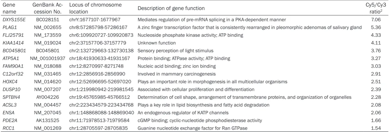
Table 1. The hypermethylation gene list selected from gastric cancer samples in DMH microarray analysis1
Gene
name GenBank Ac-cession No. Locus of chromosome location Description of gene function Cy5/Cy3 ratio2 DXYS155E BC028151 chrY:1677107-1677967 Mediates regulation of pre-mRNA splicing in a PKA-dependent manner 7.06
PLAG1 NM_002655 chr8:57285798-57286167 A zinc finger transcription factor that is consistently rearranged in pleomorphic adenomas of salivary gland 5.36
FLJ25791 NM_173559 chr6:109920727-109920873 Nucleoside phosphate kinase activity; ATP binding 4.33
KIAA1414 NM_019024 chr2:37157706-37157779 Unknown function 4.11
BC045801 BC045801 chr2:132729663-132730138 Sensory perception of light stimulus 3.76
ATP5A1 NM_001001937 chr18:41930633-41931167 Protein binding; ATPase activity; ATP binding 3.27
FAM90A1 NM_018088 chr12:8270997-8271748 Nucleic acid binding; zinc ion binding 3.03
C12orf32 NM_031465 chr12:2856916-2856990 Involved in mammary carcinogenesis 2.91
HOXC4 NM_014620 chr12:52696695-52697020 Plays an important role in morphogenesis in all multicellular organisms 2.51
DUSP10 NM_007207 chr1:219980942-219981545 Associated with cellular proliferation and differentiation 2.39
SPTBN4 AY004226 chr19:45765985-45766512 Determination of cell shape, arrangement of transmembrane proteins, and organization of organelles 2.28
ACSL3 NM_004457 chr2:223434579-223434768 Plays a key role in lipid biosynthesis and fatty acid degradation 2.08
ENSA NM_207045 chr1:148868088-148869040 An endogenous regulator of KATP channels 2.06
PDE2A AK131525 chr11:71978513-71979584 cGMP binding; cyclic-nucleotide phosphodiesterase activity 1.66
RCC1 NM_001269 chr1:28705597-28705835 Guanine nucleotide exchange factor for Ran GTPase 1.54
1DMH microarray: The differential methylation hybridization (DMH) microarray is a high-throughput microarray technique designed to identify changes in DNA methylation patterns as our previous reports
Sequenom, San Diego, CA, USA), and mass spectra were collected using a MassARRAY compact MALDI-TOF analyzer (Sequenom). Mass spectra methylation ratios were generat-ed using EpiTYPER (ver. 4.0; Sequenom).
Immunohistochemical staining of tissue micro-array
Gastric carcinoma and adjacent non-tumor tis-sue microarray (TMA) sections with stage and grade information were purchased from Biomax, Inc. (Rockville, MD, USA). We used the following tissue microarray sections, containing different numbers of cases and cores: ST1004
cally significant differences. A P-value < 0.05 was considered statistically significant.
Results
Identification of hypermethylation genes from
gastric cancer by DMH microarray
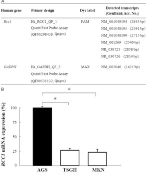
[image:5.629.100.394.86.437.2]The DMH microarray hybridization results showed that hundreds of gene spots were obvi-ously more hypermethylated in gastric tumor genomes than their adjacent non-tumor tis-sues. Yellow spots (Cy5:Cy3 = 1.0) represent equal amounts of bound DNA from each ampli-con, an indication of no methylation differences Figure 1. Validation of RCC1 mRNA expression in gastric cancer cell lines by
quantitative reverse transcription PCR (qRT-PCR). A. Primer and probe designs of RCC1 and GAPDH genes for qRT-PCR analysis. B. Quantitative data of relative
RCC1 mRNA expression levels in three different differentiation states of gastric cancer cell lines. The results are shown as the mean ± SD of three independent experiments. Each bar represents the relative RCC1 expression percentage nor-malized to the GAPDH housekeeping gene and compared to the AGS cell line.
*P=0.002 (Kruskal Wallis test).
(55 cases with 100 cores) and ST801a (40 cases with 80 cores). The sections were in duplicate cores per case. The TMA sections were processed, stained, and analyzed essentially as described previously [35]. In this study, the total num-ber of exam samples in the TMA sections included 85 pairs of human gastric can-cer tissues with adjacent non-tumor tissue plus 10 samples of normal stom-ach tissue. According to the manufacturer’s instruc-tions, the specimens were stained with an anti-RCC1 antibody (GeneTex Inc., Irvine, CA, USA), and the nuclei were counterstain- ed with hematoxylin [36]. Positive staining was de- tected as a brown color that was present in more than 25% of the cells.
Statistical analysis
statisti-between gastric tumor and non-tumor genomes. Selection of genes was based on the criteria described above. We conducted a con-firmation study to determine whether the cutoff ratio (≥1.5) could accurately identify hyper-methylation. These newly collected genes are consistent hypermethylation in all of the eight gastric cancer genomes as shown in Table 1. Upon further examination, we selected the
RCC1 gene to examine in greater details by quantitative RT-PCR, protein expression, MALDI-TOF mass spectrometry of methylation status analysis, and also the tumor tissue array assays.
mRNA expression of RCC1 in different human gastric cancer cell lines
To explore the role of RCC1 in gastric carcino-ma, we first examined the mRNA expression levels of RCC1 in various gastric cancer cell lines. The mRNA expression levels of RCC1
were quantitatively analyzed in the three gas-tric cancer cell lines using real-time PCR (Figure 1A). As shown in Figure 1B, the AGS (100%) cell
line expressed relatively higher levels of RCC1
mRNA than did the TSGH9201 (25%) and MKN45 (20%) cell lines. The relative expres-sion of RCC1 mRNA in the AGS cell line was sta-tistically significantly higher than that in the TSGH9201 and MKN45 cell lines (P < 0.05).
Protein expression levels of RCC1 in different gastric cancer cell lines
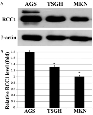
In western blot analysis, RCC1 expression was observed in all of the analyzed cell lines (Figure 2A). After being normalized to a β-actin internal control, the AGS cell line (1.8-fold) expressed more RCC1 protein than did the TSGH9201 (1.3-fold) and MKN45 (1.0-fold) cell lines, which was statistically significant (P < 0.05) and is shown in Figure 2B. These protein expression results are in concordance with the measured RNA expression, meaning that RCC1 mRNA and protein expression was higher in the AGS cell line than in the TSGH9201 and MKN45 cell lines. These gastric cancer cell lines exhib-ited different types of cell differentiation: the AGS cell line was established from a well-differ-entiated gastric epithelial cancer [37]; the TSGH9201 cell line was established from a human signet ring gastric carcinoma with a moderately differentiated cell type [38], and the MKN45 cell line was established from a poorly differentiated liver metastasis of a gas-tric carcinoma patient [39]. These results sug-gested that RCC1 expression levels may be cor-related with the differentiation status of gastric cancer cells.
MALDI-TOF mass spectrometry analysis of CpG site methylation in the RCC1 gene in different human gastric cancer cell lines
Epigenetic mechanisms, especially DNA meth-ylation, are associated with gastric carcinoma development and progression [40, 41]. We therefore analyzed the methylation patterns of the RCC1 gene in the three gastric cancer cell lines. The locations of CpG islands in the human
[image:6.629.99.296.79.323.2]RCC1 gene were predicted by EpiDesigner soft-ware, and our data showed that a typical CpG island was found within the RCC1 gene promot-er that extended to the exon 1 region, between -49 to 466 nucleotides (Figure 3A). A total of 24 potential CpG sites (Figure 3B) were ana-lyzed by MALDI-TOF mass spectrometry, and 20 CpG sites could be detected (Figure 3C). The results showed that significantly different Figure 2. Protein expression levels of RCC1 analyzed
in three different gastric cancer cell lines by west-ern blotting. A. Protein expression levels of RCC1 and β-actin in three gastric cancer cell lines (AGS,
TSGH9201, and MKN45) as determined by western
methylation levels of the RCC1 gene were found in these three gastric cell lines at a specific location in the 9th CpG site (P < 0.05).
The expression of RCC1 gene was higher in the AGS cell, which corresponded to a lower per-centage (62%) of methylation at the 9th CpG site. However, the RCC1 gene exhibited higher methylation levels (87% and 100%) at the 9th CpG site in the TSGH9201 and MKN45 cell lines, which corresponded to lower RCC1 pro-tein expression in both (Figure 3D). Using a transcription factor prediction system, we fur-ther found that fur-there were three specific tran-scription factor binding motifs, called heat shock factor 1 (HSF1), transcription factor IIB
(TFIIB), and nuclear factor-X3 (NF-X3), located around the 9th CpG site. DNA methylation at the 9th CpG site of the RCC1 gene may lead to interference in RCC1 gene regulation and reduce its expression in gastric cancer cells.
Loss of RCC1 expression is frequently found in gastric carcinomas
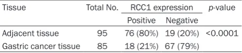
[image:7.629.96.530.80.538.2]and cytoplasms of 85 pairs of gastric cancer tissues and adjacent non-tumor tissues plus 10 normal stomach tissues. If 20% of an image’s total area showed positive staining for RCC1 in the nucleus and cytoplasm, it was scored as RCC1 positive (+). In adjacent non-tumor tissues, positive RCC1 staining was located in the regenerative compartment of the gastric epithelium, known as the gastric mucous neck region (Figure 4A-C; left panels). Moreover, lymphocytes within gastric mucosa always showed strong RCC1 staining. Positive RCC1 expression was detected in 21% (18/85) of gastric cancer tissues and in 80% (76/95) of adjacent non-tumor tissues (Table 2). The expression of RCC1 in gastric carcinoma tis-sues (Figure 4A-C; right panels) was significant-ly lower than in adjacent non-tumor tissues (P < 0.001).
Correlation analysis of RCC1 expression and clinicopathological characteristics of gastric carcinoma
We further analyzed the relationships between the clinical characteristics of gastric carcinoma and RCC1 expression using the Pearson chi-square and Fisher’s exact tests as summarized
in Table 3. Tumor differentiation (P < 0.01) and depth of invasion (P < 0.05) were significantly correlated to RCC1 expression. However, there were no significant correlations between RCC1 expression and other clinicopathologic fea-tures, including age, gender, stage, lymph node metastasis, and distant metastasis (P > 0.05). Discussion
In this study, we demonstrated that RCC1 downregulation was common both in gastric cancer cell lines and in clinical gastric carcino-mas. A loss of RCC1 expression was significant-ly correlated with tumorigenesis, cell differenti-ation, and depth of tumor invasion.
[image:8.629.102.306.79.379.2]sive imbalance between mucosal cell prolifera-tion and apoptosis due to the activaprolifera-tion of oncogenes and the inactivation of tumor sup-pressor genes [42, 43].
RCC1 was first identified during premature chromosomal condensation in BHK cells and was found to affect mammalian cell cycle pro-gression [44]. Cell lines with mutant forms for RCC1 display either cell cycle arrest or an immature chromatin structure phenotype at different cell cycle boundaries. RCC1 encodes a bulky nuclear protein that associates with chromosomes and belongs to a class of highly conserved proteins present in all eukaryotic species [45]. In recent years, functional studies of the RCC1 protein have shown that it is a gua-nine nucleotide exchange factor (GEF) that acts on the nuclear Ras-like G protein Ran [20]. Ras and Ran function as biological switches, and RCC1 may carry out its biological functions through Ran, which is essential for nuclear pore transport function [21]. In particular, this sug-gests the presence of two functional domains in RCC1: one to receive upstream signals and the other to transfer them into downstream events. A loss of RCC1 induces premature initi-ation of mitosis, resulting in G1 arrest with micronuclei possessing mitotic condensed chromosomes [46]. However, little is known about the relationship between RCC1 and carcinomas.
This study is the first to identify a relationship between RCC1 and gastric carcinoma. We found that the transcription and translation of RCC1 were significantly different among three different types of differentiated gastric cancer cell lines (AGS, TSGH9201, and MKN45). The AGS cell line was established from a well-differ-entiated gastric epithelial cancer cell [37], and the TSGH9201 cell was established from a human signet ring gastric carcinoma with a moderately differentiated cell type [38]; how-ever, the MKN45 cell line was established from
region of RCC1 were analyzed by MALDI-TOF mass spectrometry. The results showed that significantly different methylation levels of the RCC1 gene were found in these three gastric cell lines at a specific location in the 9th CpG site. This might explain how the hypermethyl-ation of the RCC1 gene at the 9thCpG site of the promoter to exon 1 region in both the TSGH9201 and MKN45 cell lines correlated to a downregulation of RCC1 expression.
When analyzing transcription factor (TF) bind-ing motifs, we further found that there were three specific TF binding motifs, called HSF1, TFIIB, and NF-X3, located within the 9th CpG site of the RCC1 promoter to exon 1 region. Promoter hypermethylation-mediated silencing of tumor suppressor genes (TSGs) is a hallmark of oncogenesis. In a previous study of nasopha-ryngeal carcinoma (NPC), a candidate TSG called oxidored-nitro domain-containing protein 1 (NOR1) was found, which was downregulated during tumorigenesis. Furthermore, a function-al NOR1 promoter that is regulated by HSF1 and nuclear respiratory factor 1 (NRF1) was identified [47]. It is known that the transcription factor TFIIB plays a central role in preinitiation complex assembly, providing a bridge between promoter-bound TFIID and RNA Polymerase II [48]. TFIIB possesses sequence-specific DNA -binding ability and interacts with a TFIIB -recognition element (BRE) that can be disrupt-ed by DNA methylation [49], which is present in many promoters, such as in the RCC1 gene in this study.
[image:9.629.99.346.105.159.2]The expression patterns of RCC1 were not only analyzed in three gastric cell lines but also in clinical gastric carcinoma and adjacent non-tumor tissue microarray (TMA) sections. The TMA chip displays a novel method for the serial analysis of gene expression in hundreds of tis-sue specimens at a time. Commonly, this meth-od has proven to be highly effective for the analysis of molecular alterations in different Table 2. Expression levels of RCC1 protein in gastric
can-cer tissue microarrays (ST801a and ST1004, Biomax US)
Tissue Total No. RCC1 expression p-value Positive Negative
Adjacent tissue 95 76 (80%) 19 (20%) <0.0001 Gastric cancer tissue 85 18 (21%) 67 (79%)
P < 0.0001 (Pearson chi-square).
stages of one particular tumor, such as in gas-tric, liver, kidney, or prostate cancers [50]. In this study, we examined the immunohistochem-ical staining of the RCC1 protein in 85 pairs of human gastric carcinoma tissues with adjacent non-tumor tissue plus 10 normal stomach tis-sues. In the gastric carcinoma tissues, we found a higher percentage of either the loss of or a reduction in RCC1 expression. In contrast, adjacent non-tumor tissues exhibited strong RCC1 expression in the majority of cases (Table 2). The significant relationship between the loss or weak expression of RCC1 and poorly dif-ferentiated carcinomas might reflect an increased selection pressure on and clonal expansion of the cells in which RCC1 expres-sion has been suppressed, which therefore may exhibit a more aggressive phenotype, such as a greater depth of invasion in gastric cancer.
In conclusion, we demonstrated for the first time that the RCC1 protein is downregulated in gastric carcinoma tissues. This loss of RCC1 expression may be due to hypermethylation of CpG islands in the RCC1 gene in a region span-ning from its promoter region to exon 1, particu-larly at the 9th CpG site. In addition, we observed strong and significant associations between RCC1 expression and degree of differ-entiation and depth of invasion, indicating that moderate or poor differentiation is associated with reduced RCC1 expression in gastric can-cer cell lines as well as in gastric carcinoma tis-sues. Therefore, our data suggest that RCC1 may play a tumor suppressor role in gastric carcinoma.
Acknowledgements
This work was supported in part by the grants TCVGH-DYU-102-8302 and TCVGH-NCHU-102- 7605 from the Taichung Veterans General Hospital, the grant MOST-104-2313-B-005-043-MY3 from the Ministry of Science and Technology, and the Ministry of Education, Taiwan, Republic of China, under the Aiming for Top University (ATU-104-S-0508) plan. The funders had no role in the study design, data collection and analysis, decision to publish, or preparation of the manuscript.
Disclosure of conflict of interest
None.
Address correspondence to: Dr. Cheng-Chung Wu, Department of General Surgery, Taichung Veterans General Hospital, No. 1650, Sec. 4, Taiwan Bou- levard, Taichung 40705, Taiwan. Tel: +886-4-2359- 2525 Ext. 5021; Fax: +886-4-23592705; E-mail: [email protected]; Dr. Chuan-Mu Chen, Department of Life Sciences, Research and Development Office, National Chung Hsing University, No. 250 Kuo-Kuang Road, Taichung 40227, Taiwan. Tel: +886-4-22856309; Fax: +886-4-22874740; E-mail: ch- [email protected]
References
[1] Bertuccio P, Chatenoud L, Livi F, Proud D, Ferlay J, Negri E, Malvezzi M and La Vecchia C. Recent pattern in gastric cancer: a global over-view. Int J Cancer 2009; 125: 666-673. [2] Brenner H, Rothenbacher D and Amdt V.
[image:10.629.99.305.117.445.2]Epidemiology of stomach cancer. Methods Mol Biol 2009; 472: 467-477.
Table 3. Relationship between RCC1 and clini-copathological characteristics of patients with gastric carcinoma
Characteristic RCC1 (+)(n=18) RCC1 (-)(n=67) P-value
Age 0.178a
<60 8 40 ≥60 10 27
Gender 1.000b
Male 13 49 Female 5 18
Tumor differentiation 0.004b
Well 0 10 Moderate 1 24 Poor 14 30 Unknown 3 3
Stage 1.000b
I/II 15 55
III 3 12
Depth of invasion 0.044a
T1/T2 9 17 T3/T4 9 50
Lymph node metastasis 0.348b
No 12 53
Yes 6 14
Distant metastasis 1.000b
No 18 65
Yes 0 2
[3] Ferlay J, Shin HR, Bray F, Forman D, Mathers C and Parkin DM. Estimates of worldwide burden of cancer in 2008: GLOBOCAN 2008. Int J Cancer 2010; 127: 2893-2917.
[4] Leung WK, Wu MS, Kakugawa Y, Kim JJ, Yeoh KG, Goh KL, Wu KC, Wu DC, Sollano J, Kachintorn U, Gotoda T, Lin JT, You WC, Ng EK and Sung JJ. Screening for gastric cancer in Asia: current evidence and practice. Lancet Oncol 2008; 9: 279-287.
[5] Hrtgrink HH, Jansen EP, van Grieken NC and van de Velde CJ. Gastric cancer. Lancet 2009; 374: 477-490.
[6] Lastraioli E, Romoli MR and Arcangeli A. Immunohistochemical biomarkers in gastric cancer research and management. Int J Surg Oncol 2012; 2012: e868645.
[7] Correa P. Human gastric carcinogenesis: a multistep and multifactorial process-First American Cancer Society Award lecture on cancer epidemiology and prevention. Cancer Res 1992; 52: 6735-6740.
[8] Lauren P. The two histological main types of gastric carcinoma: diffuse and so-called intes-tinal-type carcinoma. Acta Pathol Microbiol Scand 1965; 64: 31-49.
[9] Nakamura K, Sugano H and Takagi K. Carcinoma of the stomach in incipient phase: its histogenesis and histological appearances. Gann 1968; 59: 251-258.
[10] Carneiro F, Huntsman DG, Smyrk TC, Owen DA, Seruca R, Pharoah P, Caldas C and Sobrinho-Simões M. Model of the early development of diffuse gastric cancer in E-cadherin mutation carriers and its implications for patient screen-ing. J Pathol 2004; 203: 681-687.
[11] Leung WK and Sung JJ. Intestinal metaplasia and gastric carcinogenesis. Aliment Pharmacol Ther 2002; 16: 1209-1216.
[12] Hwang JJ, Lee DH, Lee AR, Yoon H, Shin CM, Park YS and Kim N. Characteristics of gastric cancer in peptic ulcer patients with Helicobacter pylori infection. World J Gas- troenterol 2015; 21: 4954-4960.
[13] Choi YJ, Kim N, Chang H, Lee HS, Park SM, Park JH, Shin CM, Kim JM, Kim JS, Lee DH and Jung HC. Helicobacter pylori-induced epitheli-al-mesenchymal transition, a potential role of gastric cancer initiation and an emergence of stem cells. Carcinogenesis 2015; 36: 553-563.
[14] Strickler JG, Zheng J, Shu Q, Burgart LJ, Alberts SR and Shibata D. p53 mutations and micro-satellite instability in sporadic gastric cancer: When guardians fail. Cancer Res 1994; 54: 4750-4755.
[15] Hara T, Ooi A, Kobayashi M, Mai M, Yanagihara K and Nakanishi I. Amplification of c-myc, K-sam, and c-met in gastric cancers: Detection
by fluorescence in situ hybridization. Lab Invest 1998; 78: 1143-1153.
[16] Yu J, Miehlke S, Ebert MP, Sundt TM 3rd, Dearani JA and Park SJ. Frequency of TPR-MET rearrangement in patients with gastric carci-noma and in first-degree relatives. Cancer 2000; 88: 1801-1805.
[17] Nakatsuru S, Yanagisawa A, Ichii S, Tahara E, Kato Y, Nakamura Y and Horii A. Somatic mu-tation of the APC gene in gastric cancer: Frequent mutations in very well differentiated adenocarcinoma and signet-ring cell carcino-ma. Hum Mol Genet 1992; 1: 559-563. [18] Ohtsubo M, Kai R, Furuno N, Sekiguchi T,
Sekiguchi M, Hayashida H, Kuma K, Miyata T, Fukushige S and Murotsu T. Isolation and char-acterization of the active cDNA of the human cell cycle gene (RCC1) involved in the regula-tion of onset of chromosome condensaregula-tion. Genes Dev 1987; 1: 585-593.
[19] Nishimoto T, Seino H, Seki N and Hori T. The human CHC1 gene encoding RCC1 (regulator of chromosome condensation) (CHC1) is local-ized to human chromosome 1p36.1. Genomics 1994; 23: 719-721.
[20] Bischoff FR and Ponstingl H. Catalysis of gua-nine nucleotide exchange on Ran by the mi-totic regulator RCC1. Nature 1991; 354: 80-82.
[21] Moore MS and Blobel G. The GTP-binding pro-tein Ran/TC4 is required for propro-tein import into the nucleus. Nature 1993; 365: 661-663. [22] Zhao H, Li Q, Wang J, Su X, Ng KM, Qiu T, Shan
L, Ling Y, Wang L, Cai J and Ying J. Frequent epigenetic silencing of the folate-metabolish-ing gene cystathionine-beta-synthase in gas-trointestinal cancer. PLoS One 2012; 7: e46214.
[23] Yan PS, Chen CM, Shi H, Rahmatpanah F, Wei SH, Caldwell CW and Huang TH. Dissecting complex epigenetic alterations in breast can-cer using CpG island microarrays. Cancan-cer Res 2001; 61: 8375-8380.
[24] Yang H, Chen CM, Yan P, Huang TH, Shi H, Burger M, Nimmrich I, Maier S, Berlin K and Caldwell CW. The androgen receptor gene is preferentially hypermethylated in follicular non-Hodgkin’s lymphomas. Clin Cancer Res 2003; 9: 4034-4042.
[25] Yan PS, Chen CM, Shi H, Rahmatpanah F, Wei SH and Huang TH. Applications of CpG island microarrays for high-throughput analysis of DNA methylation. J Nutr 2002; 132: 2430S-2434S.
[26] Livak KJ and Schmittgen TD. Analysis of rela-tive gene expression data using real-time quantitative PCR and the 2(-Delta Delta C(T)). Methods 2001; 25: 402-408.
Chong KY and Chen CM. Lung tumorigenesis induced by human vascular endothelial growth factor (hVEGF)-A165 overexpression in trans-genic mice and amelioration of tumor forma-tion by miR-16. Oncotarget 2015; 6: 10222-10238.
[28] Hung TH, Hsu SC, Cheng CY, Choo KB, Tseng CP, Chen TC, Lan YW, Huang TT, Lai HC, Chen CM and Chong KY. Wnt5A regulates ABCB1 ex-pression in multidrug-resistant cancer cells through activation of the non-canonical PKA/β-catenin pathway. Oncotarget 2014; 5: 12273-12290.
[29] Lai CW, Chen HL, Lin KY, Liu FC, Chong KY, Cheng WT and Chen CM. FTSJ2, a heat shock-inducible mitochondrial protein, suppresses cell invasion and migration. PLoS One 2014; 9: e90818.
[30] Chen HL, Huang JY, Chu TW, Tsai TC, Hung CM, Lin CC, Liu FC, Wang LC, Chen YJ, Lin MF and Chen CM. Expression of VP1 protein in the milk of transgenic mice: a potential oral vaccine protects against enterovirus 71 infection. Vaccine 2008; 26: 2882-2889.
[31] Tung YT, Tsai TC, Kuo YH, Yen CC, Sun JY, Chang WH, Chen HL and Chen CM. Comparison of solid-state-cultured and wood-cultured Antrodia camphorata in anti-inflammatory ef-fects using NF-κB/luciferase inducible trans-genic mice. Phytomedicine 2014; 21: 1708-1716.
[32] Shi H, Yan PS, Chen CM, Rahmatpanah F, Lofton-Day C, Caldwell CW and Huang TH. Expressed CpG island sequence tag microar-ray for dual screening of DNA hypermethylation and gene silencing in cancer cells. Cancer Res 2002; 62: 3214-3220.
[33] Lai ZL, Tsou YA, Fan SR, Tsai MH, Chen HL, Chang NW, Cheng JC and Chen CM. Methylation-associated gene silencing of RARB in areca carcinogens induced mouse oral squamous cell carcinoma. Biomed Res Int 2014; 2014: 378358.
[34] Lian F, Sreedharan S, Arnold RS, Master VA, Ogan K, Pattaras JG, Roberts DL and Petros JA. von Hippel-Lindau exonic methylation analysis using MALDI-TOF mass spectrometry. J Urol 2014; 192: 1528-1533.
[35] Sun Y, Li JY, He JS, Zhou LX and Chen K. Tissue microarray analysis of multiple gene expres-sion in intestinal metaplasia, dysplasia and carcinoma of the stomach. Histopathology 2005; 46: 505-514.
[36] Chen CM, Chen HL, Hsiau TH, Hsiau AH, Shi H, Brock GJ, Wei SH, Caldwell CW, Yan PS and Huang TH. Methylation target array for rapid analysis of CpG island hypermethylation in multiple tissue genomes. Am J Pathol 2003; 163: 37-45.
[37] Zang J, Wang P, Gao S, Xiao D, Zhang J and Wang K. Differential proteins expression be-tween gastric cancer and normal cell lines. Life Sci J 2008; 5: 28-32.
[38] Shyu RY, Jiang SY, Wang CC, Wu MF, Harn HJ, Chang TM and Yeh MY. Establishment and characterization of TSGH9201, a human gas-tric carcinoma cell line that is growth inhibited by epidermal growth factor. J Surg Oncol 1995; 58: 17-24.
[39] Liu YF, Yang A, Liu W, Wang C, Wang M, Zhang L, Wang D, Dong JF and Li M. NME2 reduces proliferation, migration and invasion of gastric cancer cells to limit metastasis. PLoS One 2015; 10: e0115968.
[40] Calcagno DQ, Gigek CO, Chen ES, Burbano RR, and Smith Mde A. DNA and histone methyla-tion in gastric carcinogenesis. World J Gastroenterol 2013; 19: 1182-1192.
[41] Tsai KW, Wu CW, Hu LY, Li SC, Liao YL, Lai CH, Kao HW, Fang WL, Huang KH, Chan WC and Lin WC. Epigenetic regulation of miR-34b and miR-129 expression in gastric cancer. Int J Cancer 2011; 129: 2600-2610.
[42] Correa P and Shiao YH. Phenotypic and geno-typic events in gastric carcinogenesis. Cancer Res 1994; 54 Suppl 7: 1941s-1943s.
[43] Farber E. Cancer development and its natural history: a cancer prevention perspective. Cancer 1988; 62 Suppl 8: 1676-1679.
[44] Nishimoto T, Eilen E and Basilico C. Premature of chromosome condensation in a ts DNA- mu-tant of BHK cells. Cell 1978; 15: 475-483. [45] Shia WY and Skeath JB. The Drosophila RCC1
homolog, Bj1, regulates nucleocytoplasmic transport and neural differentiation during Drosophila development. Dev Biol 2004; 270: 106-121.
[46] Nishitani H, Ohtaubo M, Yamashita K, Iida H, Pines J, Yasudo H, Shibata Y, Hunter T and Nishimoto T. Loss of RCC1, a nuclear DNA-binding protein, uncouples the completion of DNA replication from the activation of cdc2 protein kinase and mitosis. EMBO J 1991; 10: 1555-1564.
[47] Li W, Li X, Wang W, Li X, Tan Y, Yi M, Yang J, McCarthy JB, Xiong W, Wu M, Ma J, Su B, Zhang Z, Liao Q, Xiang B and Li G. NOR1 is an HSF1- and NRF1-regulated putative tumor suppres-sor inactivated by promoter hypermethylation in nasopharyngeal carcinoma. Carcinogenesis 2011; 32: 1305-1314.
[48] Evans R, Fairley JA and Roberts SG. Activator-mediated disruption of sequence-specific DNA contacts by the general transcription factor TFIIB. Genes Dev 2001; 15: 2945-2949. [49] Glait-Santar C and Benayahu D. SVEP1
[50] Richter J, Wagner U, Kononen J, Fijan A, Bruderer J, Schmid U, Ackermann D, Maurer R, Alund G, Knönagel H, Rist M, Wilber K, Anabitarte M, Hering F, Hardmeier T, Schönenberger A, Flury R, Jäger P, Fehr JL,
Table S1. MALDI-TOF MS analysis of amplicon products of RCC1 gene
Human gene Forward primer sequence (5’→3’) Reverse primer sequence (5’→3’) Amplicon size (bp)
RCC1