Review Article
Microenviromental change after synthetic E-selectins
interfere in ischemia-reperfusion in rats and its
contribution to endogenetic/exogenous
never stem cells
Wei-Hua Lin1*, Feng Xu1, Long Bao1, Shi-Ying Zheng2, Wen-Ming Shen2,3*
Departments of 1Emergency Surgery, 2Cardio-Thoracic Surgery, The First Affiliated Hospital of Soochow University,
Suzhou 215006, Jiangsu Province, China; 3The Affiliated Wujin People’s Hospital of Jiangsu University, Changzhou
213000, Jiangsu Province, China. *Co-first authors.
Received July 13, 2015; Accepted August 25, 2015; Epub September 1, 2015; Published September 15, 2015
Abstract: Objective: To explore the special significances in advantages of using anti-inflammatory drugs, such as amelioration of growing conditions and the promotion of cell growth. Methods: Utilizing anti-adhesive effects of synthetic E-selectins, we observed the changes of inflammatory cytokines (TNF-α, IL-1β) contented in brain tissues and rat serums in rats hind cerebral ischemia-reperfusion models. Both growth and expression of endogenetic/ exogenous neurological stem cells were detected after ameliorated local microenvironment. Results: The contents of TNF-α and IL-1β were decreased in brain tissues and rat serums after applying synthetic E-selectins. Expression of exogenous neurological stem cells was enhanced. Animal neurological functions improved. Conclusion: Anti-inflammatory therapy in early stage could enhance proliferation of stem cells so that it has vital significations in treating cerebrovascular diseases.
Keywords: Ischemia-reperfusion, inflammatory cytokines, synthetic e-selectins, endogenetic/exogenous never stem cells
Introduction
In the mechanism of ischemia-reperfusion inju
-ry, the inflammatory response plays a very important role. After the ischemia-reperfusion
injury, the endogenous stem cells in the state
of body rest are activated and begin to migrate, proliferate as well as differentiate towards the ischemic injury area, for the purpose of com
-pensating injured neurological function. How-ever, the self-repair capacity of central nervous
system is limited, which is related to the reason that ischemic microenvironment with local
inflammatory is not conducive to the prolifera
-tion and survival of newborn cells. Selectins play a role of promoting activation, aggregation and leakage of leukocyte in inflammatory response after ischemia reperfusion as well as infiltration in the ischemic area. Synthetic E-selectin is a kind of selectins antagonist, which can inhibit selectins-mediated leukocyte
and endothelial adhesion and have a good
anti-inflammatory property. In this study, by observ
-ing the changes of the 2 inflammatory cytokines-TNF-α and IL-1β in rats’ serum with ischemia-reperfusion injury after synthetic E-selectins, we mainly research the impact of changes of local microenvironment with ischemia reperfu
-sion injury after being applied the synthetic anti-inflammatory E selectins on the endoge -nous and exoge-nous stem cells and
neurologi-cal function in experimental animals. Moreover, we also make a preliminary study on the signifi
-cance of using inhibitors inflammation to improve environment of stem cells as well as
promote their growth condition. Materials and methods
Reagents: Synthetic E-selectins (Shanghai Bio-
chemical Research Institute); rat IL-1β, TNF-α ELISA kit (Shanghai Langke Bioengineering Co.,
anti-BrdU monoclonal antibody (Becton Dic-kinson Company).
Experimental animal grouping: 170 healthy
adult SD rats, male or female, weighing
280-320 g. They are randomly divided into 5 groups, namely (1) sham operation group 46; (2)
Ischemia-reperfusion group 46; (3) Treatment group of synthetic E-selectins 46; (4) simple
hAMSCs transplantation group 16; (5) hAMSCs
synthetic graft + E-selectins treatment group 16. In the 5 groups of experimental animals, we should randomly select 5 rats for blood sam -pling in (1)-(3) group at the ischemic time point
of 2 h, 6 h, 12 h, 24 h, 48 h and 72 h, in which the 48 h-group can be used to make the brain
homogenate plasma specimen. The remaining 16 rats in each group as well as the (4), (5)
groups will be used for neurological function
scoring. (1)-(3) groups mainly detect the growth
condition of endogenous stem cells and BrdU
intraperitoneal injection time in each group:
after ischemia for about 1 w, continuously injur
-ing for 6 d and 1 d before be-ing killed, a dose of 50 mg/kg for each time; synthetic E-selectins should be injected in the tail vein after the isch
-emia-reperfusion model being made and
of 1 d, 3 d, 1 w, 2 w and 4 w after ischemia
(Table 1) to conduct neurological evaluation.
The highest score is 18 point, for 1-6 is mild
injury, 7-12 is the moderate injury and 13-18 is the severe injury.
Collection of plasma
Draw blood using cardiac puncture for 3-4 ml in rats in (1)-(3) group at different time points (2 h, 6 h, 12 h, 24 h, 48 h, 72 h) after ischemia-reperfusion, natural solidify at room tempera
-ture after 10-20 min, centrifuge 20 min with the rotate speed of 2000-3000 r/min. Then carefully collect the supernatant, package, and place it in the -70°C refrigerator for
examin-ation.
Preparation for the brain tissue homogenate
Rats (1)-(3) group with ischemic 48 h will be
killed by the overdose anesthesia after the
heart puncture blood. Then we should remove
their brains quickly, separate the bilateral corti -cal on ice plate, clean it using ice saline rinse,
dry it by filter paper, accurately weigh its brain
[image:2.612.90.344.96.191.2]tissue, and add the 10% homogenate with 9 Table 1. Change of plasma IL-1β content in different
groups (x ± s, ng/ml)
Sham control group IR group SeS group IR 2 h 2.706±0.022 2.732±0.026 2.704±0.015 IR 6 h 2.694±0.011 3.406±0.076* 2.794±0.031▲ IR 12 h 2.678±0.023 4.144±0.053* 2.960±0.043*,▲ IR 24 h 2.676±0.024 4.648±0.089* 3.050±0.047*,▲ IR 48 h 2.684±0.018 4.804±0.072* 3.204±0.070*,▲ IR 72 h 2.666±0.056 3.234±0.072* 2.926±0.046*,▲
Note: SeS: synthetic E-selectins; IR: ischemia-reperfusion. *compared
with the sham control group, P<0.05; ▲Compared with the ischemia-reperfusion group, P<0.05.
before the rats regaining conscious
-ness, with a dose of 10 mg/kg. The
(4), (5) groups detect the growth
con-dition of exogenous stem cells: hAM
-SCs has been labeled BrdU before
transplantation, so there is no need to
conduct intraperitoneal injection of BrdU in order to avoid confusion with
endogenous stem cells, which may
impact the results of experimental
observation. hAMSCs migration path should choose the tail vein
transplan-tation and the time is 24 h after isch
-emia-reperfusion. This study was con -ducted in accordance with the de-
claration of Helsinki.
Establishment of an animal model
This experiment uses the Longa’s suture method to make the artery ischemia-reperfusion model in rats’
brain. For those rats participating in
neurological function score in each experimental animals function score
group, we will change to use the
modi-fied neurological severity score
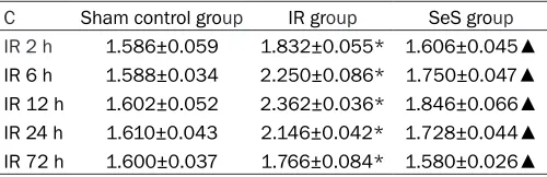
(mNSS) respectively at the time points Table 2. Changes of plasma TNF-α content in different
groups (x ± s, ng/ml)
C Sham control group IR group SeS group IR 2 h 1.586±0.059 1.832±0.055* 1.606±0.045▲ IR 6 h 1.588±0.034 2.250±0.086* 1.750±0.047▲ IR 12 h 1.602±0.052 2.362±0.036* 1.846±0.066▲ IR 24 h 1.610±0.043 2.146±0.042* 1.728±0.044▲ IR 72 h 1.600±0.037 1.766±0.084* 1.580±0.026▲
Note: IR: ischemia-reperfusion; SeS: synthetic E-selectins; *compared
[image:2.612.91.341.269.350.2]times physiological saline according to its vol-ume and weight ratio, 4°C, 12000 r/min,
centri-fuging for 5 min. Finally carefully collect the supernatant and place it in -70°C refrigerator for examination.
ELISA detects the content of IL-1β, TNF-α in the supernatant and plasma of the brain tis-sue
All the operations should be strictly in accor-dance with the instructions steps in the rat
IL-1β, TNF-α ELISA kits and determinate the content of IL-1β, TNF-α in rats’ plasma and
brain tissue supernatants.
Preparation for the pathological specimens: After experimental animals in each group finish
the mNSS score respectively in 2 w, 4 w, we
should randomly select 8 killed rats. First, embed them in paraffin, regard the infarct locus as the neutral infarction, slice it to coro
-nal shape with the slice thickness of 12 μm, and apply the immunofluorescence staining to detect the fate of implanted cells. Conduct the HE staining, BrdU immunohistochemistry, and immunofluorescence staining of endogenous
stem cells on the sham surgery group,
isch-emia-reperfusion group, and the synthesized
E-selectins group while treatment group.
24-48 h, then decreasing gradually. Compared
with the sham control group, it has significant difference. The IL-1β content of synthetic E-selectins group is significantly lower and has a significantly difference compares with isch
-emia-reperfusion model group (P<0.05).
Plasma TNF-α content change: plasma TNF-α content change of rats in each group at differ -ent time points (2 h, 6 h, 12 h, 24 h, 48 h, 72 h)
after the ischemia-reperfusion is shown in
Table 2, whose results show that the plasma
IL-1β content will rise at 2 h after rats’ focal ischemia-reperfusion and peak at 24-48 h, and
then decrease at 24 h gradually. Compared
with the sham control group, it had significant difference. The TNF-α content of synthetic E-selectins group is significantly lower and has a significantly difference compares with isch
-emia-reperfusion model group (P<0.05).
IL-1β, TNF-α content of brain tissue: at the time point of 48 h is as shown in Table 3. The results
show that after 48 h of rats’ focal ischemia-reperfusion, the IL-1β, TNF-α content in brain tissue has been significantly increased, and the difference is significant (P<0.05) compared
with sham control group. After using the syn
-thetic E-selectins, the IL-1β, TNF-α content has been significantly decreased and the differ -Table 3. Content of IL-1β, TNF-α in brain tissue at the 48th h after
Ischemia-reperfusion (x ± s, ng/g)
Sham control group IR group SeS group
IL-1β content 0.357±0.019 1.266±0.044* 0.648±0.033*,▲
TNF-α content 0.693±0.012 1.265±0.042* 0.856±0.025*,▲
Note: IR: ischemia-reperfusion; SeS: synthetic E-selectins; *compared with the
sham control group, P<0.05; ▲Compared with the ischemia-reperfusion group,
[image:3.612.91.366.97.137.2]P<0.05.
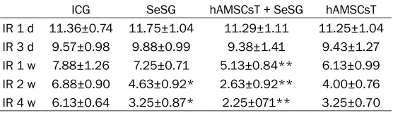
Table 4. Neurologic function score of rats at different time points
ICG SeSG hAMSCsT + SeSG hAMSCsT
IR 1 d 11.36±0.74 11.75±1.04 11.29±1.11 11.25±1.04 IR 3 d 9.57±0.98 9.88±0.99 9.38±1.41 9.43±1.27 IR 1 w 7.88±1.26 7.25±0.71 5.13±0.84** 6.13±0.99 IR 2 w 6.88±0.90 4.63±0.92* 2.63±0.92** 4.00±0.76 IR 4 w 6.13±0.64 3.25±0.87* 2.25±071** 3.25±0.70 ICG: ischemia control group; SeSG: synthetic E-selectins group; hAMSCsT + SeSG: hAMSCs transplantation + synthetic E-selectins group hAMSCsT: hAMSCs transplantation group; IR: ischemia-reperfusion. Note: *compared with the
ischemia control group, P<0.05; **compared with the pure amniotic stem cell transplantation group, P<0.05.
Statistical processing
Use SPSS10.0 software to con -duct the statistical analysis. Indicate the measurement data
as χ ± s and analyze the t test and ANOVA of lines between any
two group. P<0.05 is considered
statistically significant.
Results
Changes of plasma IL-1β con
-tent: plasma IL-1β content change of rats in each group: at different time points (2 h, 6 h, 12 h, 24 h, 48 h, 72 h) after the ischemia-reperfusion is shown
in Table 1, whose results show
[image:3.612.91.369.205.285.2]ence is significant (P<0.05) compared with the
ischemia-reperfusion group.
Functional score for animals: in each group is
as shown in Table 4. In the 3rd d after ischemia-reperfusion, the neurologic dysfunction in all
groups has been improved in varying degrees,
but there is no significant difference among the
groups (P>0.05); at the 1st W after ischemia-reperfusion, the neurologic dysfunction in all groups has been further improved, but there is no significant difference between the synthetic
E-selection group and the ischemia control group (P>0.05), while the score in hAMSCs
transplantation + synthetic E-selectins group has been significantly lowered compared with
pure hAMSCs transplantation group, and the
difference is significant (P<0.05). At the 2nd W after ischemia-reperfusion, the score in all
groups expect the ischemia control group all
suffers a further decrease, and the neurologic dysfunction of ischemic control group only has
a little improvement compared with it at 1
week, while the score of synthesized E-selectins treatment group is significantly lower than the
ischemia control group (P<0.05), the score of hAMSCs transplantation + synthetic E-selectins group is significantly lower than pure hAMSCs
transplantation group (P<0.05).
Detection of immunofluorescence
(Figure 1A, 1B) The immunofluorescence result
[image:4.612.90.526.70.270.2]shows that in the 2nd w, 4th w after ischemia-Figure 1. A: Tail vein transplantation group + synthetic E-selectins group (fluorescence microscopy 100×). There gathers large number of green fluorescence positive cells in the ischemia area. B: Tail vein transplantation group (fluorescence microscopy 100×). There scatters some green fluorescence positive cells in the ischemia area.
[image:4.612.89.528.330.480.2]reperfusion, there are no green fluorescence
positive cells appearing in the synthetic E-selectins treatment group, sham operation
group and ischemia-reperfusion group; while there are green fluorescence positive cells
(transplanted hAMSCs) appearing in the
hAM-SCs transplantation + synthetic E-selectins
treatment group and the pure hAMSCs trans-plantation group.
BrdU immunohistochemical staining: If the
brown-yellow material appears, it will be
posi-tive and BrdU-posiposi-tive material is in the nucle -us. The experimental result shows that there
are no BrdU-positive cells in sham operation group, ischemia-reperfusion group and syn -thetic E-selectins group.
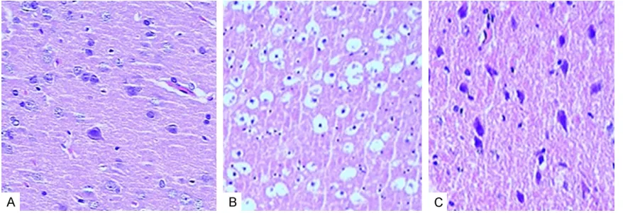
HE staining result (Figure 2A-C)
The neuron in the brain of rats in sham opera -tion group has a clear outline and complete structure, mostly shaped as conical, star as well as spindle. The tissue space surrounding cells is normal. The nucleolus is large and
round, and its color is lighter and uniform,
located in the central cell and rich in Nissl bod-ies, besides, its nucleolus is clear; the neuron
death in central area of necrosis can be observed in ischemia-reperfusion groups,
whose shape is not easily to distinguish, besides, its cell structure disappears, its
cyto-plasm empty bubble-like degenerates, its cells are sparse with disorder arrangement; part of the cytoplasmic autolyzes, part of the particles drop out, Nissl bodies significantly reduce, nucleus size shrinks, pycnosis stain, and the
structure is unclear; the neuron ischemic change in the synthetic E-selectins treatment
group is significantly reduced.
Discussion
Cerebral ischemia-reperfusion may induce a wide range of inflammatory reactions, on one hand, it may increase the impairment of brain function; on the other hand, and it may inhibit the regeneration of brain tissue. Once the isch -emic brain injury occurs, the intrinsic repair
mechanisms of the body starts and the injured
tissue as well as the surrounding normal tissue
will secrete some cell factors through the forms of paracrine and/or autocrine to promote the growth of survive neuron axon after ischemic
injury and establish a new synaptic connection,
for the purpose of restoring part functions [1,
2]. It is regrettable that in the central nervous
system, this self-repair capacity is very limited, which is mainly because ischemic local inflam
-matory microenvironment is not conducive for the proliferation and survival of new cells. A series of studies show that the local inflamma
-tory reaction of brain tissue after the ischemia-reperfusion is one of the main reasons that cause the brain injury [3]. Adhesion molecules and cytokines generated by ischemia and reperfusion in the early period constitute the basis of ischemic injury changing to the inflam
-matory injury [4]. Many studies show that when the cerebral ischemia-reperfusion injures the body, the synthesis mechanism of TNF-α, IL-1β is activated, resulting in the TNF-α, IL-1β con
-tent rising. The co-expression of two factors--TNF-α and IL-1β has played an important role in regulating the inflammatory response induced by cerebral ischemia [5, 6]. TNF-α and IL-1β
may be involved in and promote cerebral
isch-emia-reperfusion injury through the following ways: 1) excessive inflammatory response: TNF-α releases and activate excessive poly
-morph nuclear leukocyte, and increase the adhesion molecule expression of leukocyte as well as endothelial cell to promote leukocyte infiltrating to the brain tissue; it also activates
the macrophages, endothelial cells and
microg-lia cells to produce inflammatory metabolites, and further promote the activated leukocytes
going into the ischemic area as well as the
sur-rounding area. The IL-1β expression also can
induce the adhesion molecules expression, and promote neutrophil to go into the central nervous system. 2) The blood-brain barrier
inju-ry: TNF-α has a direct toxic effect on the capil -laries, leading to small artery spasm, increas-ing vascular permeability, and openincreas-ing the
blood-brain barrier. The synergic action of TNF-α and IL-1β can increase blood-brain bar
-rier injury and increase peripheral leukocyte infiltration [7, 8]. 3) The apoptosis promotion: in the case of cerebral ischemia, TNF-α and IL-1β can promote the injury and apoptosis of neu -rons through activating multiple apoptosis pathway such as Bcl-2, cathepsin B. 4)
Stimulation of excitatory amino acids and the release of free radicals, inducing cerebral isch
-emia-reperfusion injury.
Our experiments will confirm that IL-1β content
-emia, and reach peak during 24~48 h, while the TNF-α content begins to increase at the 2nd h after cerebral ischemia-reperfusion, reaches peak during 6~12 h, gradually decreasing at
24th h. At the 48th h, the content of IL-1β and TNF-α in brain tissue is significantly higher than
that in sham ischemic control group, which
sug-gests that there appears secondary inflamma
-tory response after cerebral ischemia-reperfu
-sion, constituting a micro-inflammatory en-vironment of local cerebral ischemia. It is not conducive to the proliferation and survival of
new neurological cells and should be
conduct-ed anti-inflammatory treatment in early stage.
The synthetic E-selectins and P-selectins
respectively have a inhibited effect on E-selectins ad well as L-selectins [9], which can block the adhesion of leukocyte on endothelial cells and improve the blood supply of infarcted brain tissue, making the brain tissue get an effective protection when being conducting ischemia reperfusion [10, 11].
In this study, after applying synthetic E-selectins, the TNF-α and IL-1β content in brain tissue and plasma has decreased significantly, and the difference has a statistical significance com
-pared with the cerebral ischemia-reperfusion
group, so we suggest that the synthetic E-selectins can inhibit the synthesis
mecha-nism of IL-1β and TNF-α, leading to decrease of IL-1β and TNF-α content. On this basis, we examine the expression of endogenous stem
cells at 2nd week after ischemia-reperfusion. Our experiments show that there are no green
fluorescence-positive cells in synthetic E-selectins treatment group, ischemia-reperfu -sion group and sham group, and there are also
no BrdU-positive cells when conducting BrdU
immuno-histochemistry staining, which may be
because there exists factors to inhibit neurons’ differentiation and survival in micro-environ
-ment or there lacks of factors to promote the neuronal differentiation and survival [12]. While
although the micro-environment can
theoreti-cally promote the proliferation and differentia
-tion of endogenous stem cells after the syn -thetic E-selectins treatment, it still cannot
overcome the negative factors, may be due to
the limited endogenous stem cells and our
lim-ited detecting ability, etc. The function score of
experimental animals in synthetic E-selectins
treatment group decreases, and the difference has a statistical significance compared with the
ischemia-reperfusion group. HE staining shows
that the ischemic change in brain tissue is
sig-nificantly reduced after applying the synthetic
E-selectins, which may be due to the increase
of local blood flow in cerebral ischemia area and the improvement of blood supply in brain tissue after the anti-adhesion E-selectins, as well as anti-inflammatory of the synthetic
E-selectins. Besides, we also detect the growth
and survival condition of the transplanted hAM
-SCs in the use of synthetic E-selectins, and find
that compared with pure transplanted hAMSCs, its growth and survival condition is much better
after using the synthetic E-selectins, and there gathers large number of green fluorescence
positive cells in the ischemic area, which shows
that the animal neurological score is further
reduced. Thus we believe that applying the
syn-thetic E-selectins can improve the inflammatory micro-environment after ischemia-reperfusion and the blood supply for infarcted brain tissue, which is conducive to the growth of exogenous
stem cells. Moreover, the transplanted hAMSCs
can also secrete a number of nutritional factors
which can improve the ischemic
microenviron-ment and promote the survival of stem cells, so the combination of synthetic E-selectins and the transplantation of exogenous neural stem cells can enhance the proliferation, differentia
-tion of neurological cells, protect the brain tis
-sue function, and promote the recovery of experimental animals’ neurological function. This is can provide clinical guidance for the application of the therapy using the stem cell
transplantation to treat cerebral ischemia in
clinic medicine in the future. However, how to improve the growth and survival condition of
endogenous as well as exogenous stem cells
after the improvement of inflammatory micro
-environment still needs to be further studied.
Disclosure of conflict of interest None.
Address correspondence to: Drs. Feng Xu and Shi-Ying Zheng, Departments of Emergency Surgery and Cardio-Thoracic Surgery, The First Affiliated Hospital of Soochow University, Suzhou 215006, Jiangsu Province, China. E-mail: [email protected] (FX); [email protected] (SYZ)
References
[2] Wieloch T, Nikolich K. Mechanisms of neural plasticity following brain injury. J Curr Opin Neurobiol 2006; 16: 258-264.
[3] Matsuo Y, Onodera H, Shiga Y, Nakamura M, Ninomiya M, Kihara T, Kogure K. Correlation between myeloper-oxidasepuantified neutro -phil accumulation and ischemic brain injury in the rat: effects of neutrophi depletion. Stroke 2003; 25: 1469-1472.
[4] Lan Haitao, Zhang Qingjun. The protective function of recombinant human erythropoietin on rats’ cerebral ischemia-reperfusion inflam -matory injury. Neurosurgery 2006; 1: 51-54. [5] Minami M, Kuraishi Y, Yabuuchi K, Yamazaki A,
Satoh M. Induction of interleukin-1 beta mRNA in rat brain after transient forebrain ischemia. J Neurochem 1992; 58: 390-392.
[6] Zhu Y, Saito K, Murakami Y, Asano M, Iwakura Y, Seishima M. Early increase in mRNA levels of proinflammatroy cytokines and their interac -tions in the mouse hippocampus after tran -sient global ischemia. Neurosci Lett 2006; 393: 122-126.
[7] Hartz AM, Bauer B, Fricker G, Miller DS. Rapid modulation of P-glycoprotein-mediated trans -port at the blood brain barrier by tumor necro-sis factor-alpha and lipopolysaccharide. Mol Pharmacol 2006; 69: 462-470.
[8] Lee NY, Kang YS. The brain-to-blood efflux transport of taurine and changes in the blood-brain barrier transport system by tumor necro-sis factor-alpha. Brain Res 2004; 1023: 141-147.
[9] Zhang SM, Zhou D. On the application of E-se -lectins synthetic to treat rats’ focal cerebral ischemia reperfusion injury. Clin Neurosci 2000; 8: 250-252.
[10] Zhang SM, Zhou D. On the adhesion molecules and cerebral ischemia. JOf Experimental Sur -gery 1999; 16: 286-287.
[11] Li JW, Zhang SM, Xie XS. The impact of E-selec -tins on the blood oxygen rate in cerebral isch-emia-reperfusion, Chinese Journal of Hemor-heology 2004; 14: 30-31.