Original Article
Dual high expression of STAT3 and cyclinD1
is associated with poor prognosis after curative
resection of esophageal squamous cell carcinoma
Haiying Li1,2*, Weiwei Xiao3*, Jiwei Ma1,2, Yong Zhang1,2, Ru Li1,2, Jiecheng Ye1,2, Xiao Wang5, Xueyun Zhong1,2,
Shaoxiang Wang4
1Department of Pathology, 2Guangdong Province Key Laboratory of Molecular Immunology and Antibody Engineer-ing, School of Medicine, 5Guangzhou Jinan Biomedicine Research and Development Center, National Engineering Research Center of Genetic Medicine, Jinan University, Guangzhou 510632, Guangdong Province, China; 3 Depart-ment of Radiation Oncology, Sun Yat-Sen University Cancer Center; State Key Laboratory of Oncology in South China; Collaborative Innovation Center for Cancer Medicine, Guangzhou, Guangdong, China; 4Institute of Molecu-lar Medicine, Shenzhen University, Shenzhen 518060, China. *Equal contributors.
Received September 11, 2014; Accepted October 31, 2014; Epub October 15, 2014; Published November 1, 2014
Abstract: Background: Signal transducer of activator of transcription 3 (STAT3) and cyclinD1 are overexpressed in various human cancers, and their overexpression positively correlates to tumor progression and poor prognosis.
However, the clinical significance of dual high expression of these two proteins in esophageal squamous cell car -cinoma (ESCC) has yet to be determined. Methods: The expression of STAT3 and cyclinD1 was analyzed in tissue microarrays containing tumor and adjacent tissue samples from 82 patients who had undergone curative resection for histologically proven ESCC. Kaplan-Meier plots and Cox proportional hazards regression model were used to ana-lyze the prognostic value of STAT3 and cyclinD1 expression. Results: We discovered that expressions of STAT3 and
cyclinD1 in cancer tissues were significantly higher than that in adjacent tissues. High expression of STAT3 and cy -clinD1 was associated with malignant behaviors. Moreover, the expression of STAT3 was positively associated with the expression of cyclinD1. High STAT3 or cyclinD1 expression alone was associated with lower overall survival (OS) rates. Furthermore, dual high expression of STAT3 and cyclinD1 expression predict even worse survival outcome in both univariate and multivariate analysis. Conclusion: STAT3 and cyclinD1 correlate with more aggressive tumor behavior in ESCC. When STAT3 and cyclinD1 are considered together, they serve as effective prognostic markers in patients with surgically resected ESCC.
Keywords: Signal transducer of activator of transcription 3, cyclinD1, esophageal squamous cell carcinoma, im -munohistochemistry, prognosis
Introduction
Recently, in the worldwide, especially China, there has been a dramatic rise in the incidence and mortality due to esophageal adenocarci-noma and squamous cell carciadenocarci-noma [1, 2] and esophageal squamous cell carcinoma (ESCC) occurs at a high frequency in China (13 per 100 000) [3]. As the dominant type of esophageal cancer in China, has a generally poor prognosis due to the lack of effective clinical methods for its early detection [4]. There is a pressing need for reliable biomarkers to identify the subset of patients with a high risk of poor survival out-comes and guide treatment.
major factor promoting an immunosuppressive environment which allows tumor growth and metastasis [8].
As a cell cycle regulator, cyclinD1 is essential for progression through G1 phase and is a can-didate of proto-oncogene [9]. It has been pro-posed cyclinD1 expression is an independent risk factor in metastasizing bladder cancer [10]. Moreover, the overexpression of cyclinD1 was an independent adverse prognostic indica-tor for overall survival and relapse-free survival in estrogen-negative breast cancer patients [11].
Gagin D et al provided the first evidence that constitutive activation of STAT3 played a caus-ative role in overexpression of cyclinD1, and STAT3 proteins was implicated in resistance to apoptosis, potentially via their ability to modu-late cyclinD1 expression in head and neck squamous cell carcinoma (HNSCC) [12]. Given that STAT3 and cyclinD1 were independent prognostic indicators in some kinds of cancers, and the close relationship between the two pro-teins, the combined impact of STAT3 and cyclinD1 on the survival time of ESCC patients have yet to be characterized with documented clinical and pathological information.
The purpose of this study was to determine the expressions of STAT3 and cyclinD1 proteins and to analyze their correlations with the clini-copathological parameters, including age, sex, histological grade, stages and tumor size in ESCC patients. Additionally, we assessed the influence of STAT3 and cyclinD1 expression on the overall survival of patients with ESCC. Materials and methods
Patients and tissue specimens
The study cohort includes paraffin-embedded ESCC samples and adjacent noncancerous tis-sues from 82 patients who underwent curative surgery between June 2003 and June 2007 in Guangzhou Cancer Hospital, China. All cases were clinically diagnosed and confirmed patho-logically as ESCC without distant metastasis at the time of diagnosis.
Written informed consent was obtained from all ESCC patients and this study was approved by the research Ethics Committee of Guangzhou Cancer Hospital. This investigation conformed to the principles outlined in the Declaration of Helsinki. Demographic and clinicopathological
data of ESCC patients were collected from medical records. None of the patients received radiotherapy or chemotherapy prior to curative surgery. The following clinicopathological pa- rameters were recorded: age, sex, histological grade, tumor size, depth of invasion, and clini-cal stage. The histopathologiclini-cal diagnosis was determined according to the World Health Organization criteria [13]: Grade I (there is a high proportion of large, differentiated, kerati-nocyte-like squamous cells and a low propor-tion of small basal-type cells); Grade II (between the Grade I and Grade III); Grade III (predomi-nantly consist of basal-type cells, which usually exhibit a high mitotic rate). Tumor staging was based on the 6th edition of the tumor-node-metastasis (TNM) classification of the Inter- national Union against Cancer.
Tissue microarray construction
All H&E stained slides from the ESCC patients were reviewed by three histopathologists (Xueyun Zhong, Jiwei Ma, Yong Zhang) and rep-resentative areas were located away from necrotic and hemorrhagic materials and were premarked in the corresponding paraffin blo- cks. A hollow needle was used to punch and remove bipartite cylinders tissue core (1.0 mm in diameter) from selected donor tissue regions. Then, the punched tissue cores were inserted into a recipient paraffin block with a precisely spaced array pattern, using an automatic tis-sue arraying instrument (Beecher Instruments, Silver Spring, Maryland, USA). Two core biop-sies (1 mm in diameter) were taken from each representative tumor tissue and peritumoral tissue to construct the tissue microarray (TMA). In accordance with the provisions of Ultra- sensitiveTM S-P Kit (Fuzhou Maixin Biotechnology
Development Co., Ltd), we disposed the TMA in proper order. The TMA we made was neat, com-plete and without loss and the histological structure target sites were well-preserved.
Immunohistochemical staining
= 6.0) for 10 minutes. The endogenous peroxi-dase activity was blocked by incubating sec-tions with 10% normalgoat serum for 1 hour at room temperature. Then the sections were incubated overnight at 4°C in a moist chamber, then purified rabbit antihuman STAT3 poly-clonal antibody (1:250, Santa Cruz Biotech, CA, USA) or mouse anti-human cyclinD1 monoclo-nal antibody (1:50, Beijing Sequoia Jinqiao Biotech Corporation). After washing with PBS, sections were incubated with biotin-labeled rabbit anti-goat secondary antibody at room temperature for 10 min followed by incubating with streptavidin-peroxidase for 10 min. Dia- minobenzidine was used as the final chromo-gen, and hematoxylin was used for counter-staining. Staining with PBS instead of primary antibody against STAT3 and cyclinD1 were used as negative control.
Immunohistochemical scoring
The protein expression level of STAT3 and cyclinD1 was evaluated by microscopic exami-nation of stained tissue sections. STAT3 and cyclinD1 expression level was determined by integrating the percentage of positive tumor
cells and the intensity of positive staining. The intensity of staining was scored as follows: neg-ative (score 0), bordering (score 1), weak (score 2), moderate (score 3), and strong (score 4). The extent of staining was scored according to the percentage of positive stained tumor cells in the field: negative (score 0), 0-25% (score 1), 26-50% (score 2), 51-75% (score 3), and 76-100% (score 4).
The product of the intensity and extent score was considered as the overall IHC score (val-ues: from 0 to 16). The staining was observed and assessed by two independent pathologists (Haiying Li and Jiwei Ma) without knowing the identity of the samples. If there was a discrep-ancy in individual evaluations, then the two pathologists reevaluated the sections together to reach a consensus.
Statistical analysis
[image:3.612.91.525.97.166.2]The data were analyzed with SPSS software version 19.0. The association between STAT3/ cyclinD1 protein expression and clinicopatho-logical features was analyzed by Chi-square test. The time to death was defined as the peri-od between curative surgery and death due to Table 1. Comparison of STAT3 and cyclinD1 protein expression in ESCC tissues and adjacent non-cancerous tissue
Group Cancer tissues Non-cancerous tissues χ2 P value*
High expression of STAT3** 54/82 (65.85%) 14/82 (17.07%) 40.20 <0.01
Low expression of STAT3 28/82 (34.15%) 68/82 (82.93%)
High expression of cyclinD1 43/82 (52.44%) 21/82 (25.61%) 12.40 <0.01 Low expression of cyclinD1 39/82 (47.56%) 61/82 (74.40%)
*Chi square test; **Signal transducers and activators of transcription 3.
Figure 1. Expressions of STAT3 and cyclinD1 in different ESCC tissues (n = 82). Representative ESCC tumor samples
[image:3.612.92.523.200.349.2]any reason. Kaplan-Meier and log-rank tests were used to compare overall survival rates, while Cox regression analysis was used for the multivariate analysis. In all statistical analyses, a P value <0.05 was considered statistically significant, and all P values were two-sided.
Results
Clinical characteristics of ESCC patients
Table 1 lists the characteristics of recruited patients (n = 82). The gender ratio of male to female was 3.56: 1. The median age was 57 years (range: from 36 to75 years). The median tumor size (maximum diameter) was 4 cm (range: from 0.8 to 12.5 cm). The number of cases in Grade II and Grade III was 76 (92.7%) was and 6 (7.3%). The depth of invasion con-sists of two types: muscular layer, 16 cases, 19.5% and serosa layer, 66 cases, 80.5%. Tumor stage was distributed as follows: I + II, 28 cases, 34.1%; III + IV, 54 cases, 65.9%. The follow-up information of 82 patients was col-lected within the range from 1 to 39 months after surgery.
STAT3 and cyclinD1 proteins expression is up-regulated in ESCC tissues compared to adja-cent noncancerous tissues
According to IHC staining, the cancer cells were found to be relatively homogenous within a tumor (excluding necrotic, hemorrhagic, and fibrotic components). Representative cases of immunohistochemical staining are shown in Figure 1. Both the expression of STAT3 and CyclinD1 were found to be mainly located in cytoplasm/nucleus and nucleus, respectively. Brown cytoplasm and nucleus immunoreactivi-ty for the STAT3 and cyclinD1 were recognized as positive staining.
[image:4.612.90.523.97.367.2]The median scores of STAT3 and cyclinD1 expression in the TMA were 8 and 4, respec-tively, according to the staining index as men-tioned above. Thus, we defined overall positivity index 0-7 as low expression and 8-16 as high expression for STAT3 at protein level; and 0-3 and 4-16 for cyclinD1, correspondingly. As shown in Table 1, high expression of STAT3 and cyclinD1 were detected in 54 and 43 out of 82 TMA cancer tissues (65.9% and 52.4%) while Table 2. Correlation analysis between clinicopathological parameters and expression of STAT3 and cyclinD1
Variable N STAT3 expression P value* CyclinD1 expression P value*
High Low High Low
Gender 0.452 0.348
Male 64 40 (62.50%) 24 (37.50%) 31 (48.40%) 33 (51.60%) Female 18 13 (72.20%) 5 (27.80%) 11 (61.10%) 7 (38.90%)
Age at surgery** 0.398 0.824
<57 40 24 (60.00%) 16 (40.00%) 21 (52.50%) 19 (47.50%)
≥57 42 29 (69.00%) 13 (31.00%) 21 (50.00%) 21 (50.00%)
Histological grade*** 0.326 0.438
II 76 48 (63.50%) 28 (36.80%) 38 (50.00%) 38 (50.00%) III 6 5 (83.30%) 1 (16.70%) 4 (66.70%) 2 (33.30%)
Tumor size** 0.582 0.009
<4 cm 28 19 (67.90%) 9 (32.10%) 9 (32.10%) 19 (67.90%)
≥4 cm 44 27 (61.40%) 17 (38.60%) 28 (63.60%) 16 (36.40%)
Depth of invasion 0.052 0.077
Muscular layer 16 7 (43.80%) 9 (56.20%) 5 (31.20%) 11 (68.80%) Serosa layer 66 46 (69.70%) 20 (30.30%) 37 (56.10%) 29 (43.90%)
Tumor stage 0.047 0.012
I-II 28 14 (50.00%) 14 (50.00%) 9 (32.10%) 19 (67.90%) III-IV 54 39 (72.20%) 15 (27.80%) 33 (61.10%) 21 (38.90%)
27.67 months or 23.09 months in patients with low expression of STAT3 or cyclinD1 compared with 17.48 months and 21.81 months in patients with high expression of STAT3 or cyclinD1, respectively (P = 0.044, 0.032).
Relationship of dual high expression of STAT3 and cyclinD1 proteins with overall survival by univariate analysis
The mean value of overall survival time was 13.51 months with dual high expression com-pared with 24.02 months and 24.59 months in the patients who carried with dual low and sin-the remaining cancer tissues and most of sin-the
adjacent non-cancerous tissues showed low expression of STAT3 and cyclinD1. Both STAT3 and cyclinD1 protein expression levels in the ESCC tissues were significantly higher than those in the adjacent non-cancerous tissues (Table 1).
Relationship between STAT3 and cyclinD1 proteins expression and ESCC patients’ clinico-pathological variables
The association among STAT3, cyclinD1 expres-sion in ESCC and clinicopathological variables
was assessed and displayed in Table 2. High expression of STAT3 was found to significantly correlate with advanced tumor stage (P = 0.047) and high ex- pression of cyclinD1 was found to significantly correlate with lager tumor size (P = 0.009) and advanced tumor stage (P = 0.012). No significant difference in STAT3 or cyclinD1 expression was observed with gender, age at surgery, histological grade, and depth of invasion (P > 0.05). Interestingly, the expression of STAT3 was positively associated with the expression of cyclinD1 (P = 0.025).
Relationship between clinico-pathological variables, STAT3 or cyclinD1 proteins expression, and overall survival by univari-ate analysis
[image:5.612.91.365.106.461.2]In univariate survival analyses, Kaplan-Meier survival curves were employed and the statis-tics were carried out by log-rank method (Table 3). Kaplan-Meier analysis demonstrated that tu- mor stage had a significant impact on overall survival as a well-known clinicopathological prognostic factor (P = 0.016). What is more, overall survival in patients with high expression of STAT3/cyclinD1 was significantly shortened than in patients with low expression of STAT3/cy- clinD1 (Figure 2). The mean value of overall survival time was Table 3. Correlation of clinicopathological features with
ex-pressions of STAT3 and cyclinD1 protein by univariate survival analysis
Patients features N Mean ± SE Median ± SE P value
Gender 0.163
Male 64 24.43±2.28 21.00±3.62 Female 18 15.72±2.46 15.00±4.25
Age at surgery 0.095
<57 40 27.19±3.11
≥57 42 17.91±1.79 17.00±3.43
Tumor size 0.749
<4 cm 28 20.48±2.47 22.00±3.73
≥4 cm 44 21.28±2.89 18.00±4.26
Histological grade 0.155
II 76 22.42±1.97 20.00±2.76 III 6 12.63±7.47 27.67±3.38
Depth of invasion 0.245
Muscular layer 16 24.43±3.28 Serosa layer 66 20.69±2.05 18.00±3.42
Tumor stage 0.016
I-II 28 24.02±1.88 25.00±4.29 III-IV 54 17.72±2.41 14.00±2.27
STAT3 protein expression 0.023
Low 28 27.67±3.38
High 54 17.48±1.58 17.00±1.80
CyclinD1 protein expression 0.032 Low 39 23.09±1.78 25.00±3.06
High 43 21.81±1.90 20.00±2.84 STAT3 and cyclinD1 protein
expression 0.008
Dual low expression 20 24.02±2.16 - 0.013#
Single high expression 31 24.59±2.36 25.00±2.18 0.045##
Dual high expression 31 13.51±2.29 9.00±2.50 0.980###
gle high expression of STAT3 and cyclinD1. Univariate analyses also indicated that the dif-ference of survival time among the ESCC patients with dual and single high expression and dual low expression of STAT3 or cyclinD1 was significant (P = 0.008), suggesting that combining with STAT3 and cyclinD1 maybe more favourable for predicting the outcome of the patients with ESCC (Table 3). Furthermore, we compared the survival time of three groups
[image:6.612.91.521.67.537.2]of ESCC patients with dual low expression of STAT3 and cyclinD1 (n = 20), high expression of STAT3 and low expression (n = 20)/high expres-sion of cyclinD1 and low expresexpres-sion of STAT3 (n = 11) and dual high expression of STAT3 and cyclinD1 (n = 31), respectively. And we found that the prognosis of patients with dual high expression of STAT3 and cyclinD1 was the poor-est (P = 0.013, 0.045). There is no significance of survival time (P = 0.980) between the Figure 2. Relationship between the tumor stages, the expressions of STAT3, cyclinD1 and survival rates by Kaplan-Meier survival curve. A. Later tumor stage had the relationship with poor prognosis (P = 0.016); B, C. High
expres-sion of STAT3 and cyclinD1 were significantly correlated with poor prognosis (P = 0.032, 0.023); D. The dual high
patients with high expression of STAT3 and low expression of cyclinD1 (n = 20) and high expres-sion of cyclinD1 and low expresexpres-sion of STAT3 (n = 11).
Prognostic significance of STAT3 and cyclinD1
protein expression in ESCC in multivariate analysis
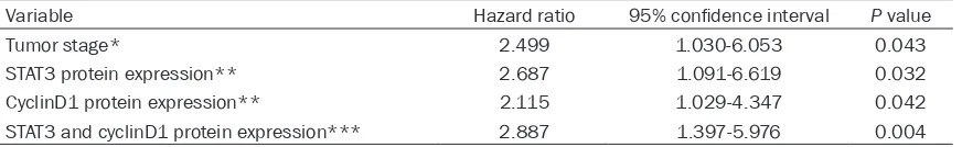
Since variables observed to have a prognostic influence by univariate analysis may covariate, the expression of STAT3 and CyclinD1 protein expression and those clinicopathological parameters that were significant in univariate analysis were further examined in multivariate analysis using Cox regression model (Table 4). The results showed that high expression of STAT3 or cyclinD1 protein were both indepen-dent prognostic factors for overall survival (STAT3 hazard ratio: 2.687, 95% confidence interval: 1.091~6.619, P = 0.032; cyclinD1 haz-ard ratio: 2.115, 95% confidence interval: 1.029~4.347, P = 0.042). More importantly, dual high expression of STAT3 and cyclinD1 pro-tein was found to be the most significant inde-pendent adverse prognostic factor for overall survival (hazard ratio: 3.223, 95% confidence interval: 1.417~7.330, P = 0.005). With regard to clinicopathological parameters, tumor stage remained to be an independent prognostic fac-tor for overall survival (hazard ratio: 2.499, 95% confidence interval: 1.030~6.053, P = 0.043). Discussion
ESCC is one of the most malignant gastrointes-tinal cancers and occurs at a high frequency rate in China and other Asian countries [14]. Despite improvements in early detection, surgi-cal techniques and chemoradiotherapy, the five-year survival rate remains low [15] and the prognosis has not been significantly improved
[image:7.612.90.526.97.164.2][16, 17]. The prediction of ESCC clinical progno-sis still depends on conventional pathologic variables such as tumor size, tumor grade, lymph node and distal metastasis status [18, 19]. Thus, it is of great clinical importance to find effective biomarkers for the prognosis of ESCC, as well as novel therapeutic strategies. We determined that high expression of STAT3 in ESCC had a highly significant relationship with advanced tumor stage (P = 0.047) and poor prognosis (P = 0.023). The JAK-STAT path-way is an important oncogenic signaling cas-cade that consists of the Janus kinase (JAK) family of nonreceptor tyrosine kinases and the STAT family of transcription factors [5] which are activated by phosphorylation of tyrosine and serine residues by upstream kinases [20]. STAT3 was first described as a DNA-binding protein activated in interleukin 6-stimulated hepatocytes and selectively interacting with an enhancer element on promoters of acute-phase genes [21]. Aberrant signaling by STAT3 is found in many types of malignancies, includ-ing multiple myeloma, head and neck cancer, breast cancer, and prostate cancer [22-26]. Moreover, STAT3 also exerts tumor-extrinsic effects, supporting tumor survival and metas-tasis apart from its tumor-intrinsic effects [27]. We also added evidence that STAT3 expression was increased compared with the surrounding normal tissue in our study and to the prognos-tic value of STAT3 in archived ESCC cohort. We also detected significant correlations between high cyclinD1 expression and lager tumor size (P = 0.009), advanced tumor stage (P = 0.012) and poor prognosis (P = 0.032). As we all know, cyclinD1 is the regulatory subunit of a dimeric holoenzyme including the cell cycledependent kinase CDK4, which promotes progression through the G1-S phase of the cell cycle. Except for regulating cell cycle, cyclinD1 Table 4. Prognostic value of tumor stage and STAT3/cyclinD1 expression for the overall survival by Cox regression
Variable Hazard ratio 95% confidence interval P value
Tumor stage* 2.499 1.030-6.053 0.043
STAT3 protein expression** 2.687 1.091-6.619 0.032
CyclinD1 protein expression** 2.115 1.029-4.347 0.042 STAT3 and cyclinD1 protein expression*** 2.887 1.397-5.976 0.004
also regulates angiogenesis, lipogenesis and mitochondrial function [28-31]. Compared with other two D-type cyclins, only overexpression of cyclinD1 is observed frequently in cancer and closely associated with malignant progression and metastatic disease [32]. As a result, it’s not surprising to find the prognostic signifi-cance of cyclinD1 in ESCC patients.
One of most notable findings of our study was the expression of STAT3 was positively associ-ated with the expression of cyclinD1 (P = 0.025). Huang C et al. demonstrated that inhib-iting SW1990 cells proliferation by RNAi against STAT3 can induce cell apoptosis and signifi-cantly reduced the levels of cyclinD1 when compared with parental and control vector-transfected cells, and significantly suppress tumor growth when it was directly injected into tumors [33]. Direct evidence of STAT3/cyclinD1 signaling axis came from CNE1 cells, breast cancer cells and hepatocellular carcinoma cells [34-36], showing that nuclear STAT3 could tar-get the CYCLIN D1 promoter directly, in turn, upregulating the CYCLIN D1 promoter activity and transcription. To date, we report about an association between STAT3 and cyclinD1 expression in ESCC tissues for the first time, which warrants further investigation.
Another significant finding in our study was that high expression of STAT3 or cyclinD1 alone, as well as dual high expression of the combination of STAT3 and cyclinD1, was significantly corre-lated with OS of ESCC patients. Interestingly, patients with dual high expression of STAT3 and cyclinD1 had a significantly shorter survival time than the patients in the other 2 groups in both univariate and multivariate analyses after we made a direct comparison of prognosis between three subgroups (dual high expres-sion, single high expression and dual low expression). It is plausible that JAK2/STAT3/ cyclinD1 pathway activation would give birth to the observed aggressive behavior of ESCC and poor outcome of ESCC patients.
In this study, we determined the prognostic value of STAT3 and cyclinD1 in ESCC. To the best of our knowledge, this is the first report demonstrating combination of STAT3 and cyclinD1 enables us to more accurately predict the true prognosis of ESCC patients. The major limitation of the present work was its retrospec-tive nature; further study is in progress to go to depth.
In summary, STAT3 and cyclinD1 correlate with more aggressive tumor behavior in ESCC. High STAT3 or cyclinD1 expression alone was associ-ated with shorter survival time. Dual high expression of STAT3 and cyclinD1 expression predicted even worse survival outcome. When STAT3 and cyclinD1 are considered together, they serve as effective prognostic markers in patients with surgically resected ESCC. From the foregoing, STAT3 and cycinD1 may be the underlying molecular markers for the diagnosis and prognosis of ESCC.
Acknowledgements
This work was sponsored by the National High Technology Research and Development Pro- gram of China (863 Program), No. 2012AA- 02A503, People’s Republic of China; the Science and Technology Innovation Key Project of Guangdong Higher Education Institutes, No. CXZD1110; Science and Technology Program of Guangzhou, No.2014J4100103, China. Disclosure of conflict of interest
None.
Address correspondence to: Xueyun Zhong, De- partment of Pathology of Medical College, Jinan University, Guangzhou 510632, Guangdong, China. Tel: +86-20-85228363; E-mail: tzxy@jnu.edu.cn; Shaoxiang Wang, Institute of Molecular Medicine, Shenzhen University, Shenzhen 518060, China. Tel: +86-20-85228363; E-mail: hackerwsx@163.com
References
[1] Zeng R, Duan L, Kong Y, Liang Y, Wu X, Wei X, Yang K. Clinicopathological and prognostic role
of MMP-9 in esophageal squamous cell carci -noma: a meta-analysis. Chin J Cancer Res 2013; 25: 637-645.
[2] Lam TK, Freedman ND, Fan JH, Qiao YL, Dawsey SM, Taylor PR, Abnet CC. Prediagnostic plasma vitamin C and risk of gastric
adenocar-cinoma and esophageal squamous cell carci -noma in a Chinese population. Am J Clin Nutr 2013; 98: 1289-1297.
[3] Parkin DM, Bray F, Ferlay J, Pisani P. Global cancer statistics, 2002. CA Cancer J Clin 2005; 55: 74-108.
[4] Chen JW, Xie JD, Ling YH, Li P, Yan SM, Xi SY, Luo RZ, Yun JP, Xie D, Cai MY. The prognostic effect of perineural invasion in esophageal
squamous cell carcinoma. BMC Cancer 2014;
[5] Darnell JE Jr. STATs and gene regulation. Sci-ence 1997; 277: 1630-1635.
[6] Xiong A, Yang Z, Shen Y, Zhou J, Shen Q. Tran-scription Factor STAT3 as a Novel Molecular Target for Cancer Prevention. Cancers (Basel) 2014; 6: 926-957.
[7] Lee SW, Ahn YY, Kim YS, Kang SB, Nam SW, Lee DS, Jeong HY, Kim JM. The Immunohisto-chemical Expression of STAT3, Bcl-xL, and MMP-2 Proteins in Colon Adenoma and Adeno-carcinoma. Gut Liver 2012; 6: 45-51.
[8] Bishop JL, Thaper D, Zoubeidi A. The Multifac-eted Roles of STAT3 Signaling in the Progres-sion of Prostate Cancer. Cancers (Basel) 2014; 6: 829-859.
[9] Wang Y, Zhu JF, Liu YY, Han GP. An analysis of cyclin D1, cytokeratin 5/6 and cytokeratin 8/18 expression in breast papillomas and papillary carcinomas. Diagn Pathol 2013; 8: 8.
[10] Seiler R, Thalmann GN, Rotzer D, Perren A, Fleischmann A. CCND1/CyclinD1 status in me-tastasizing bladder cancer: a prognosticator and predictor of chemotherapeutic response. Mod Pathol 2014; 27: 87-95.
[11] Umekita Y, Ohi Y, Sagara Y, Yoshida H. Overex-pression of cyclinD1 predicts for poor progno-sis in estrogen receptor-negative breast can-cer patients. Int J Cancan-cer 2002; 98: 415-418.
[12] Gagarin D, Yang Z, Butler J, Wimmer M, Du B,
Cahan P, McCaffrey TA. Genomic profiling of acquired resistance to apoptosis in cells de -rived from human atherosclerotic lesions: po-tential role of STATs, cyclinD1, BAD, and Bcl-XL. J Mo Cell Cardiol 2005; 39: 453-465.
[13] Kleihues P, Sobin LH. World Health
Organiza-tion classificaOrganiza-tion of tumors. Cancer 2000; 88:
2887.
[14] Gao H, Wang L, Cui S, Wang M. Combination of meta-analysis and graph clustering to identify prognostic markers of ESCC. Genet Mol Biol 2012; 35: 530-537.
[15] Kato K, Hida Y, Miyamoto M, Hashida H, Shino-hara T, Itoh T, Okushiba S, Kondo S, Katoh H. Overexpression of caveolin-1 in esophageal
squamous cell carcinoma correlates with
lymph node metastasis and pathologic stage. Cancer 2002; 94: 929-933.
[16] Parkin DM, Bray FI, Devesa SS. Cancer burden in the year 2000. The global picture. Eur J Can-cer 2001; 37 Suppl 8: S4-66.
[17] Koshy M, Esiashvilli N, Landry JC, Thomas CR Jr, Matthews RH. Multiple management dalities in esophageal cancer: combined mo-dality management approaches. Oncologist 2004; 9: 147-159.
[18] Lin DC, Du XL, Wang MR. Protein alterations in ESCC and clinical implications: a review. Dis Esophagus 2009; 22: 9-20.
[19] Ashida A, Boku N, Aoyagi K, Sato H, Tsubosa Y, Minashi K, Muto M, Ohtsu A, Ochiai A, Yoshida
T. Expression profiling of esophageal squa -mous cell carcinoma patients treated with
de-finitive chemoradiotherapy: clinical implica -tions. Int J Oncol 2006; 28: 1345-1352.
[20] Ihle JN. STATs and MAPKs: obligate or opportu-nistic partners in signaling. Bioessays 1996; 18: 95-98.
[21] Ai T, Wang Z, Zhang M, Zhang L, Wang N, Li W, Song L. Expression and prognostic relevance of STAT3 and cyclin D1 in non-small cell lung cancer. Int J Biol Markers 2012; 27: e132-138.
[22] Grandis JR, Drenning SD, Chakraborty A, Zhou
MY, Zeng Q, Pitt AS, Tweardy DJ. Requirement
of Stat3 but not Stat1 activation for epidermal growth factor receptor- mediated cell growth In vitro. J Clin Invest 1998; 102: 1385-1392.
[23] Catlett-Falcone R, Landowski TH, Oshiro MM, Turkson J, Levitzki A, Savino R, Ciliberto G, Moscinski L, Fernandez-Luna JL, Nunez G. Constitutive activation of Stat3 signaling con-fers resistance to apoptosis in human U266 myeloma cells. Immunity 1999; 10: 105-115.
[24] Buettner R, Mora LB, Jove R. Activated STAT signaling in human tumors provides novel mo-lecular targets for therapeutic intervention. Clin Cancer Res 2002; 8: 945-954.
[25] Epling-Burnette PK, Liu JH, Catlett-Falcone R, Turkson J, Oshiro M, Kothapalli R, Li Y, Wang JM, Yang-Yen HF, Karras J. Inhibition of STAT3 signaling leads to apoptosis of leukemic large granular lymphocytes and decreased Mcl-1 ex-pression. J Clin Invest 2001; 107: 351-362.
[26] Barton BE, Murphy TF, Adem P, Watson RA, Ir-win RJ, Huang HF. IL-6 signaling by STAT3 par-ticipates in the change from hyperplasia to neoplasia in NRP-152 and NRP-154 rat pros-tatic epithelial cells. BMC Cancer 2001; 1: 19.
[27] Bournazou E, Bromberg J. Targeting the tumor microenvironment: STAT3 signaling. JAK-STAT 2013; 2: e23828.
[28] Wang C, Li Z, Lu Y, Du R, Katiyar S, Yang J, Fu M, Leader JE, Quong A, Novikoff PM. Cyclin D1 repression of nuclear respiratory factor 1 inte-grates nuclear DNA synthesis and mitochon-drial function. Proc Natl Acad Sci U S A 2006; 103: 11567-11572.
[29] Sakamaki T, Casimiro MC, Ju X, Quong AA, Katiyar S, Liu M, Jiao X, Li A, Zhang X, Lu Y. Cy-clin D1 determines mitochondrial function in vivo. Mol Cell Biol 2006; 26: 5449-5469.
[30] Wang C, Pattabiraman N, Zhou JN, Fu M, Saka-maki T, Albanese C, Li Z, Wu K, Hulit J, Neu-meister P. Cyclin D1 repression of peroxisome proliferator-activated receptor gamma expres-sion and transactivation. Mol Cell Biol 2003; 23: 6159-6173.
vascular endothelial growth factor-stimulated growth of vascular endothelial cells: implica-tion of tumor vascularizaimplica-tion. Clin Can Res 2006; 12: 4720-4729.
[32] Li Z, Wang C, Prendergast GC, Pestell RG. Cy-clin D1 functions in cell migration. Cell Cycle 2006; 5: 2440-2442.
[33] Huang C, Yang G, Jiang T, Cao J, Huang KJ, Qiu ZJ. Down-regulation of STAT3 expression by vector-based small interfering RNA inhibits pancreatic cancer growth. World J Gastroen-terol 2011; 17: 2992-3001.
[34] Xu Y, Shi Y, Yuan Q, Liu X, Yan B, Chen L, Tao Y, Cao Y. Epstein-Barr Virus encoded LMP1 regu-lates cyclin D1 promoter activity by nuclear EGFR and STAT3 in CNE1 cells. J Exp Clin Can-cer Res 2013; 32: 90.
[35] Saxena NK, Vertino PM, Anania FA, Sharma D. Leptin-induced growth stimulation of breast cancer cells involves recruitment of histone acetyltransferases and mediator complex to CYCLIN D1 promoter via activation of Stat3. J Biol Chem 2007; 282: 13316-13325.