Original Article
Ectopic expression of AP-2α transcription factor
suppresses glioma progression
Wenjing Su
1*, Juan Xia
1,2*, Xueqin Chen
1, Miao Xu
1, Ling Nie
1, Ni Chen
1, Jing Gong
1, Xinglan Li
1, Qiao Zhou
11Laboratory of Pathology, State Key Laboratory of Biotherapy and Department of Pathology, West China Hospital,
West China Medical School, Sichuan University, Chengdu 610041, China; 2Suining Central Hospital, Suining
629000, China. *Equal contributors.
Received September 27, 2014; Accepted November 26, 2014; Epub December 1, 2014; Published December 15, 2014
Abstract: The transcriptional factor AP-2α is a tumor suppressor gene and is downregulated in various neoplasms including glioma. Although the level of AP-2α is negatively associated with the grade of human glioma, the specific functions of AP-2α in glioma are still unknown. In this study, we experimentally showed that artificial overexpression of AP-2α in glioma T98G and U251 cells significantly downregulated the mRNA levels of Bcl-xl, Bcl-2, c-IAP2 and survivin, together with upregulation of the Hrk mRNA levels. Reintroduction of AP-2α also induced downregulation of the protein levels of survivin and VEGF in glioma cells. In biological assays with T98G and U251 cells, AP-2α re -duced tumor cell growth, increased cell death, attenuated cell migration and endothelial tube formation. The AP-2α transcription factor may play an important role in suppressing glioma progression.
Keywords: AP-2 alpha, glioma, cell growth, apoptosis, invasion
Introduction
AP-2α, which is located in the short arm of chro
-mosome 6 adjoining to the HLA gene, is a
mem-ber of the transcription factor activator
pro-tein-2 (AP-2) family {Britto, 2007 #42} {Britto,
2007 #42}. AP-2α binds to CG-rich sequences
such as 5’-GCCN
3GGC, 5’-GCCN
4GGC and 5’-
GCCN
3/4GGG via a DNA-binding domain within
the COOH-terminal half of the protein and plays
key roles in transcriptional regulation [1].
In addition to its important function in physio
-logical processes such as embryo develop
-ment, AP-2α acts as a tumor suppressor gene,
inhibiting the initiation and progression of a
variety of malignancies. AP-2α expression was
decreased in many neoplasms such as glioma
[2-4], prostate cancer [5], breast cancer [6-9],
colon cancer [10], melanoma [11, 12], ovarian
cancer [13] and renal carcinoma [14].
AP-2α has a pivotal role in regulating the expres
-sion of key genes, the products of which are
involved in tumor initiation and progression. For
example, AP-2α regulates genes that are
invo-lved in proliferation (c-MYC), cell cycle regula
-tion (HER-2 and p21WAF1), apoptosis (c-KIT,
Bcl-2, and FAS/APO-1), cell adhesion(MCAM/
MUC18 and E-cadherin), and tumor invasion/
angiogenesis (MMP-9, PAI-I, and PAR-1)
[15-19].
Decreased AP-2α level was associated with
grade of glioma [20]. However, the specific
effects of AP-2α on glioma cells were not clearly
understand. In the present study, we artificially
overexpressed AP-2α in glioma cells, and inves
-tigated its effects on glioma cell growth,
apop-tosis, migration and endothelial tube formati-
on.
Materials and methods
Cell lines, tissue samples and general reagents
Human glioma cells T98G, U251,
adenovirus-immortalized human embryonic kidney epithe
-lial cell HEK-293, and human umbilical vein
endothelial cells (HUVEC) were all from the
American Type Culture Collection and were cul
-tured in DMEM with 10% FCS (Life Technologies,
TakaRa, Japan). Archived formalin-fixed, paraf
AP-2α suppresses glioma progression
normal brain (n = 2), were also used. All tissue
samples were from West China Hospital and
were collected and used according to the
ethi-cal guidelines and procedures approved by the
institutional supervisory committee. Tris base,
EDTA, Tween 20 and dithiothreitol were from
Amresco (Solon, OH). PMSF, aprotinin and
pep-statin were from Roche Diagnostics (Mannheim,
Germany).
RT-PCR and real-time qPCR
Total RNA was isolated using the TRIzol reagent
(Invitrogen/Life Technologies, TakaRa, Japan).
cDNA was synthesized from the isolated RNA
by RT and amplified by PCR. PCR primer
sequences and product lengths were as
fol-lows. AP-2α: 5’-ACT CCT TAC CTC ACG CCA TC-3’,
5’-ATA GGG ATG GCG GAG ACG-3’, 136 bp; actin:
5’-TGG AGA AAT CTG GCA CCA C-3’, 5’-GAG GCG
TAC AGG GAT AGC AC-3’, 190 bp; Bcl-xl: 5’-
CTG
TGC GTG GAA AGC GTA G-3’, 5’-CTCGGCTG-
CTGCATTGTTC-3’, 159 bp; Bcl-2: 5’-GTC ATG
TGT GTG GAG AGC GTC-3’, 5’-GAG TCT TCA GAC
AGC
CAG G-3’, 193 bp; Hrk: 5’-CTA GGC GAC
GAG CTG CAC CAG-3’, 5’-GCA CAG CCA AGG CCA
GTA GGT G-3’, 102 bp; c-IAP2: 5’-AGG GAA GAG
GAG AGA GAA AGA GC-3, 5’-CGG CAG TTA GTA
GAC TAT CCA GG-3’, 133 bp; Survivin: 5’-GCA
GTT TGA AGA ATT AAC CCT TG-3’, 5’-CAC TTT
CTC CG CAG TTT CCT C-3’, 121 bp. PCR
prod-ucts were separated by agarose gel electropho
-resis and visualized by the fluorescent dye
GoldView (Beijing SBS Genetech, Beijing, China)
under UV light. Real time PCR was carried out
as described [21].
Western blot
The primary antibodies used were as follows:
AP-2α (rabbit monoclonal, 1:500, Santa Cruz
Biotechnology, USA), VEGF (mouse Polyclonal,
1:500, Boster, China), Survivin (rabbit poly
-clonal, 1:500, R&D, USA), GAPDH (mouse
monoclonal, 1:10000, Kangcheng, China).
Horseradish peroxidase-labeled secondary
antibodies were from Zymed Laboratories
(South San Francisco, CA). Western blotting
was carried out as previously described [22].
Recombinant adenoviral vectors for
overex-pression of AP-2α
The coding sequence of AP-2α (transcript vari
-ant 3, 1321 bp) was cloned from HEK-293 cells
by RT-PCR with the following primers: 5’-CTC
GAG CCG CGA TGT CCA TAC TTG C-3’, 5’-AAG
CTT GCC TCA CTT TCT GTG CTT CTC-3’. The
ade-novirus vector for AP-2α was constructed as
described [21] and named AD-AP2α. The
pAdTrack-CMV vector was used as control
(AD-control). The titers and multiplicity of infec
-tion (MOI) were determined according to the
manufacturer’s protocols.
MTT assays
T98G and U251 cells transfected with AD-AP2α
or AD-control were incubated in 96-well tissue
culture plates for the indicated times. MTT
(Sigma Aldrich, USA) was then added to each
well and incubated for 4 hours. Then the
super-natant was removed, replaced with 100 μl
dimethylsulfoxide and incubated for 10 min in
dark. The absorbance at 570 nm (A570) was
measured by spectrophotometer (Bio-Tek,
USA).
Migration assays
Transwell migration assays were used to exam
-ine the effect of AP-2α on glioma cell migration.
T98G or U251 cell were transfected with
AD-AP2α (AD-control as negative control). 24 h
[image:2.612.89.287.73.246.2]later, cells were isolated and 1 × 10
5cells
sus-pended in serum-free media were placed in the
Figure 1. AP-2α expression in glioma. AP-2α expresupper compartment of 5 mm-pore transwells
(Corning Costar, Lowell, MA). Cells were allowed
to migrate for 24 h and the transwell slides
were finally fixed with paraformaldehyde and
stained with crystal violet.
Tube formation assay
Tube formation assay was performed as previ
-ously described [23]. Conditioned media were
obtained by incubating the cells transfected
with AD-AP2α or AD-control in DMEM without
serum for 24 h. The 96-well plate was coated
with 50 µl growth factor-reduced matrigel (BD
Biosciences). A total of 4 × 10
5HUVEC cells was
resuspended in 100 µl of conditioned media
with 1% FCS and seeded on matrigel-coated
wells. HUVEC cells were incubated for 18 h to
allow formation of tube-like structures. Total
tube lengths formed were measured and
com-pared from three different viewing fields at 10 ×
magnification using an inverted microscope
(Nikon TMS). Tube formation assays were per
-formed in triplicate.
Terminal deoxynucleotidyl
transferase-mediat-ed biotinylattransferase-mediat-ed dUTP nick end-labeling
Terminal deoxynucleotidyl transferase-mediat
-ed dUTP nick end labeling (TUNEL) was per
-formed by using in situ cell death detection kit
(Roche, USA) as previously described [22].
Statistical analysis
All experiments were repeated at least thrice.
PASW Statistics 18.0 software package was
used for statistical analysis.
P
value less than
0.05 was considered significant statistically.
Results
AP-2α expression was significantly decreased
in human glioma cells
RT-PCR and western blot confirmed significant
reduction of AP-2α level in human glioma cells
(T98G, U251) and glioma tissues (G1, G2) as
[image:3.612.96.521.71.358.2]AP-2α suppresses glioma progression
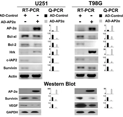
Artificial overexpression of AP-2α decreased
mRNA levels of Bcl-xl, Bcl-2, c-IAP2 and
sur-vivin, increased mRNA levels of Hrk, and
de-creased protein levels of survivin and VEGF
To examine the effects of AP-2α on glioma cells,
recombinant adenovirus for overexpression of
AP-2α (AD-AP2α, MOI 50, 96 hours) was con
-structed (
Figure 2B
). RT-PCR, Q-PCR and
west-ern blot confirmed the efficient expression of
AP-2α in AD-AP2α infected T98G and U251
cells (
Figure 3
).
Artificial overexpression of AP-2α in T98G and
sion of Bcl-2, Bcl-xl, c-IAP2 and survivin, togeth
-er with increased Hrk mRNA level, and west-ern
blot displayed repression of survivin and VEGF
after AD-AP2α treatment (
Figure 3
). Incon-
spicuous alterations were detected in the
lev-els of other molecules such as Bik, BNIP3,
c-IAP1 and CASP8 in T98G and U251 overex
-pressing AD-AP2α (data not shown).
AP-2α inhibited cell growth, promoted apopto
-sis, inhibited cell migration and HUVEC tube
formation ability in glioma
[image:4.612.95.518.71.473.2]Having confirmed the contributions of AP-2α to
AP-2α suppresses glioma progression
to show the biological effects of artificial AP-2α
overexpression in glioma cells. Concomitant
with AD-AP2α mediated AP-2α overexpression,
glioma cells showed significantly reduced cell
growth (
Figure 4A
), increased cell apoptosis
(
Figure 4B
) and decreased cell migration
(
Figure 4C
). Microtubule formation of HUVEC
cells was decreased when stimulated with
supernatant from glioma cells re-expressing
AP-2α (AD-AP2α) (
Figure 4C
).
Discussion
The AP-2 family consists of five highly homolo
-gous members, namely AP-2α, AP-2β, AP-2γ,
AP-2δ and AP-2ε [24], the expression of which
is cell-type specific. The amine terminus of the
AP-2α protein is responsible for transactivation
activity, and a basic and helical region in the
COOH terminus is responsible for dimerization
and DNA binding [25, 26].
AP-2α mediates programmed gene expression
in embryonic morphogenesis and cell differen
-tiation [27]. AP-2α knockout mice die perina
-tally with severe multiple congenital defects
involving face, skull, and sensory organs. In situ
hybridization showed that mouse embryos spe
-cifically expressed AP-2α in ectoderm-derived
tissues, including craniofacial, gonad, kidney,
and skin [27, 28].
Also, AP-2α has been shown to be a tumor sup
-pressor gene, the loss of which is linked to
malignant transformation, tumor progression
and elevated risk of metastasis in such tumors
as melanoma [29-32], prostate cancer [33,
34], breast cancer [35, 36], pancreatic cancer
[37, 38], colorectal cancer [39] and ovarian
cancer [13]. Notably, loss of AP-2α was strongly
correlated with higher grade in human glioma,
and appeared to be the most frequent
molecu-lar change in astrocytic tumor progression from
lower to higher grade, although no significant
effect of AP-2α on survival was observed [20].
In this research, significant reductions of AP-2α
were also detected in glioma cells and tissues,
which is in consistence with studies before.
However, the role of AP-2α in the development/
progression of glioma has not been fully
defined. It has been reported that there are
multitudinous genes regulated by AP-2α such
as those involved in cell proliferation (Hoxa, IGF
receptor type I and IGF-binding proteins 3/5)
[40-43], cell cycle regulation (p21WAF1/CIP17
and HER-2) [44, 45], apoptosis (c-KIT) [30, 46,
47], cell adhesion (E-cadherin and MCAM/
MUC18) [48, 49], and cell migration/angiogen
-esis (MMP-2, and PAR-1) [48, 50, 51]. Our data
showed that AP-2α inhibited the expression of
the anti-apoptotic genes Bcl-xl, Bcl-2, c-IAP2
and surviving and the pro-angiogenic gene
VEGF, whereas promoted the expression of the
pro-apoptotic gene HRK, thus enhanced glioma
cell apoptosis and inhibited HUVEC tube forma
-tion ability. These finding are compatible with
our earlier works [47, 52, 53]. Except for this,
we also detected the influences of AP-2α on a
series of genes involving in cell proliferation
and migration (data not shown) but failed to get
meaningful results. Nonetheless, AP-2α
induced attenuated glioma cell proliferation
and migration in biological experiments, the
mechanism of which has not been well
under-stood yet.
In summary, the results of this study showed
that AP-2α, which is dramatically down-regulat
-ed in glioma, suppresses the progression of
glioma by regulating a series of tumor-associat
-ed genes. These findings may sh-ed some new
lights on the AP-2α dysfunction in glioma pro
-gression and suggest AP-2α a potential thera
-peutic target in glioma.
Acknowledgements
This work was supported by grants from the
Natural Science Foundation of China (NSFC,
81272848, 81272820, 81101529, 81101628,
81302225) and Postdoctoral Fund of China
(20100480076, 201104643, 2013M531970,
2014T70876).
Disclosure of conflict of interest
None.
Address correspondence to: Dr. Qiao Zhou, La- boratory of Pathology, State Key Laboratory of Biotherapy and Department of Pathology, West China Hospital, West China Medical School, Sichuan University, Chengdu 610041, China. Tel: +86-28-8516-4027; Fax: +86-28-+86-28-8516-4027; E-mail: zhouqiao@mcwcums.com
References
[1] Mohibullah N, Donner A, Ippolito JA, Williams T. SELEX and missing phosphate contact analy -ses reveal flexibility within the AP-2 [alpha] pro -tein: DNA binding complex. Nucleic Acids Res 1999; 27: 2760-9.
[2] Britto R, Umesh S, Hegde AS, Hegde S, Santosh V, Chandramouli BA, Somasundaram K. Shift in AP-2alpha localization characterizes astro-cytoma progression. Cancer Biol Ther 2007; 6: 413.
[3] Heimberger AB, McGary EC, Suki D, Ruiz M, Wang H, Fuller GN, Bar-Eli M. Loss of the AP-2α transcription factor is associated with the grade of human gliomas. Clin Cancer Res 2005; 11: 267.
[4] Britto R, Umesh S, Hegde AS, Hegde S, Santosh V, Chandramouli BA, Somasundaram K. Shift in AP-2alpha localization characterizes astro-cytoma progression. Cancer Biol Ther 2007; 6: 413-8.
[5] Ruiz M, Troncoso P, Bruns C, Bar-Eli M. Activator protein 2α transcription factor expression is associated with luminal differentiation and is lost in prostate cancer. Clin Cancer Res 2001; 7: 4086.
[6] Turner BC, Zhang J, Gumbs AA, Maher MG, Kaplan L, Carter D, Glazer PM, Hurst HC, Haffty BG, Williams T. Expression of AP-2 transcrip -tion factors in human breast cancer correlates with the regulation of multiple growth factor signalling pathways. Cancer Res 1998; 58: 5466.
[7] Friedrichs N, Jäger R, Paggen E, Rudlowski C, Merkelbach-Bruse S, Schorle H, Buettner R. Distinct spatial expression patterns of AP-2alpha and AP-2gamma in non-neoplastic hu-man breast and breast cancer. Mod Pathol 2004; 18: 431-438.
[8] Pellikainen JM and Kosma VM. Activator pro -tein-2 in carcinogenesis with a special refer-ence to breast cancer-A mini review. Int J Cancer 2007; 120: 2061-2067.
[9] Gee JM, Robertson JF, Ellis IO, Nicholson RI, Hurst HC. Immunohistochemical analysis re -veals a tumour suppressor-like role for the transcription factor AP-2 in invasive breast cancer. J Pathol 1999; 189: 514-520.
[10] Ropponen KM, Kellokoski JK, Pirinen RT, Moisio KI, Eskelinen MJ, Alhava EM, Kosma
VM. Expression of transcription factor AP-2 in colorectal adenomas and adenocarcinomas; comparison of immunohistochemistry and in situ hybridisation. J Clin Pathol 2001; 54: 533. [11] Tellez CS, Davis DW, Prieto VG, Gershenwald
JE, Johnson MM, McCarty MF, Bar-Eli M. Quantitative analysis of melanocytic tissue ar -ray reveals inverse correlation between activa -tor protein-2α and protease-activated recep -tor-1 expression during melanoma progres-sion. J Invest Dermatol 2006; 127: 387-393. [12] Baldi A, Santini D, Battista T, Dragonetti E,
Ferranti G, Petitti T, Groeger AM, Angelini A, Rossiello R, Baldi F, Natali PG, Paggi MG. Expression of AP-2 transcription factor and of its downstream target genes c-kit, E-cadherin and p21 in human cutaneous melanoma. J Cell Biochem 2001; 83: 364-372.
[13] Anttila MA, Kellokoski JK, Moisio KI, Mitchell PJ, Saarikoski S, Syrjänen K, Kosma VM. Expression of transcription factor AP-2alpha predicts survival in epithelial ovarian cancer. Br J Cancer 2000; 82: 1974-83.
[14] Oya M, Mikami S, Mizuno R, Miyajima A, Horiguchi Y, Nakashima J, Marumo K, Mukai M, Murai M. Differential expression of activator protein-2 isoforms in renal cell carcinoma. Urology 2004; 64: 162-167.
[15] Yu L, Hitchler MJ, Sun W, Sarsour EH, Goswami PC, Klingelhutz AJ, Domann FE. AP-2α Inhibits c-MYC Induced Oxidative Stress and Apoptosis in HaCaT Human Keratinocytes. J Oncol 2009; 2009: 780874.
[16] Wajapeyee N, Britto R, Ravishankar HM, Somasundaram K. Apoptosis induction by acti -vator protein 2α involves transcriptional re -pression of Bcl-2. J Biol Chem 2006; 281: 16207.
[17] Jean D, Gershenwald JE, Huang S, Luca M, Hudson MJ, Tainsky MA, Bar-Eli M. Loss of AP-2 Results in Up-regulation ofMCAM/MUC18 and an Increase in Tumor Growth and Metastasis of Human Melanoma Cells. J Biol Chem 1998; 273: 16501.
[18] Schwartz B, Melnikova VO, Tellez C, Mourad-Zeidan A, Blehm K, Zhao YJ, McCarty M, Adam L, Bar-Eli M. Loss of AP-2α results in deregula -tion of E-cadherin and MMP-9 and an increase in tumorigenicity of colon cancer cells in vivo. Oncogene 2007; 26: 4049-4058.
[19] Tellez C, McCarty M, Ruiz M, Bar-Eli M. Loss of activator protein-2α results in overexpression of protease-activated receptor-1 and corre-lates with the malignant phenotype of human melanoma. J Biol Chem 2003; 278: 46632. [20] Heimberger AB, McGary EC, Suki D, Ruiz M,
AP-2α suppresses glioma progression
[21] Chen X, Gong J, Zeng H, Chen N, Huang R, Huang Y, Nie L, Xu M, Xia J, Zhao F, Meng W, Zhou Q. MicroRNA145 targets BNIP3 and sup -presses prostate cancer progression. Cancer Res 2010; 70: 2728-38.
[22] Chen N, Chen X, Huang R, Zeng H, Gong J, Meng W, Lu Y, Zhao F, Wang L, Zhou Q. BCL-xL is a target gene regulated by hypoxia-inducible factor-1 {alpha}. J Biol Chem 2009; 284: 10004-12.
[23] Chan KC, Ko JM, Lung HL, Sedlacek R, Zhang ZF, Luo DZ, Feng ZB, Chen S, Chen H, Chan KW, Tsao SW, Chua DT, Zabarovsky ER, Stanbridge EJ, Lung ML. Catalytic activity of Matrix metalloproteinase-19 is essential for tu-mor suppressor and anti-angiogenic activities in nasopharyngeal carcinoma. Int J Cancer 2011; 129: 1826-37.
[24] Eckert D, Buhl S, Weber S, Jäger R, Schorle H. The AP-2 family of transcription factors. Genome Biol 2005; 6: 246.
[25] Williams T and Tjian R. Characterization of a dimerization motif in AP-2 and its function in heterologous DNA-binding proteins. Science 1991; 251: 1067-71.
[26] Williams T and Tjian R. Analysis of the DNA-binding and activation properties of the hu-man transcription factor AP-2. Genes Dev 1991; 5: 670-82.
[27] Schorle H, Meier P, Buchert M, Jaenisch R, Mitchell PJ. Transcription factor AP-2 essential for cranial closure and craniofacial develop-ment. Nature 1996; 381: 235-8.
[28] Zhang J, Hagopian-Donaldson S, Serbedzija G, Elsemore J, Plehn-Dujowich D, McMahon AP, Flavell RA, Williams T. Neural tube, skeletal and body wall defects in mice lacking tran -scription factor AP-2. Nature 1996; 381: 238-41.
[29] Karjalainen JM, Kellokoski JK, Eskelinen MJ, Alhava EM, Kosma VM. Downregulation of transcription factor AP-2 predicts poor survival in stage I cutaneous malignant melanoma. J Clin Oncol 1998; 16: 3584-91.
[30] Baldi A, Santini D, Battista T, Dragonetti E, Ferranti G, Petitti T, Groeger AM, Angelini A, Rossiello R, Baldi F, Natali PG, Paggi MG. Expression of AP-2 transcription factor and of its downstream target genes c-kit, E-cadherin and p21 in human cutaneous melanoma. J Cell Biochem 2001; 83: 364-72.
[31] Tellez CS, Davis DW, Prieto VG, Gershenwald JE, Johnson MM, McCarty MF, Bar-Eli M. Quantitative analysis of melanocytic tissue ar -ray reveals inverse correlation between activa -tor protein-2alpha and protease-activated re-ceptor-1 expression during melanoma progres-sion. J Invest Dermatol 2007; 127: 387-93. [32] Berger AJ, Davis DW, Tellez C, Prieto VG,
Gershenwald JE, Johnson MM, Rimm DL,
Bar-Eli M. Automated quantitative analysis of acti -vator protein-2alpha subcellular expression in melanoma tissue microarrays correlates with survival prediction. Cancer Res 2005; 65: 11185-92.
[33] Ruiz M, Troncoso P, Bruns C, Bar-Eli M. Activator protein 2alpha transcription factor expression is associated with luminal differentiation and is lost in prostate cancer. Clin Cancer Res 2001; 7: 4086-95.
[34] Lipponen P, Aaltomaa S, Kellokoski J, Ala-Opas M, Kosma V. Expression of activator protein 2 in prostate cancer is related to tumor differen-tiation and cell proliferation. Eur Urol 2000; 37: 573-8.
[35] Pellikainen J, Kataja V, Ropponen K, Kellokoski J, Pietiläinen T, Böhm J, Eskelinen M, Kosma VM. Reduced nuclear expression of transcrip-tion factor AP-2 associates with aggressive breast cancer. Clin Cancer Res 2002; 8: 3487-95.
[36] Gee JM, Robertson JF, Ellis IO, Nicholson RI, Hurst HC. Immunohistochemical analysis re -veals a tumour suppressor-like role for the transcription factor AP-2 in invasive breast cancer. J Pathol 1999; 189: 514-20.
[37] Jonckheere N, Fauquette V, Stechly L, Saint-Laurent N, Aubert S, Susini C, Huet G, Porchet N, Van Seuningen I, Pigny P. Tumour growth and resistance to gemcitabine of pancreatic cancer cells are decreased by AP-2alpha over -expression. Br J Cancer 2009; 101: 637-44. [38] Hirono S, Yamaue H, Hoshikawa Y, Ina S, Tani
M, Kawai M, Ushijima M, Matsuura M, Saiki Y, Saiura A, Yamamoto J, Miki Y, Noda T. Mole-cular markers associated with lymph node me -tastasis in pancreatic ductal adenocarcinoma by genome-wide expression profiling. Cancer Sci 2010; 101: 259-66.
[39] Ropponen KM, Kellokoski JK, Pirinen RT, Moisio KI, Eskelinen MJ, Alhava EM, Kosma VM. Expression of transcription factor AP-2 in colorectal adenomas and adenocarcinomas; comparison of immunohistochemistry and in situ hybridisation. J Clin Pathol 2001; 54: 533-8.
[40] Ding X, Yang Z, Zhou F, Wang F, Li X, Chen C, Li X, Hu X, Xiang S, Zhang J. Transcription factor AP-2alpha regulates acute myeloid leukemia cell proliferation by influencing Hoxa gene ex -pression. Int J Biochem Cell Biol 2013; 45: 1647-56.
[41] DiGiovanni J, Kiguchi K, Frijhoff A, Wilker E, Bol DK, Beltrán L, Moats S, Ramirez A, Jorcano J, Conti C. Deregulated expression of insulin-like growth factor 1 in prostate epithelium leads to neoplasia in transgenic mice. Proc Natl Acad Sci U S A 2000; 97: 3455-60.
-enosine 3’, 5’-monophosphate regulate IGF-binding protein-3 gene expression by transcrip -tional and posttranscrip-tional mechanisms in mammary epithelial cells. Endocrinology 2000; 141: 4583-91.
[43] Duan C and Clemmons DR. Transcription fac-tor AP-2 regulates human insulin-like growth factor binding protein-5 gene expression. J Biol Chem 1995; 270: 24844-51.
[44] Zeng YX, Somasundaram K and el-Deiry WS. AP2 inhibits cancer cell growth and activates p21WAF1/CIP1 expression. Nat Genet 1997; 15: 78-82.
[45] Bosher JM, Williams T and Hurst HC. The devel -opmentally regulated transcription factor AP-2 is involved in c-erbB-2 overexpression in hu -man mammary carcinoma. Proc Natl Acad Sci U S A 1995; 92: 744-7.
[46] Huang S, Jean D, Luca M, Tainsky MA, Bar-Eli M. Loss of AP-2 results in downregulation of c-KIT and enhancement of melanoma tumorige-nicity and metastasis. EMBO J 1998; 17: 4358-69.
[47] Xu M, Chen X, Chen N, Nie L, Li X, Li Q, Zeng H, Zhou Q. Synergistic silencing by promoter methylation and reduced AP-2alpha transacti -vation of the proapoptotic HRK gene confers apoptosis resistance and enhanced tumor growth. Am J Pathol 2013; 182: 84-95.
[48] Zhang Z, Zhang L, Jia L, Cui S, Shi Y, Chang A, Zeng X, Wang P. AP-2alpha suppresses inva-sion in BeWo cells by represinva-sion of matrix me -talloproteinase-2 and -9 and up-regulation of E-cadherin. Mol Cell Biochem 2013; 381: 31-9.
[49] Jean D, Gershenwald JE, Huang S, Luca M, Hudson MJ, Tainsky MA, Bar-Eli M. Loss of AP-2 results in up-regulation of MCAM/MUC18 and an increase in tumor growth and metastasis of human melanoma cells. J Biol Chem 1998; 273: 16501-8.
[50] Gille J, Swerlick RA and Caughman SW. Transforming growth factor-alpha-induced transcriptional activation of the vascular per-meability factor (VPF/VEGF) gene requires AP-2-dependent DNA binding and transactivation. EMBO J 1997; 16: 750-9.
[51] Tellez C, McCarty M, Ruiz M, Bar-Eli M. Loss of activator protein-2alpha results in overexpres-sion of protease-activated receptor-1 and cor-relates with the malignant phenotype of hu -man melanoma. J Biol Chem 2003; 278: 46632-42.
[52] Liu X, Chen N, Wang X, He Y, Chen X, Huang Y, Yin W, Zhou Q. Apoptosis and proliferation markers in diffusely infiltrating astrocytomas: profiling of 17 molecules. J Neuropathol Exp Neurol 2006; 65: 905-13.