Original Article
Effects of subchronic exposure to lead acetate and
cadmium chloride on rat’s bone: Ca and Pi contents,
bone density, and histopathological evaluation
Hongke Lu1, Guiping Yuan2, Zhongqiong Yin1, Shujun Dai1, Renyong Jia1, Jiao Xu1, Xu Song1, Li Li1, Cheng Lv1
1College of Veterinary Medicine, Sichuan Agricultural University, Ya’an 625014, China; 2Analytical & Testing
Cen-ter, Sichuan University, Chengdu 610064, China
Received November 29, 2013; Accepted December 28, 2013; Epub January 15, 2014; Published February 1, 2014
Abstract: This study aims to investigate the effects of low-dose subchronic exposure to lead acetate (Pb(NO3)2) and cadmium chloride (CdCl2·2.5H2O) on bone in rats. The rats were assigned randomly to a control group and three experimental groups that were given the mixture of Pb(NO3)2 and CdCl2·2.5H2O by gastric gavage at doses of 0 mg/ kg body weight (b.w.) (Group I, to serve as a control), 29.96 mg/kg b.w. (Group II, 29.25+0.71), 89.88 mg/kg b.w. (Group III, 87.74+2.14), and 269.65 mg/kg b.w. (Group IV, 263.23+6.42) for at least 90 consecutive days. Calcium (Ca) and phosphorus (Pi) contents in the bone were determined. Bone mineral density (BMD) was measured at the tibia and femur region by dual-energy X-ray absorbsiometry. The histopathology of bone was evaluated by light microscope, scanning electron microscope, and transmission electron microscope. The BMD of rats in the
experi-mental group was significantly lower and the contents of Ca and Pi were decreased than those in the control group.
The histopathological evaluation showed that co-induction of Pb and Cd results in bone microstructure damage,
especially to trabecular bone, marrow cavity, collagen fiber, and osteoblast. In general, results indicate that combin -ing Pb with Cd induces bone damage and increases the risk of osteoporosis.
Keywords: Lead and cadmium, bone mineral density, calcium and phosphorus, bone microstructure, subchronic, rats
Introduction
The environment has been filled with pollutants
over the years. People are frequently exposed to two or more kinds of contaminants. These pollutants are recognized for being great harm to public health worldwide; they are natural or anthropogenic sources of pollution to the envi-ronment [1]. Lead (Pb) and cadmium (Cd) are widely distributed in the environment because of natural and human-related activities [2, 3]. Exposure to Pb and Cd can lead to damage to several organs, including liver, kidney, and bone [4-6]. Therefore, investigating the effects of the co-administration of Pb and Cd is necessary. Most animal studies about Pb and Cd toxicity are limited to individual exposure to the harm of bone. Pb mainly accumulates in bones by replacing calcium, and the bones contain approximately 90% of the total amount of Pb in the body [7, 8]. The long-term effects of Pb
In our previous research, lead acetate (Pb (NO3)2) and cadmium chloride (CdCl2·2.5H2O) were used in an equitoxic mixture ratio design.
The fixed mixture ratio was based on the single
toxicant LD50-value (median lethal dose value) for an oral acute study [13]. Moreover, the result shows that the acute toxicity of Pb and Cd can lead to bone damage. In this study, peo-ple may be exposed to Pb and Cd from indus-trial sources because of the presence of these elements in the natural environment. Thus, we chose low-dose exposure via the subchronic experiment. The experimental model of rats treated with relatively low Pb and Cd levels are based on the mixture toxicant LD50-value of 2696.54 mg/kg our oral acute study for a 90-day period as a model of Pb and Cd-induced subchronic toxicological evaluation.
The purpose of this study is to evaluate the effect of subchronic Pb and Cd co-exposure on bone and its mechanism by examining the con-tent of Ca and Pi in bone and BMD, and through histopathological evaluation.
Materials and methods
Chemicals and animals
Analytical grade Pb (NO3)2 and CdCl2·2.5H2O (AR>99.0%) were obtained from Sigma Aldrich (St. Louis, MO, USA). The study was carried out with Sprague-Dawley rats with an initial body weight of approximately 150 g. All rats were obtained from Chengdu Dossy Experimen- tal Animals Co., Ltd. (License No. SCXK (Sichu- an) 2008-24, China) and kept in the animal house at the Sichuan Agriculture University (Ya’an, China). The animals were housed under controlled conventional conditions (tempera-ture 25±3°C, relative humidity of 35% to 60%, 12-hour light-dark cycle) and allowed free access to a standard rat chow and drinking water during the experiment. The experimental protocol was approved by the Local Ethics Committee for Animal Experiments in Sichuan Agricultural University (China) for the care and use of laboratory animals.
Experimental protocol
All animals took 7 days to adapt to the experi-mental conditions prior to the experiment. The rats were divided randomly into four groups, that is, one control group and three
experimen-tal groups; each group has 20 rats. Experimenexperimen-tal groups were treated with a mixture of Pb (NO3)2 and CdCl2·2.5H2O solution at dose of 29.96 mg/kg body weight (b.w.) (Group II, 29.25+0.71), 89.88 mg/kg b.w. (Group III, 87.74+2.14), and 269.65 mg/kg b.w. (Group IV, 263.23+6.42) by gastric gavage for at least 90 consecutive days. The control group was treated with a solution free of Cd and Pb. In each case, the volume of the reagent, which was administered to the rats by gavage is 10 mL/kg b.w. Each rat was
marked with a unique identification number by
trinitrophenol.
After the exposure period (90 days) of the experiment, six rats were randomly selected from each group. All rats were anesthetized with diethyl ether. Rats that fasted were
sacri-ficed by femur and tibia dissection under ether
anesthesia. Left femur and tibia were isolated from soft tissues for the measurement of BMD, as well as Ca, Pi, Pb, and Cd content. The right femur was obtained for bone histopathology.
Analysis of Ca, Pi, Pb, and Cd contents in bone
The bone sample was dried to constant weight (dry weight) at 80°C for 48 hours. The dried sample was ground into powder and placed in
10-mL conical flasks containing 5 ml of nitric
acid overnight. The sample was digested with 2 ml of 30% H2O2 with a microwave system every other day. Ca and phosphate (Pi) contents in the preparations of bone were measured by inductively coupled plasma mass spectrometry (ICP-MS; SPECTRO ARCOS; Spectro Analytic Instruments Gmbh, Germany). The bone sam-ple was then analyzed for Pb and Cd content with atomic absorption spectrometry (AAS, SPECTRAA 220FS; Varian Associates Inc., USA).
Bone densitometry
BMD was measured at the tibia and left femur region by dual-energy X-ray absorbsiometry (DEXA) with a Lunar® DPX-L (USA) densitometer with small animal software. All samples mea-sured three times the average record. All densi-tometrical parameters were analyzed by the same examiner on the same day.
Bone histopathological evaluation
analysis. The right upper femur was fixed in 4%
paraformaldehyde for three days at 4°C and
decalcified in 10% ethylenediaminetetraacetic
(EDTA) acid at room temperature for three days. The femur was dehydrated through a series of ascending ethanol solutions (40% to 100%), cleared with dimethylbenzene, enclosed in
par-affin, and sliced with 5-mm thickness with aro -tary microtome (Leica RM 2255, Leica Instruments Ltd., Germany) for HE stain. Finally, photos were taken with a microscope for analy-sis (Nikon 80i, Nikon Corporation, Japan).
ers, and bone matrix were observed under transmission electron microscope (TEM) (JEM-1010, Joel, Japan).
Statistical analysis
All statistical analyses were performed with SPSS 17.0 software. Data were expressed as mean ± SE (x ± s) for the number of experiment.
The significance level of P<0.05(*), P<0.01(**) or P<0.001(***) were represented as aster -isks. The statistical analyses were carried out with ANOVA, and the analysis of group differ-Table 1. Effects of subchronic co-exposure to lead acetate and
cadmium chloride on Ca and Pi contents in bone of rats
Groups Doses (mg/kg/day) Ca (mg/g) Pi (mg/g)
Group I 0 103.50±3.41 67.50±2.95
Group II 29.96 96.83±2.60 63.33±1.26
Group III 89.88 93.67±2.30* 60.17±2.24*
[image:3.612.90.371.96.163.2]Group IV 269.65 89.17±1.33** 59.00±2.11* Group I: Control group; Group II: Low dose group; Group III: Intermediate dose group; Group IV: High dose group. Data are shown as means ± SE (n=6). Statisti-cally significant differences are indicated by *p<0.05, and **p<0.01, vs. control.
Figure 1. The content of Pb and Cd in the bone. Group I: Control group; Group II: Low dose group; Group III: Intermediate dose group; Group IV: High dose group. Data are shown as means ± SE (n=6). Three asterisks indicates
statistically significant difference (P<0.001) vs. control.
Scanning electron microscopy observation: The femur frag-ments were cut in small squares with a saw and kept in 2.5% glutaraldehyde after wa- shing in a buffer solution for a few minutes to remove any debris. The bone was mounted on the scanning electron microscope (SEM) stubs with carbon tape and was carbon-coated. The sample was stud-ied and photographed with a Hitachi S520 SEM (Tokyo, Japan).
Transmission electron micros-copy observation: The cancel-lous bone from femur
metaph-ysis of rats was fixed in 2.5%
glutaraldehyde at 4°C for 24
hours and decalcified in EDTA
acid for three weeks at room temperature. The bone was
cleared with PBS, fixed in 1%
osmium tetroxide, dehydrated through a series of ascending ethanol solutions, embedded in epoxy resin 618, and sliced for uranyl acetate and chro-matic acid Pb double staining. The compact bone of the same size from femoral shaft was
fixed in 2.5% glutaraldehyde
for 24 hours at 4°C, cleared with PBS, soaked in saturated sucrose solution overnight, and cut into frozen ultrathin section by microtome (Leica EM6/UC6, Germany). Ultimate- ly, the changes in osteocyte
ultrastructure, collagenous
fib-Table 2. Effects of subchronic co-exposure to lead acetate and cadmium chloride on bone density (BMD) of rats
Groups Doses (mg/kg/day) Femur (g/cm2) Tibia (g/cm2)
Group I 0 0.183±0.004 0.150±0.003
Group II 29.96 0.174±0.005 0.137±0.003
Group III 89.88 0.155±0.006** 0.134±0.007*
[image:3.612.91.371.215.363.2] [image:3.612.91.372.457.522.2]ences was performed by the Student-Newman-Keul’s test.
Result
Ca and Pi content in bone
At the 90th day, Ca and Pi content in bone were determined by ICP-MS (Table 1). The Ca and Pi contents in the bone of vehicle-control group (Groups I) were higher than those in other groups. Pb and Cd co-exposure for Groups III
and Group IV were decreased (P<0.05 and P<0.01, respectively) by Ca. Pb and Cd
co-exposure administration (Groups III to IV)
caused a significant (P<0.01) decrease in Pi.
Femur accumulation of the Pb and Cd
Pb and Cd contents were determined by AAS, and the results are shown in Figure 1. In control group (Groups I), the Pb and Cd content in bone is relatively low. Co-exposure to Pb and Cd caused an obvious increase in the accumula-tion of these elements. Pb and Cd contents in the bone gradually increased with increased doses of ingested Pb and Cd. The content of Pb and Cd in bone is much higher in exposure
less and thinner trabecular bones than the rats in the control group; the trabecular bones are also fractured. Moreover, the marrow cavity of test rats expanded into irregular shapes. The density of the cortical bone of test rats is less than that of the ones in the control group (Figure 2B-D).
Under SEM, the bone trabeculars from the dis-tal of femur in the control group were numerous and displayed uniformity and solid reticulation (Figure 3A). However, the bone trabeculars became few and thin as their spacing increases because of exposure to Cd and Pb for 90 days, and their lacunae were in every size and every shape, coupled with destroyed solid reticula-tion (Figure 3B). At high magnification, the sur -face of bone trabeculars of rats in the control
group is smooth, and the collagen fibers dis -posed tightly and orderly (Figure 3C, 3D). However, the surface of bone trabecular of rats from the experimental group is less smooth than that in the control group, and the bone lacunae, which mostly were orbicular or elliptic-spatulate and shallower, were formed (Figure 3E). Moreover, the collagen fiber of bone tra -becular is in chaos and loose structure and prone to breakage (Figure 3F).
Figure 2. Effects of subchronic co-exposure to lead acetate and cadmium chloride on the structure of rats bone under the optical microscope for 90 d.
A: The control group shows the normal structure of trabecular (↑) (HE×100);
B-D: The trabecular bone was less, thinner (↖) and fractured (↑), marrow
cavity (↑) are expanded into irregular shapes in the experimental group (HE×100).
group (Groups II to IV) than in
control group (p<0.001).
BMD of the femur and tibia
The BMD of the femur and tibia was determined by DEXA (Table 2). Compared with the control group, the BMD of rats that have been exposed to Pb and Cd (Group III and Group IV)
was significantly decreased (P <0.01) in the femur. The BMD
in the tibia in Group III and
Group IV (P<0.05 and P<0.01,
respectively) was decreased in the 90-day period.
Bone histopathological find -ings
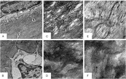
Figure 3. Effects of subchronic co-exposure to lead acetate and cadmium chloride on the structure of rats bone under the scanning electron microscopy for 90 d. A: The control group shows the bone trabeculas from distal of
femur were more numerous (↑) (×50); B: The femoral bone trabecula were few, thin, and developed local fracture (↗) (×55); C: The surfaces of bone trabeculas from control group were smooth (↑) (×600); D: The collagen fibers
disposed tight and orderly (↘) (×3000); E: The femoral trabecular partly showed erosive bone destruction and
there were lacunae on the surface of collagen fibers (↘) (×500); F: The collagen fiber of trabecular fiber arranged
in disorder, loose structure and breakage (↘) (×3000).
Under TEM, the control group has rich organ-elles and evenly distributed chromatin (Figure 4A). The ultrastructure changes showed fewer organelles from osteoblast, focal cytolysis, con-densation, and margination of chromatin,
cou-pled with greater bone lacunae and more floc in
experimental group (Figure 4B). The collagen
fiber from the control group appeared in an
orderly arrangement, and hydroxyapatite crys-tal, which is tiny acicular, regularly clings to the
fiber (Figure 4C, 4D). By contrast, the collagen
fiber is in chaos and loose structure and has
shown signs of breakage and dissolution, cou-pled with disappeared transversus striation,
after the decalcification (Figure 4E). Moreover,
the experimental group, without decalcifica -tion, showed decreased amount of hydroxyapa-tite crystal and disorder (Figure 4F).
Discussion
This study is the first attempt to report the equi -toxic mixture ratio of Pb and Cd as the
experi-mental animal model method for subchronic toxicity experiments. Moreover, this animal model may help detect the early events of chronic Pb and Cd intoxication with relatively low and environmentally realistic concentra-tions. The results indicated that the co-expo-sure to Pb and Cd, which accumulate in the body, can induce bone damage via change in Ca and Pi content in bone and decrease in BMD and bone morphology lesions.
result in a significant decrease in Ca and Pi con -tent, as indicated by a marked increase in Pb and Cd in bone with the increasing doses of ingested Pb and Cd. Therefore, these results indicate that Pb and Cd exposure can impair calcium and phosphorus absorption, displace calcium with these metals, and decrease bone mineralization, which interferes with the
nor-mal ossification.
BMD is an indicator for diagnosing osteoporo-sis; this parameter provides information regard-ing the quantity of mineral in bone, which is only one component of bone strength [18].
Research has shown that calcium deficiency
can reduce BMD and weaken bone strength
and long-term calcium deficiency can lead to
osteoporosis [19]. Moreover, Pb exposure can inhibit the development of bone and reduce BMD because the change in the distribution of elements in the bones [20]. This mineral imbal-ance can cause osteoporosis because of Cd exposure [21]. Exposure to Cd may be an
impor-tant contributing factor to lower BMD and osteopenia, which result in the development of osteoporosis [12]. In the present study, the
BMD was significantly decreased in Pb and
Cd-exposed rats. With repeated
administra-tion, the content of Ca and Pi deficiency in
bone, which indicates co-exposure to Pb and Cd, results in an increased risk of osteoporosis.
Osteoporosis can cause incremental bone fra-gility and increased fracture risk [22, 23]. Moreover, bone microstructure is an important factor in understanding the mechanisms of bone fragility. In this study, co-exposure to Pb and Cd can induce bone microstructure dam-age. Pb plays an important role in the develop-ment of bone pathologies [24]. Moreover, Pb can cause decreased trabecular bone of rats and irregularity in the primary bone marrow cavity [14]. When Cd accumulates in the body, it can induce damage to the cancellous bone
[image:6.612.91.521.72.342.2]microstructure [25]. Collagen fibers constitute
Figure 4. Effects of subchronic co-exposure to lead acetate and cadmium chloride on the structure of rats bone under the transmission electron microscopy for 90 d. A: The control group had rich organelles and evenly distributed
chromatin (↑) (×2500); B: The osteoblast showed fewer organelles, focal cytolysis, condensation and margination of chromatin, coupled with greater bone lacunae and more floc (↑) (×2500); C, D: Collagen fiber from control group appeared in an orderly arrangement (↑) (×5000), and hydroxyapatite crystal, which appeared to be tiny acicular, regularly cling to the fiber (↑) (×65000); E: Collagen fiber arranged in disorder, loose structure, breakage, and dis
an organic matrix where minerals are incorpo-rated while creating the bone tissue. Moreover,
collagen fibers are responsible for bone elastic -ity. In addition, the osteoblast participates in
ossification. The results showed that trabecular bone becomes fewer, the collagen fiber of bone
trabecular is arranged in a disorderly and loose structure, which is prone to breakage, and the osteoblast showed fewer organelles, focal cytolysis. Therefore, these results indicate that co-exposure to Pb and Cd interferes with the
normal ossification and increased fracture risk,
thus leading to osteoporosis.
In summary, exposure to Pb and Cd can induce bone damage and leads to an increased risk of osteoporosis. The effects produced by the combined treatment of metals were not only the inhibition of Ca and Pi deposit and the reduction of bone density, but also severe bone morphology lesions.
Acknowledgements
This work was partly supported by the National Natural Science Foundation of China and the Sichuan Youth Science and Technology Innovation Research Team for waterfowl dis-ease prevention and control (Grant No. 31101860 and 2013TD0015).
Disclosure of conflict of interest
None.
Address correspondence to: Dr. Guiping Yuan, Ana- lytical & Testing Center, Sichuan University, Chengdu 610064, China; Dr. Zhongqiong Yin, College of Veterinary Medicine, Sichuan Agricultural University, Ya’an 625014, China. Tel: +86 835 2885614; Fax: +86 835 2885302; E-mail: ygp_04@163.com (GP Yuan); yinzhongq@163.com (ZQ Yin)
References
[1] Lorenzon S, Francese M, Smith VJ, Ferrero EA. Heavy metals affects the circulating haemo-cyte number in the shrimp Palaemon elegans.
Fish Shellfish Immunol 2001; 11: 459-472.
[2] Bajpai R, Upreti DK. Accumulation and toxic ef-fect of arsenic and other heavy metals in a contaminated area of West Bengal, India, in the lichen Pyxine cocoes (Sw.) Nyl. Ecotoxicol Environ Saf 2012; 83: 63-70.
[3] Mol S. Levels of Heavy Metals in Canned Boni-to, Sardines, and Mackerel Produced in Turkey. Biol Trace Elem Res 2011; 143: 974-982.
[4] Gangoso L, Alvarez-Lloret P, Rodríguez-Navarro AA, Mateo R, Hiraldo F, Donázar JA. Long-term effects of lead poisoning on bone mineraliza-tion in vultures exposed to ammunimineraliza-tion sourc-es. Environ Pollut 2009; 157: 569-574. [5] Dai S, Yin Z, Yuan G, Lu H, Jia R, Xu J, Song X,
Li L, Shu Y, Liang X, He C, Lv C, Zhang W.
Quan-tification of metallothionein on the liver and
kidney of rats by subchronic lead and cadmi-um in combination. Environ Toxicol Pharmacol 2013; 36: 1207-1216.
[6] Trzcinka-Ochocka M, Jakubowski M, Szymczak W, Janasik B, Brodzka R. The effects of low en-vironmental cadmium exposure on bone den-sity. Environ Res 2010; 110: 286-193. [7] Berglund M, Ãkesson A, Bjellerup P, Vahter M.
Metal–bone interactions. Toxicol Lett 2000; 112-113: 219-225.
[8] Nilsson U, Attewell R, Christoffersson JO, Schutz A, Ahlgren L, Skerfving S, Mattsson S. Kinetics of lead in bone and blood after end of occupational exposure. Basic Clin Pharmacol Toxicol 1991; 68: 477-484.
[9] Theppeang K, Glass TA, Bandeen-Roche K, Todd AC, Rohde CA, Links JM, Schwartz BS. As-sociations of Bone Mineral Density and Lead Levels in Blood, Tibia, and Patella in Urban-Dwelling Women. Environ Health Perspect 2008; 116: 784-790.
[10] Honda R, Tsuritani I, Noborisaka Y, Suzuki H, Ishizaki M. Urinary cadmium excretion is cor-related with calcaneal bone mass in Japanese women living in an urban area. Environ Res 2003; 91: 63-70.
[11] Chen X, Zhu GY, Shao CL, Jin TY, Tan MG, Gu SZ, Zhang YY, Xiao HF. Effects of cadmium on bone microstructure and serum tartrate-resis-tant acid phosphatase 5b in male rats. Exp Biol Med 2011; 9: 1-8.
[12] Smith E, Gancarz D, Rofe A, Kempson IM, We-ber J, Juhasz AL. Antagonistic effects of cad-mium on lead accumulation in pregnant and non-pregnant mice. J Hazard Mater 2012; 199-200: 453-456.
[13] Lu HK, Dai SJ, Yin ZQ, Yuan GP, Wang C. Study on damage of bone in rat induced by experi-mental acute combined exposure to lead and cadmium. Chin Vet Sci 2012; 42: 1278-1282. [14] Hu XQ, Mi MT. Effect of lead on calcium ab-sorption and bone development in weanling rats. Acta Acad Med Militaris Tertiae 2007; 29: 402-405.
[15] Brzóska MM, Rogalska J, Moniuszko-Jakoniuk J. The concentration of vitamin D metabolites in the serum of cadmium-exposed female rats. Osteoporosis Int 2003; 14 Suppl 6: 22-23. (ab-stract).
changes in males exposed to lead in the Bue-nos Airesarea (Argentina). Pharmacol Res 2000; 42: 599-602.
[17] Blumenthal NC, Cosma V, Skyler D, LeGeros J, Walters M. The effect of cadmium on the for-mation and properties of hydroxyapatite in vi-tro and its relation to cadmium toxicity in the skeletal system. Calcif Tissue Int 1995; 56: 316-322.
[18] Friedman AW. Important determinants of bone strength: beyond bone mineral density. J Clin Rheumatol 2006; 12: 70-7.
[19] Liu XL, Xu HQ, Xiong ZG, ZhouU AQ, Wang XY. Study on intervention of infants with calcium
deficiency. Chin J Child Health Care 2006; 14:
503-505.
[20] Lai JQ, Zhou J, Yin SA, Zhao XF. Effect of iron and zinc levels in diet on development of bone in growing rats exposed to lead. J Hygiene Res 2004; 33: 461-463.
[21] Noël L, Guérin T, Kolf-Clauw M. Subchronic di-etary exposure of rats to cadmium alters the metabolism of metals essential to bone health. Food Chem Toxicol 2004; 42: 1203-1210.
[22] Shin M, Paek D, Yoon C. The relationship be-tween the bone mineral density and urinary cadmium concentration of residents in an in-dustrial complex. Environ Res 2011; 111: 101-109.
[23] Bruyere O, De Cock C, Mottet C, Neuprez A, Malaise O, Reginster JY. Low dietary calcium in European postmenopausal osteoporotic wom-en. Public Health Nutr 2009; 12: 111-114. [24] Khazzani H, Allali F, Bennani L, Ichchou L, El
Mansouri L, Abourazzak FE, Abouqal R, Hajjaj-Hassouni N. The relationship between physical performance measures, bone mineral density, falls, and the risk of peripheral fracture: a cross-sectional analysis. BMC Public Health 2009; 9: 297.