Visualization of Diffusive Hydrogen in Low Alloy Steel by means
of Hydrogen Microprint Technique at Elevated Temperatures
Keitaro Horikawa
1, Hiroaki Okada
1;*, Hidetoshi Kobayashi
1and Wataru Urushihara
21
Department of Mechanical Science and Bioengineering, School of Engineering Science, Osaka University, Toyonaka 560-8531, Japan
2Surface Design & Corrosion Research Section, Materials Research Laboratory, Kobe Steel, Ltd.,
Kobe 651-2271, Japan
Hydrogen diffusion behavior of low alloy steel was visualized by means of hydrogen microprint technique. Effects of exposure time and temperature on the availability of hydrogen microprint technique were also examined. It was found that silver particles, which representing the emission site of hydrogen atoms, were distributed almost uniformly in the matrix after hydrogen charging, and that were arranged at the periphery of second phase particles such as MnS and Al2O3. Area density of the silver particles was clearly increased when the exposure time after cathodic hydrogen charging increased at room temperature or the exposure temperature was higher than room temperature, which was in agreement with the result obtained from the thermal desorption analysis. From a series of the heating experiments, it was also shown that hydrogen microprint technique would be applicable at temperatures below 120C. After the long time exposure in the hydrogen-charged specimens, preferential accumulation of silver particles around the Al2O3and MnS particles was identified. This suggested that hydrogen atoms were diffused not through the inside of the second phase particles but through the interface between the second phase particles and matrix phase. [doi:10.2320/matertrans.MRA2008325]
(Received September 16, 2008; Accepted January 14, 2009; Published March 4, 2009)
Keywords: low alloy steel, hydrogen, diffusion, hydrogen microprint, thermal desorption analysis
1. Introduction
In recent years, against problems on the global warming and the exhaustion of fossil fuels, a number of systems using hydrogen as a source of energy have been developed in the industries. To utilize hydrogen gas effectively as energy sources, it becomes important to evaluate the interaction between hydrogen gas and metallic materials, particularly steels, which are industry available. It is well known that hydrogen atoms dissociated by the interaction with steels and atmosphere can diffuse into the materials and might induce hydrogen embrittlement. Thus far, the information with related to the local hydrogen diffusion path inside steels and the interaction between diffusive hydrogen and second phase particles has not been experimentally clarified. In order to detect the path of hydrogen diffusion in the metallic materials, several kinds of hydrogen visualizing methods have been proposed such as hydrogen microprint technique (HMT),1)silver decoration method2)and secondary ion mass spectroscopy (SIMS).3)Among them, HMT is a promising method to visualize diffusive hydrogen as a distribution of silver particles with high spatial resolution. Several kinds of papers have dealed with the availability of HMT,4)however,
the applied temperature for HMT is almost limited at room temperature. Thus far, it seems to be the only report5)that
HMT was performed at temperatures differed from room temperature. It will be worthwhile to study the availability of HMT at high temperatures from the viewpoint of the industrial use, because hydrogen embrittlement of steels including delayed fracture is caused after a long time exposure at various temperatures.6)In this study, effects of exposure time and temperature on the availability of HMT were studied for the hydrogen-charged low alloy steel.
2. Experimental Procedure
[image:1.595.304.551.387.414.2]The material used in the present study was a low alloy steel plate whose chemical composition and tensile properties at room temperature are shown in Table 1 and Table 2. The rectangle shaped specimens with a size of 10mm10mm in side and 2.0 mm in thickness were cut from the plate. Hydrogen diffusion tests, together with HMT were per-formed. Prior to HMT, both sides of the specimen surfaces were polished using #240, #800, #1200 SiC papers and buffed with a solution containing alumina powders (particle size: 0.3mm). One side of the specimen surface was covered with a protective film. Hydrogen was then charged cathodi-cally for the specimens in a 3 mass% NaCl-0.1 mass% KSCN solution at a current density of 10 A/m2 for 70 h, or 5000 A/m2for 3 h at room temperature from the backside of the one surface covered with the protective film. Within 5 minutes after hydrogen charging, the protective film was removed and the specimen surfaces were covered with a liquid nuclear emulsion (Ilford L-4, diluted by pure water, 2 times) composed of gelatin and silver bromide crystals
Table 1 Chemical composition of the low alloy steel (mass%).
C Si Mn Cr Mo S P Fe
0.48 0.29 0.94 1.11 0.27 49 70 Bal.
massppm
Table 2 Tensile properties of the low alloy steel (""_¼6:7103s1).
Yield stress (MPa) Tensile Strength (MPa) Fracture Strain (%)
803 1062 18.8
[image:1.595.305.549.464.503.2]using a wire loop method, then dried for 30 minutes. Some specimens were then kept at room temperature by changing the exposure to air time between 5 minutes and 96 h. In order to examine the availability of HMT at higher temperatures, some other specimens were exposed at 60C, 120C, 180C
and 260C, respectively on the hot stage for 5 minutes after
hydrogen charging. The measurement of surface temperature of specimen was carried out by using a K-type thermocouple attached directly at the specimens. After the heat treatments, the specimens were dipped into formalin (40 mass% HCHO water solution) for 3 seconds to harden the gelatin layer, and immersed in a fixing solution (15 mass% Na2S2O3 water solution) for 5 minutes to remove the remaining silver bromide particles that had not reacted with hydrogen. During the tests, 10 mass% water solution of NaNO2 was used to dilute the emulsion and to make the fixing solution to prevent any corrosion in aqueous solutions. Arrangement of the silver particles was observed with a scanning electron microscope (SEM) equipped with an energy dispersive X-ray spectrom-eter (EDXS). The amount of hydrogen contained in the specimen after hydrogen charging was also measured by the thermal deposition analysis (TDA) using gas chroma-tography with a heating rate of 100C min1 until the temperature reached 500C. High purity argon carrier gas of
99.999% purity was passed through a quartz glass tube at a flow rate of 20 mL min1. The amount of hydrogen gas was measured at 1 minute intervals during heating a sample weighed about 1 g.
3. Results
3.1 Hydrogen absorption by chathodic hydrogen charg-ing
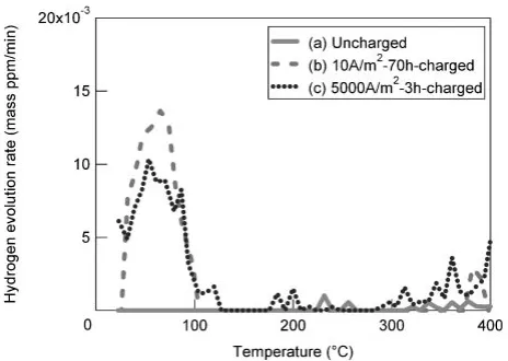
The TDA curves for the specimens with and without hydrogen charging are shown in Fig. 1. In the specimen without hydrogen charging, thermal desorption of hydrogen was not observed below 400C. On the other hand, the
thermal desorption of hydrogen appeared as a single peak around 60–70C in the specimen hydrogen charged with a
current density of 10 A/m2 for 70 h. The similar evolution peak was also observed in the specimen hydrogen charged
with a current density of 5000 A/m2 for 3 h. This indicates that almost the same amount of hydrogen remained to the specimens in the charging conditions both of 10 A/m2 for 70 h and 5000 A/m2for 3 h. In these two conditions, almost all the diffusive hydrogen were evolved below 120C from
the specimens. The diffusive hydrogen concentration was calculated by integrating the relation between hydrogen desorption rate and heating time below 400C. The hydrogen
concentration was calculated about 0.4 massppm, which exceeded the maximum solubility of hydrogen in ferrite iron,7)both in the hydrogen charged specimens, 10 A/m2for 70 h and 5000 A/m2for 3 h, while that was 0.01 massppm in the uncharged specimen.
3.2 Effect of testing condition on the availability of HMT
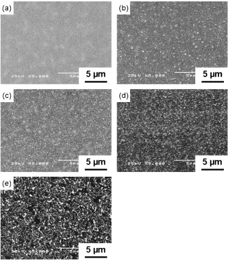
Figure 2 shows the HMT images of specimen after hydrogen charging (10 A/m2 for 70 h) exposed at room temperature, adjacent to the second phase particles. When the exposure time was 5 minutes, few silver particles were observed at the matrix as well as around the second phase particles such as MnS (Fig. 2(a), (b)), Al2O3(Fig. 2(c)), and Al2O3MgO (Fig. 2(d)). This indicates that the number of hydrogen atoms diffused to the specimen surfaces was relatively small in this exposure condition. A similar behav-ior was also observed in the specimen hydrogen charged with 5000 A/m2 for 3 h, exposed for 5 minutes at room temper-ature. On the other hand, when the exposure time was 96 h at room temperature after hydrogen charging (10 A/m2for 70 h and 5000 A/m2 for 3 h), a number of silver particles were visible in the matrix as shown in Fig. 3. This suggests that hydrogen atoms diffused into the thickness direction were increased as the exposure time was prolonged from 5 minutes to 96 h. In addition, not all the second phase particles, however, preferential accumulation of silver particles around Al2O3particles was clearly identified as shown in Fig. 3(d). Figure 4 shows the effect of exposure temperature after hydrogen charging (5000 A/m2 for 3 h) on HMT, each specimens were exposed to air for 5 minutes at temperatures ranging from room temperature to 260C. It is revealed that
the area density of the silver particles in the matrix was increased when the exposure temperature was higher than room temperature. It is assumed that the increase of the area density of the silver particles by heating represents the increase of the number of hydrogen atoms diffused to the specimen surface, which is in accord with the TDA results as indicated before. However, when the exposure temperature became higher than 120C, the shape of the observed silver
particles was partially changed from spherical one to irregular one. Particularly at 260C, condensation of silver
particles arose partly as shown in Fig. 4(e). The reason for this change is unknown at present, however, the gelatin layers contained in the nuclear emulsion might be damaged by heating at higher temperatures above 120C.
Figure 5 shows HMT images around Al2O3particles in the hydrogen-charged specimens (5000 A/m2 for 3 h) exposed for 5 minutes at temperatures ranging from room temperature to 260C. In the specimen exposed to air at high temperatures between 60C and 120C, silver particles were not observed directly on the second phase particles, similarly to the case of
Fig. 1 Hydrogen desorption rate vs. temperature. (a): uncharged, (b): hydrogen charged at 10 A/m2 for 70 h, (c): hydrogen charged at 5000 A/m2for 3 h.
[image:2.595.53.285.582.747.2]the specimen exposed to air at room temperature for 96 h as indicated in Fig. 3(d). These results are in accord well with the previous reports8,9) explaining that hydrogen atoms
trapped inside second phase particles are non-diffusive hydrogen, and that the second phase particles do not work as the diffusion path in the steels.
Fig. 2 HMT images after hydrogen charging (10 A/m2for 70 h) in the low alloy steel exposed for 5 minutes at room temperature, adjacent to the particles (a) MnS, (b) magnified image of (a), (c) Al2O3and (d) Al2O3MgO.
[image:3.595.112.485.71.352.2] [image:3.595.93.506.396.709.2]4. Discussion
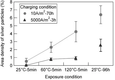
To estimate the difference of the amount of hydrogen diffusion among the different testing conditions, the area density of silver particles to the entire observed area was measured in the SEM micrographs. The relationship between the area density of silver particles and the difference of hydrogen charging as a function of exposure to air conditions is summarized in Fig. 6. It is clear that the area density of silver particles in the matrix increases as the increase of the exposure temperature. It is also found that the area density of silver particles exposed at room temperature for 96 h is about ten times as high as that exposed at room temperature for 5 minutes. Using the reported10)diffusion coefficient of
hydro-gen in low alloy steel at room temperature (25C), 60C and
120C, the mean diffusion distance of hydrogen in the low
alloy steel exposed for 5 minutes is calculated to be 1.1 mm, 1.5 mm and 1.8 mm, respectively. On the other hand, the diffusion distance is calculated to be 38 mm when the specimen was exposed at 25C for 96 h. This means that the
exposure condition of 25C for 96 h after hydrogen charging
is enough to diffuse hydrogen atoms to the thickness distance of the specimen. In this study, the direct comparison between the area density of silver particles and the diffused hydrogen
atoms would be impossible since the initial condition of hydrogen atom distribution in the specimens after hydrogen charging is not the same. However, temperature dependency of the calculated diffusion distance correlates well with that of the increase of area density of the silver particles on the specimens revealed by HMT as shown before in Fig. 4.
In connection with hydrogen diffusion behavior, Fig. 6 also indicates that the area density of silver particles in the charging condition of 10 A/m2for 70 h becomes higher than that of 5000 A/m2for 3 h under the same exposure condition after hydrogen charging. However, based on the TDA results as previously shown in Fig. 1, the hydrogen concentrations in the charging operations between 10 A/m2 for 70 h and 5000 A/m2for 3 h are almost the same (0.4 massppm). Thus, it is assumed that the difference of the gradient of hydrogen concentration in the thickness direction inside the specimen would affect the difference of the number of hydrogen atoms diffused to the specimen surface in the following exposure operation. Otsuka et al.11) have pointed out the possibility that the hydrogen atoms can be trapped by the local stress field generated by the difference of the thermal expansion coefficient between the second phase particles and the matrix phase and by the microvoids induced by large contraction of the particles using tritium autoradiography. However, it
Fig. 4 Effect of exposure temperature after hydrogen charging (5000 A/m2for 3 h) on HMT images exposed for 5 minutes at (a) 25C, (b) 60C, (c) 120C, (d) 180C and (e) 260C in the matrix.
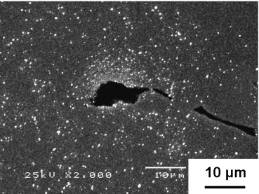
[image:4.595.131.464.71.452.2]remains controversial whether the particle, particularly MnS or the interface works as permanent trapping sites or as diffusion paths at room temperature.8,9,11,12)To elucidate the interaction between the particles and hydrogen atoms more clearly, HMT was also conducted for the specimen, in which the hydrogen charging with a very high current density of 1105A/m2 for 3 h was applied, as shown in Fig. 7. The
preferential accumulation of silver particles was clearly identified around MnS particles after the exposure for a long time (96 h) at room temperature. This indicates that the interface between the second phase particles and the matrix phase works as the preferential hydrogen diffusion path in the low alloy steel. However, in this case too, no silver particles were visible directly on the MnS particles. It is thus assumed that hydrogen atoms trapped inside MnS particles are essentially non-diffusive hydrogen at room temperature in the same way as presumed before,13)even when the hydrogen
concentration in the specimen is very high.
5. Summary
Hydrogen diffusion behavior around the second phase particles in low alloy steel was visualized using HMT. The results obtained were summarized as follows: (1) The area density of the visualized hydrogen atoms in the matrix phase increased when the specimen was exposed at room tem-perature for a long time (96 h) or exposed at higher temperatures for a short time (5 minutes). (2) Even after the exposure for a long time at room temperature or high temperatures for a short time, accumulation of silver particles was observed on the matrix and at the periphery
Fig. 5 Effect of exposure temperature after hydrogen charging (5000 A/m2for 3 h) on HMT images exposed for 5 minutes at (a) 25C, (b) 60C, (c) 120C, (d) 180C and (e) 260C, adjacent to an Al
2O3particle.
[image:5.595.135.462.70.445.2] [image:5.595.71.268.503.639.2]of the second phase particles, while not observed directly on the particles, such as Al2O3 or MnS. (3) HMT can be applicable at the high temperatures below 120C without
the damage of silver particles.
Acknowledgements
The work was supported by the Ministry of Education, Culture, Sports, Science and Technology of Japanese Government, Grant-in-Aid for Scientific Research (C), 20560652, 2008.
REFERENCES
1) J. Ovejero-Garcia: J. Mater. Sci.20(1985) 2623–2629.
2) T. Schober and C. Dieker: Metall. Trans.14A(1983) 2440–2442.
3) K. Takai, Y. Homma, K. Izutsu and M. Nagumo: J. Japan Inst. Metals
60(1996) 1155–1162.
4) K. Ichitani, S. Kuramoto and M. Kanno: Corros. Sci.45(2003) 1227– 1241.
5) K. Koyama, G. Itoh and M. Kanno: J. Japan Inst. Metals62(1998) 790– 795.
6) M. Nagumo, M. Nakamura and K. Takai: Metall. Trans.32A(2001) 339–347.
7) Y. Fukai, K. Tanaka and Y. Uchida: Suiso To Kinzoku, (Uchida Rokakuho, Tokyo, 1998) pp. 25–54.
8) J. Y. Lee and S. M. Lee: Surf. Coat. Technol.28(1986) 301–314. 9) D. L. Tuyen and B. E. Wilde: Corrosion39(1983) 258–265. 10) H. Hagi: J. Japan Inst. Metals57(1993) 864–869.
11) T. Otsuka, H. Hanada, H. Nakashima, K. Sakamoto, M. Hayakawa, K. Hashizume and M. Sugisaki: Fusion Sci. Technol.48(2005) 708–711. 12) A. M. Brass, J. Chene and A. Boutry-Forveille: Corros. Sci.38(1996)
569–585.
13) K. Kusabiraki, T. Kubo, T. Ooka, M. Matsuyama and K. Watanabe: J. Japan Inst. Metals51(1987) 174–180.
Fig. 7 A HMT image of the specimen exposed at room temperature for 96 h, in which the strong hydrogen charging with a very high current density of1105A/m2for 3 h was applied.
[image:6.595.114.483.72.349.2]