ORIGINAL RESEARCH ARTICLE
ANATOMICAL VARIATIONS IN DORSALIS PEDIS ARTERY AND ITS BRANCHES WITH CLINICAL
1,*
Dr. Preeti Awari
1
Assitant Professor, Department of Anatomy, Dr.D.Y.
Dr. D.Y. Patil Vidyapeeth Pimpri, Pune
2
Director of Academics, Dr.D.Y.
Dr. D.Y. Patil Vidyapeeth Pimpri, Pune
ARTICLE INFO ABSTRACT
50 feet were dissected to see the course and branching pattern of dorsalis pedis artery. The following variations of the artery were seen. The dorsalis pedis artery was deviated laterally in 4% of feet. It was continuation of perforating branch of perone
40% of feet. In 8% feet the medial tarsal artery was absent. In 12% feet there were two medial tarsal arteries. There were two lateral tarsal arteries in 14% of feet and three lateral tarsal arteries in feet. Knowledge of these variations can help the radiologists in diagnosing peripheral vascular diseases by colour Doppler. Awareness about the arterial variations in the foot can help microvascular surgeons to do infrapopliteal interventions in cri
surgeries.
Copyright © 2016, Dr. Preeti Awari andDr. Vatsalaswamy
permits unrestricted use, distribution and reproduction in any medium, provided the original work is properly cited.
INTRODUCTION
To diagnose patients with peripheral arterial disease, palpation of peripheral arterial pulse is one of the simplest screening In lower extremity, pulsations of dorsalis pedis artery are taken to evaluate the arteriosclerotic diseases. In the era of microvascular surgery the dorsalis pedis artery is used as stem for myocutaneous flap in reconstructive and plastic surgeries
(Mir Wajahath Ali et al., 1996). Knowing the normal anatomy
and anatomical variations in the origin, course and branching pattern of the dorsalis pedis artery can increase succ
these all surgeries. With increasing incidence of diabetic foot, peripheral arterial diseases and accidental injuries to foot a need may arise for a vascular surgery. A detailed anatomical knowledge about the arteries of the foot and its variations is needed in such situations. The main artery to supply dorsum of the foot is dorsalis pedis artery as it also contributes to plantar arch (Rajeshwari et al., 2013). Hence this study is undertaken to trace the course of Dorsalis pedis artery, its branching pattern and variations if any.
*Corresponding author: Dr. Preeti Awari,
Assitant Professor, Department of Anatomy, Dr.D.Y. Patil Medical College, Hospital and Research centre, Dr. D.Y. Patil Vidyapeeth Pimpri, Pune, India
ISSN: 0975-833X
Article History:
Received 10th July, 2016 Received in revised form 05th August, 2016
Accepted 25th September, 2016
Published online 30th October,2016
Key words:
Dorsalis Pedis Artery, Lateral Tarsal Artery, Medial Tarsal Artery, Arcuate Artery.
Citation: Dr. Preeti Awari andDr. Vatsalaswamy, P.
International Journal of Current Research, 8, (10), 40692
ORIGINAL RESEARCH ARTICLE
ANATOMICAL VARIATIONS IN DORSALIS PEDIS ARTERY AND ITS BRANCHES WITH CLINICAL
CORRRELATIONS
Dr. Preeti Awari and
2Dr. Vatsalaswamy, P.
, Department of Anatomy, Dr.D.Y. Patil Medical College, Hospital and Research centre,
Dr. D.Y. Patil Vidyapeeth Pimpri, Pune, India
, Dr.D.Y. Patil Medical College, Hospital and Research centre,
Dr. D.Y. Patil Vidyapeeth Pimpri, Pune, India
ABSTRACT
50 feet were dissected to see the course and branching pattern of dorsalis pedis artery. The following variations of the artery were seen. The dorsalis pedis artery was deviated laterally in 4% of feet. It was continuation of perforating branch of peroneal artery in 4% of feet. Arcuate artery was absent in 40% of feet. In 8% feet the medial tarsal artery was absent. In 12% feet there were two medial tarsal arteries. There were two lateral tarsal arteries in 14% of feet and three lateral tarsal arteries in
Knowledge of these variations can help the radiologists in diagnosing peripheral vascular diseases by colour Doppler. Awareness about the arterial variations in the foot can help microvascular surgeons to do infrapopliteal interventions in critical limb ishaemia and also in reconstructive surgeries.
Dr. Vatsalaswamy.This is an open access article distributez under the Creative Commons Att
and reproduction in any medium, provided the original work is properly cited.
To diagnose patients with peripheral arterial disease, palpation of peripheral arterial pulse is one of the simplest screening test. In lower extremity, pulsations of dorsalis pedis artery are taken to evaluate the arteriosclerotic diseases. In the era of microvascular surgery the dorsalis pedis artery is used as stem for myocutaneous flap in reconstructive and plastic surgeries the normal anatomy and anatomical variations in the origin, course and branching pattern of the dorsalis pedis artery can increase success rate of . With increasing incidence of diabetic foot, pheral arterial diseases and accidental injuries to foot a need may arise for a vascular surgery. A detailed anatomical knowledge about the arteries of the foot and its variations is needed in such situations. The main artery to supply dorsum of s dorsalis pedis artery as it also contributes to plantar Hence this study is undertaken to trace the course of Dorsalis pedis artery, its branching
Assitant Professor, Department of Anatomy, Dr.D.Y. Patil Medical College, Hospital and Research centre, Dr. D.Y. Patil Vidyapeeth
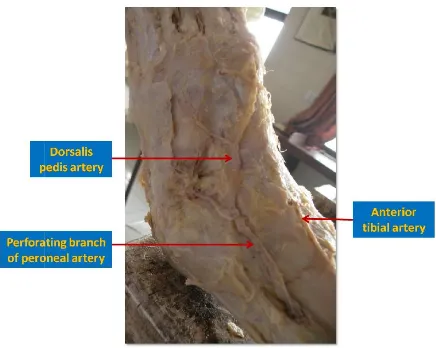
Dorsalis Pedis Artery (DPA) is the continuation of the anterior tibial artery. The dorsalis pedis artery
the malleoli and runs anteromedially till it reaches the
proximal end of 1st interosseous space where it gives out the
first metatarsal artery and continues on plantar aspect.
Branches of Dorsalis pedis artery
The two tarsal arteries lateral (LTA) and medial (MTA) arise as the dorsalis pedis artery crosses navicular. The lateral runs laterally under the extensor digitorum brevis and supplies it. Lateral tarsal artery anastomoses with branches of arcuate anterior lateral malleolar and lateral plantar arteries. Two or three medial tarsal arteries can be found on the medial border of foot and join the medial malleolar network
2005). The arcuate artery (AA) runs laterally across the bases of the lateral four metatarsal, deep to the extensor tendons on the dorsum of the foot. The arcuate artery gives rise to 2
& 4th dorsal metatarsal arteries
normal anatomy and variations in these arteries can help the radiologists in diagnosing peripheral vascular diseases by colour Doppler. Awareness about the arterial variations in foot can help microvascular surgeons to
Available online at http://www.journalcra.com
International Journal of Current Research
Vol. 8, Issue, 10, pp.40692-40696, October, 2016
INTERNATIONAL
Vatsalaswamy, P.2016. “Anatomical variations in dorsalis pedis artery and its branches with clinical corrrelations 40692-40696.
z
ANATOMICAL VARIATIONS IN DORSALIS PEDIS ARTERY AND ITS BRANCHES WITH CLINICAL
Hospital and Research centre,
Patil Medical College, Hospital and Research centre,
50 feet were dissected to see the course and branching pattern of dorsalis pedis artery. The following variations of the artery were seen. The dorsalis pedis artery was deviated laterally in 4% of feet. It al artery in 4% of feet. Arcuate artery was absent in 40% of feet. In 8% feet the medial tarsal artery was absent. In 12% feet there were two medial tarsal arteries. There were two lateral tarsal arteries in 14% of feet and three lateral tarsal arteries in 8% of Knowledge of these variations can help the radiologists in diagnosing peripheral vascular diseases by colour Doppler. Awareness about the arterial variations in the foot can help microvascular tical limb ishaemia and also in reconstructive
under the Creative Commons Attribution License, which
is the continuation of the anterior tibial artery. The dorsalis pedis artery starts midway between the malleoli and runs anteromedially till it reaches the interosseous space where it gives out the first metatarsal artery and continues on plantar aspect.
pedis artery
eries lateral (LTA) and medial (MTA) arise as the dorsalis pedis artery crosses navicular. The lateral runs laterally under the extensor digitorum brevis and supplies it. omoses with branches of arcuate, malleolar and lateral plantar arteries. Two or three medial tarsal arteries can be found on the medial border of foot and join the medial malleolar network (Keith et al., . The arcuate artery (AA) runs laterally across the bases etatarsal, deep to the extensor tendons on
the dorsum of the foot. The arcuate artery gives rise to 2nd, 3rd
arteries (Gray, 2008). Knowledge of normal anatomy and variations in these arteries can help the peripheral vascular diseases by colour Doppler. Awareness about the arterial variations in foot can help microvascular surgeons to increase clinical
INTERNATIONAL JOURNAL OF CURRENT RESEARCH
effectiveness in infrapopliteal interventions in critical limb ishaemia.
MATERIALS AND METHODS
50 lower limbs from 7 female and 18 male cadavers were procured from the department of Anatomy of Dr. D Y Patil Medical College and Research centre, Pimpri, Pune, Maharashtra, India. These cadavers were embalmed with 10% formalin and fixed. Dorsum of foot was dissecte
dissection procedure in Cunningham’s manual edition.
This is a descriptive type of study
Skin of dorsum of the foot was reflected. After reflecting the deep fascia and extensor retinaculum the dorsalis pedis artery was dissected and cleaned. Variations in origin of dorsalis pedis artery were noted. Branches of dorsalis pedis artery were exposed after reflecting the extensor digitorum brevis muscle. Any variations in course of Dorsalis Pedis Artery were noted. Any variations in origin and number of medial tarsal, lateral tarsal and arcuate arteries were noted.
Name Variation if any Dorsalis pedis artery Deviated laterally (fig.1)
[image:2.595.151.504.350.786.2]Perforating branch of Peroneal artery continues as dorsalis pedis artery (fig.2)
Figure 1.
Figure 2.
40693 Dr. Preeti Awari andDr. Vatsalaswamy, Anatomical variations in dorsalis pedis artery and its branches with clinical corrrelations
infrapopliteal interventions in critical limb
from 7 female and 18 male cadavers were procured from the department of Anatomy of Dr. D Y Patil Medical College and Research centre, Pimpri, Pune, Maharashtra, India. These cadavers were embalmed with 10% formalin and fixed. Dorsum of foot was dissected as per dissection procedure in Cunningham’s manual-I, Fifteenth
Skin of dorsum of the foot was reflected. After reflecting the deep fascia and extensor retinaculum the dorsalis pedis artery d cleaned. Variations in origin of dorsalis pedis artery were noted. Branches of dorsalis pedis artery were exposed after reflecting the extensor digitorum brevis muscle. Any variations in course of Dorsalis Pedis Artery were noted. in and number of medial tarsal, lateral
RESULTS
Lower limbs of 7 female and 18 male cadavers were dissected to study the dorsalis pedis artery and its branches. Observations were noted separately for male and female as well as for left and right sides.
DISCUSSION
In all lower limbs dorsalis pedis artery was present but the artery itself and its branches showed lot of variations.
Variations in Dorsalis pedis artery
The dorsalis pedis artery was seen deviated laterally in 4% of feet. In 2008, Ebrahim M. El. Saeed
cases there was lateral deviation of dorsalis pedis artery (Ebrahim et al., 2008). Present study correlates with the study of Ebrahim M. El. Saeed et al
of dorsalis pedis artery laterally.
[image:2.595.56.549.356.392.2]will be useful in deciding whether the absence of pulse in dorsalis pedis artery is due to thrombosis of the artery abnormal course. Non palpable dorsalis pedis pulse with impaired blood flow as visualized by arteriography is one of
Table 1. Variations in Dorsalis pedis artery
Incidence
Deviated laterally (fig.1) 4%
[image:2.595.193.411.600.774.2]Perforating branch of Peroneal artery continues as dorsalis pedis artery (fig.2) 4%
Figure 1. Abnormal course of Dorsalis pedis artery
Figure 2. Abnormal origin of Dorsalis pedis artery
Dr. Vatsalaswamy, Anatomical variations in dorsalis pedis artery and its branches with clinical corrrelations
Lower limbs of 7 female and 18 male cadavers were dissected to study the dorsalis pedis artery and its branches. Observations were noted separately for male and female as
In all lower limbs dorsalis pedis artery was present but the artery itself and its branches showed lot of variations.
Variations in Dorsalis pedis artery
The dorsalis pedis artery was seen deviated laterally in 4% of feet. In 2008, Ebrahim M. El. Saeed et al found that in 5% cases there was lateral deviation of dorsalis pedis artery . Present study correlates with the study
et al. (2008) regarding the deviation
of dorsalis pedis artery laterally. Knowledge of this variation will be useful in deciding whether the absence of pulse in dorsalis pedis artery is due to thrombosis of the artery of abnormal course. Non palpable dorsalis pedis pulse with impaired blood flow as visualized by arteriography is one of
Leg (Right or Left) Sex Both Female
Left Both
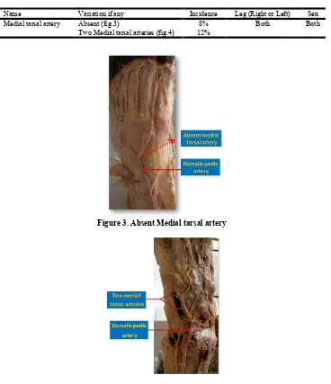
Name Variation if any Medial tarsal artery Absent (fig.3)
[image:3.595.118.486.74.501.2]Two Medial tarsal arteries
[image:3.595.206.375.90.491.2]Figure 4.
Table 3
Name Variation if any
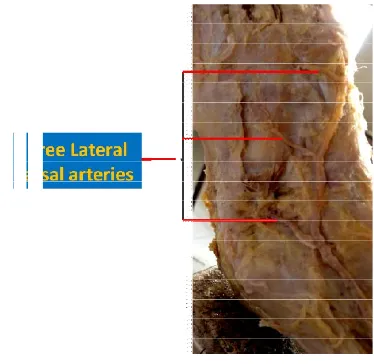
Lateral tarsal artery Two lateral tarsal arteries (fig.5) Three lateral tarsal arteries
Figure 5.
40694 International Journal of Current
Table 2. Variations in medial tarsal artery
Variation if any Incidence Leg (Right or Left)
Absent (fig.3) 8% Both
Two Medial tarsal arteries (fig.4) 12%
Figure 3. Absent Medial tarsal artery
Figure 4. Variation in number of Medial tarsal artery
Table 3. Variations noted in the lateral tarsal artery
Variation if any Incidence Leg (Right or Left) Two lateral tarsal arteries (fig.5) 14% Both
Three lateral tarsal arteries (fig.6) 8%
Figure 5. Variation in number of Lateral tarsal artery
International Journal of Current Research, Vol. 08, Issue, 10, pp.40692-40696, October
Leg (Right or Left) Sex Both
Leg (Right or Left) Sex Both
[image:3.595.236.363.342.490.2] [image:3.595.122.482.542.755.2]the contraindications for flap procedure (Christen Krag and Per
Riegels- Nielsen, 1982). In present study DPA was seen as
continuation of perforating branch of peroneal artery in 4% of feet. The anterior tibial artery was hypoplastic and the arcuate artery was absent. The Yamada et al. (1993
variation in 6.7% of legs and Mir Ali (1996)
cases. Present study correlates with the study done by Yamada
et al. (1993) regarding the origin of dorsalis pedis artery from
perforating branch of peroneal artery.
Variations in Medial tarsal artery
In 8% feet the medial tarsal artery was absent.
there were two medial tarsal arteries. In 2008, Ebrahim M. El. Saeed et al found that two medial tarsal arteries were present in 90% cases (Ebrahim et al., 2008).The forefoot skin defect can be repaired using the flap supplied by the branch of medial tarsal artery (Tan et al., 2012).
[image:4.595.201.388.55.237.2]40695 Dr. Preeti Awari andDr. Vatsalaswamy, Anatomical variations in dorsalis pedis artery and its branches
[image:4.595.162.436.288.562.2]Figure 6.
Table 4
Name Arcuate artery
Christen Krag and Per In present study DPA was seen as continuation of perforating branch of peroneal artery in 4% of feet. The anterior tibial artery was hypoplastic and the arcuate
1993)found the same
)found it in 1% of
cases. Present study correlates with the study done by Yamada regarding the origin of dorsalis pedis artery from
In 8% feet the medial tarsal artery was absent. In 12% feet there were two medial tarsal arteries. In 2008, Ebrahim M. El. found that two medial tarsal arteries were present in .The forefoot skin defect can epaired using the flap supplied by the branch of medial
Variations in lateral tarsal artery
There were two lateral tarsal a
Ebrahim M. El. Saeed et al found double lateral tarsal arteries in 70% feet (Ebrahim, 2008)
arteries in 8% of feet. Knowledge of variation in the lateral tarsal artery will help the surgeon to choose LTA pedicled extensor digitorum brevis flap for
lateral tarsal artery flaps are also introduced to cover the defect in head and neck oncologic resection. Wang C, Wang Q, Li G and Yang D used this flap for hypopharyngeal reconstruction (Wang et al., 2015).
Variations in Arcuate artery
In present study arcuate artery was absent in 40% of feet. Yamada et al. (1993) found the same variation in
Vijayalaxmi (1996)found in 6% and rajeshwari
Dr. Vatsalaswamy, Anatomical variations in dorsalis pedis artery and its branches
Figure 6. Variation in number of Lateral tarsal artery
Table 4. Variations noted in arcuate artery
Variation if any Incidence Leg (Right or Left) Sex
Absent(Fig.7) 40% Both Both
Figure 7. Absent Arcuate artery
Variations in lateral tarsal artery
There were two lateral tarsal arteries in 14% of feet. In 2008, found double lateral tarsal arteries There were three lateral tarsal Knowledge of variation in the lateral tarsal artery will help the surgeon to choose LTA pedicled extensor digitorum brevis flap for reconstructive surgeries. The lateral tarsal artery flaps are also introduced to cover the defect in head and neck oncologic resection. Wang C, Wang Q, Li G and Yang D used this flap for hypopharyngeal reconstruction
In present study arcuate artery was absent in 40% of feet. found the same variation in 33%,
found in 6% and rajeshwari (2013)found
The perfusion to dorsum of foot in such cases was done either by enlarged lateral tarsal artery or by deep plantar arch. Hollinshead mentions about small communications between branches of lateral tarsal artery and the arcuate artery. When the arcuate artery is rudimentary or missing; lateral tarsal artery may give rise to more lateral dorsal metatarsal arteries (Hollinshead, 1958). Dorsalis pedis artery is the main artery that supplies the foot as it also contributes to the formation of deep plantar arch. In the era of microvascular surgeries, dorsalis pedis artery and its branches have got immense importance in flap surgeries. Surgeons are still trying to bring to light the full potential of dorsalis pedis artery in various reconstructive surgeries. Hence this study will definitely aid in further such endeavours.
Acknowledgements
We thank the scholars whose articles are included in references of this manuscript. We also thank to the authors, editors and publishers of the articles, journals and books from where the literature for the manuscript has been reviewed.
REFERENCES
Christen Krag and Per Riegels- Nielsen, The dorsalis pedis flap for lower leg recobstruction, Acta Orthop Scand, 53, 1982,487-497
Ebrahim, M. El. Saeed, Amal Abd monsif, Madiha A. El-Sayed, Nancy M. Aly, Naser A. Gezlan Anatomical study of the Dorsalis Pedis Artery and its surgical importance in reconstructive surgery. Alexandra Bulletin Fac, Med., 2008; 44 No.2, 557-571
Gray’ Anatomy -The Anatomical Basis of Clinical Practice. Standring, Elsevier Churchill Livingstone, 2008, 40th edition, pg 1456
Hollinshead, W. H. 1958. Anatomy for Surgeons. The back
and limbs. 3rd ed. Hoeber- Harper. 807,808
Keith, L. Moore, Arthur F Dalley, Anne M R Agur Clinically
Oriented Anatomy,6th edition, Wolters Kluwer, 2005, pg.
586,602,619,620,617.
Mir Wajahath Ali, A M Mohajir Dorsalis Pedis Artery : Variations and clinical significance J. Indian Medical
Association, vol. 94, no. 11, Nov., 1996
Mir Wajahath Ali, A M Mohajir Dorsalis Pedis Artery: Variations and clinical significance J. Indian Medical
Association, vol. 94,no. 11, Nov., 1996
Rajeshwari, M.S., Roshankumar, B.N. 2013. Vijayakumar An Anatomical Study on Dorsalis Pedis Artery, Int J Anat
Res., Vol 1(2):88-92. ISSN 2321- 4287
Tan, W., Guli Zhaer, A., Huang, W., Jiang, X. 2012. Applied anatomy of the reverse pedicled island skin flap with arterial arch at the superior border of abductor hallucis muscle for repairing forefoot defect, Nan Fang Yi Ke Da
Xue Xue Bao., 32 :11,1592-6
Wang, C., Wang, Q., Li, G. and Yang, D. lateral tarsal artery flap: an option for hypopharyngeal reconstruction in patients with hypopharyngral carcinomas after surgery, Int
J Clin Exp Med., 2015 April 15 :8(4);4855-61
Yamada, T., Gloviczki, P., Bowe, R T.C., Naessens, J.M., Carmichael, S.W. 1993. Variations of the arterial anatomy of the foot. American Journal of Surgery, 166:2,130-5.