
1386
https://doi.org/10.1107/S2056989019011757 Acta Cryst.(2019). E75, 1386–1388research communications
Received 15 August 2019 Accepted 26 August 2019
Edited by M. Weil, Vienna University of Technology, Austria
Keywords:crystal structure; redetermination; low temperature; high resolution data set.
CCDC reference:1949371
Supporting information:this article has supporting information at journals.iucr.org/e
New refinement of the crystal structure of
Zn(NH
3)
2Cl
2at 100 K
Trpimir Ivsˇic´,aDavid Wenhua Bib* and Arnaud Magrezb
aLaboratory of Physics of Complex Matter, Ecole Polytechnique Fe´de´rale de Lausanne, Switzerland, andbCrystal Growth Facility, Ecole Polytechnique Fe´de´rale de Lausanne, Switzerland. *Correspondence e-mail: [email protected],
The crystal structure of [ZnCl2(NH3)2], diamminedichloridozinc, was
re-investigated at low temperature, revealing the positions of the hydrogen atoms and thus a deeper insight into the hydrogen-bonding scheme in the crystal packing. In comparison with previous crystal structure determinations [MacGillavry & Bijvoet (1936). Z. Kristallogr. 94, 249–255; Yamaguchi & Lindqvist (1981).Acta Chem. Scand.35, 727–728], an improved precision of the structural parameters was achieved. In the crystal, tetrahedral [Zn(NH3)2Cl2]
units (point-group symmetry mm2) are linked through N—H Cl hydrogen bonds into a three-dimensional network.
1. Chemical context
Zn(NH3)2Cl2 is found in discharged zinc–air batteries. It is
formed by dissolution of the zinc electrode in an ZnCl2–
NH4Cl electrolyte. At high Zn 2+
concentrations, the cations subsequently undergo complex formation with NH3and Cl
(Clarket al., 2017). Moreover, in the last century, Zn(NH3)nCl2
phases with n = 0.167–6 have been intensively investigated since these materials appear to be by-products of hydro-cracking of heavy-oil fractions. The monoammine salt withn= 1 and the diammine salt with n = 2 are the most studied members in this family. Thermal stability, thermomechanical and thermogravimetric behaviour are well documented for these systems (Gardneret al., 1989).
The crystal-structure model of Zn(NH3)2Cl2was to this day
incomplete, lacking the positions of hydrogen atoms. The crystal structure of Zn(NH3)2Cl2was originally determined by
MacGillavry & Bijvoet (1936) without refinement of atomic positions [COD number ID 1010197; ICSD number 26136; PDF4 number 01-074-0506]. This incompleteness was rather normal considering the X-ray techniques and measurement conditions at that time. An improved structure model was obtained decades later from a room-temperature data set collected on a four-circle diffractometer by Yamaguchi & Lindqvist (1981), however without hydrogen-atom positions [PDF4 number 04-015-4717].One more record of this phase in PDF4 [number 00-024-1435] can be found, however with a very limited X-ray powder diffraction pattern range from 5–63/2, which is unsuitable for Rietveld refinement.
Large single crystals of Zn(NH3)2Cl2 were obtained as a
side product of a chemical vapour transport (CVT) reaction, which allowed the reinvestigation of the crystal structure at low temperature with a particular focus on determination of the H-atom positions.
2. Structural commentary
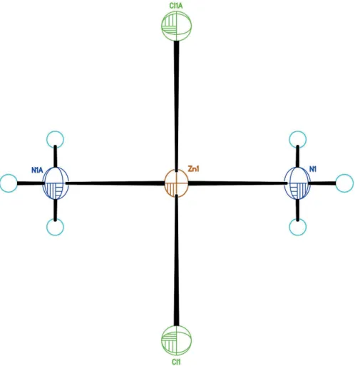
As shown in Fig. 1, the new structure refinement of Zn(NH3)2Cl2 (100 K data) confirms the former model by
Yamaguchi & Lindqvist (1981). The small shrinkage of the unit cell at low temperature is mainly due to lattice dynamics. However, the bond lengths within the Zn(NH3)2Cl2
tetra-hedron refined from 100 K data are similar to the ones obtained from room-temperature data, but with higher precision. This confirms that the tetrahedron is a very rigid building block of the structure (Fig. 2). Two independent H atoms, one in a general position (H1A) and one on a mirror plane (H1B), bond to the nitrogen atom with refined bond lengths of N1—H1A = 0.825 (17) A˚ and N1—H1B = 0.74 (4) A˚ , respectively. Hydrogen-bonding interactions of weak-to-medium strengths (Table 1) between the NH3group
and the Cl ligands of adjacent tetrahedra lead to formation of a three-dimensional network, as shown in Fig. 3.
3. Synthesis and crystallization
Zn(NH3)2Cl2 was obtained during the course of a chemical
vapour transport experiment. Prior to use, a silica tube sealed at one end (Ø = 15 mm, wall thickness = 2 mm) was heated under vacuum to 1073 K overnight. Zinc oxide (317 mg) and previously dried ammonium chloride (190 mg) were pressed into pellets and put into the pretreated silica tube. After evacuation to 106mbar, the tube was sealed and placed into a horizontal tube furnace. The temperature at the source was slowly heated to 1173 K and maintained at this value for the whole duration of growth. At the low-temperature zone (873 K), a colourless powder mixed with high-quality single crystals of Zn(NH3)2Cl2 with an irregular morphology was
collected after complete cooling to room temperature. Since
research communications
[image:2.610.47.296.71.331.2]Acta Cryst.(2019). E75, 1386–1388
Figure 2
Comparison of the [ZnN2Cl2] tetrahedra from the room temperature data (left; Yamaguchi & Lindqvist, 1981) and the present 100 K data (right). Bond lengths for the room-temperature data were calculated with
DIAMOND(Brandenburg, 2006).
Table 1
Hydrogen-bond geometry (A˚ ,).
D—H A D—H H A D A D—H A
N1—H1A Cl1i 0.825 (17) 2.806 (18) 3.4552 (9) 137.0 (17)
N1—H1A Cl1ii 0.825 (17) 2.897 (18) 3.5505 (9) 137.6 (16) N1—H1B Cl1iii 0.74 (4) 2.95 (3) 3.5556 (10) 141 (1)
N1—H1B Cl1iv 0.74 (4) 2.95 (3) 3.5556 (10) 141 (1) Symmetry codes: (i) xþ1;yþ1;zþ1; (ii) xþ1
2;yþ1;z 1 2; (iii) xþ1
2;yþ 1 2;zþ
3 2; (iv)x
1 2;y;zþ
3 2.
Figure 1
The tetrahedral [Zn(NH3)2Cl2] group. H atoms (cyan circles) are plotted with arbitrary size. [Symmetry code: (A) 1x,1
[image:2.610.313.560.458.716.2]2y,z.].
Table 2
Experimental details.
Crystal data
Chemical formula [ZnCl2(NH3)2]
Mr 170.34
Crystal system, space group Orthorhombic,Imma
Temperature (K) 100
a,b,c(A˚ ) 7.7077 (2), 8.0226 (2), 8.4526 (3)
V(A˚3) 522.67 (3)
Z 4
Radiation type MoK
(mm1) 5.56
Crystal size (mm) 0.260.180.16
Data collection
Diffractometer Rigaku SuperNOVA
Absorption correction Analytical (CrysAlis PRO; Rigaku OD, 2019)
Tmin,Tmax 0.277, 0.454
No. of measured, independent and observed [I> 2(I)] reflections
2303, 708, 678
Rint 0.017
(sin/)max(A˚
1) 0.833
Refinement
R[F2> 2(F2)],wR(F2),S 0.015, 0.036, 1.04
No. of reflections 708
No. of parameters 25
H-atom treatment All H-atom parameters refined max,min(e A˚
3
) 0.50,0.55
[image:2.610.45.295.592.702.2]the thermal stability of Zn(NH3)2Cl2 is limited as it already
decomposes at 463 K (Gardner et al., 1989), it is very likely that Zn(NH3)2Cl2crystallized at the very end of the cooling
process.
4. Refinement
Crystal data, data collection and structure refinement details are summarized in Table 2. Unlike the two previous refine-ments in space groupImam, the unit cell was chosen to be in the standard setting (Imma) for space group No. 74. Three reflections (020, 002, 004) affected by the incident beam-stop were omitted. The two H atoms were refined freely.
Acknowledgements
DWB is grateful to the help of Dr Rosario Scopelliti in ISIC of EPFL. We would like to express our great appreciation to Professor La´szlo´ Forro´ in LPMC of EPFL for the fruitful discussions and support.
Funding information
Funding for this research was provided by: Swiss National Science Foundation (SNF) Sinergia network "NanoSkyrmio-nics" (grant No. CRSII5-171003).
References
Brandenburg, K. (2006).DIAMOND. Crystal Impact GbR, Bonn, Germany.
Clark, S., Latz, A. & Horstmann, B. (2017).ChemSusChem,10, 4735– 4747.
Gardner, P. J., Pang, P. & Preston, S. (1989).Thermochim. Acta,138, 371–374.
MacGillavry, C. H. & Bijvoet, J. M. (1936).Z. Kristallogr.94, 249–255. Rigaku OD (2019). CrysAlis PRO. Rigaku Oxford Diffraction,
Yarnton, England.
Sheldrick, G. M. (2015a).Acta Cryst.A71, 3–8. Sheldrick, G. M. (2015b).Acta Cryst.C71, 3–8. Westrip, S. P. (2010).J. Appl. Cryst.43, 920–925.
Yamaguchi, T. & Lindqvist, O. (1981).Acta Chem. Scand.35, 727–728.
1388
Ivsˇic´et al. [ZnCl2(NH3)2] Acta Cryst.(2019). E75, 1386–1388
[image:3.610.48.571.69.317.2]research communications
Figure 3
supporting information
sup-1 Acta Cryst. (2019). E75, 1386-1388
supporting information
Acta Cryst. (2019). E75, 1386-1388 [https://doi.org/10.1107/S2056989019011757]
New refinement of the crystal structure of Zn(NH
3)
2Cl
2at 100
K
Trpimir Iv
š
i
ć
, David Wenhua Bi and Arnaud Magrez
Computing details
Data collection: CrysAlis PRO (Rigaku OD, 2019); cell refinement: CrysAlis PRO (Rigaku OD, 2019); data reduction:
CrysAlis PRO (Rigaku OD, 2019); program(s) used to solve structure: SHELXT (Sheldrick, 2015a); program(s) used to
refine structure: SHELXL2018 (Sheldrick, 2015b); molecular graphics: DIAMOND (Brandenburg, 2006); software used
to prepare material for publication: publCIF (Westrip, 2010).
Diamminedichloridozinc
Crystal data
[ZnCl2(NH3)2] Mr = 170.34
Orthorhombic, Imma a = 7.7077 (2) Å
b = 8.0226 (2) Å
c = 8.4526 (3) Å
V = 522.67 (3) Å3 Z = 4
F(000) = 336
Dx = 2.165 Mg m−3
Mo Kα radiation, λ = 0.71073 Å Cell parameters from 1597 reflections
θ = 3.6–37.4°
µ = 5.56 mm−1 T = 100 K
Irregular, colourless 0.26 × 0.18 × 0.16 mm
Data collection
Rigaku SuperNOVA diffractometer
Radiation source: micro-focus sealed X-ray tube
ω scans
Absorption correction: analytical (CrysAlis PRO; Rigaku OD, 2019)
Tmin = 0.277, Tmax = 0.454
2303 measured reflections
708 independent reflections 678 reflections with I > 2σ(I)
Rint = 0.017
θmax = 36.3°, θmin = 3.5° h = −12→8
k = −13→12
l = −12→14
Refinement
Refinement on F2
Least-squares matrix: full
R[F2 > 2σ(F2)] = 0.015 wR(F2) = 0.036 S = 1.04 708 reflections 25 parameters 0 restraints
Primary atom site location: structure-invariant direct methods
Hydrogen site location: difference Fourier map
All H-atom parameters refined
w = 1/[σ2(F
o2) + (0.0188P)2]
where P = (Fo2 + 2Fc2)/3
(Δ/σ)max = 0.001
Δρmax = 0.50 e Å−3
Δρmin = −0.55 e Å−3
Extinction correction: SHELXL2018 (Sheldrick, 2015b),
Fc*=kFc[1+0.001xFc2λ3/sin(2θ)]-1/4
supporting information
sup-2 Acta Cryst. (2019). E75, 1386-1388
Special details
Geometry. All esds (except the esd in the dihedral angle between two l.s. planes) are estimated using the full covariance matrix. The cell esds are taken into account individually in the estimation of esds in distances, angles and torsion angles; correlations between esds in cell parameters are only used when they are defined by crystal symmetry. An approximate (isotropic) treatment of cell esds is used for estimating esds involving l.s. planes.
Fractional atomic coordinates and isotropic or equivalent isotropic displacement parameters (Å2)
x y z Uiso*/Ueq
Zn1 0.500000 0.250000 0.61168 (2) 0.01163 (6)
Cl1 0.500000 0.47954 (3) 0.76915 (3) 0.01375 (6)
N1 0.28237 (15) 0.250000 0.47949 (13) 0.01603 (17)
H1A 0.274 (3) 0.3281 (19) 0.416 (2) 0.045 (4)*
H1B 0.206 (5) 0.250000 0.533 (4) 0.090 (11)*
Atomic displacement parameters (Å2)
U11 U22 U33 U12 U13 U23
Zn1 0.01002 (9) 0.01352 (9) 0.01134 (9) 0.000 0.000 0.000 Cl1 0.01288 (11) 0.01346 (11) 0.01490 (11) 0.000 0.000 −0.00253 (7) N1 0.0136 (4) 0.0211 (4) 0.0134 (4) 0.000 −0.0016 (3) 0.000
Geometric parameters (Å, º)
Zn1—Cl1i 2.2722 (3) N1—H1Aii 0.825 (17)
Zn1—Cl1 2.2722 (3) N1—H1A 0.825 (17)
Zn1—N1i 2.0155 (11) N1—H1B 0.74 (4)
Zn1—N1 2.0155 (11)
Cl1i—Zn1—Cl1 108.282 (14) Zn1—N1—H1Aii 115.0 (13)
N1—Zn1—Cl1 108.951 (17) Zn1—N1—H1A 115.0 (13)
N1i—Zn1—Cl1i 108.951 (17) Zn1—N1—H1B 109 (3)
N1—Zn1—Cl1i 108.951 (17) H1A—N1—H1Aii 99 (2)
N1i—Zn1—Cl1 108.950 (17) H1A—N1—H1B 109 (2)
N1i—Zn1—N1 112.66 (7) H1B—N1—H1Aii 109 (2)
Symmetry codes: (i) −x+1, −y+1/2, z; (ii) x, −y+1/2, z.
Hydrogen-bond geometry (Å, º)
D—H···A D—H H···A D···A D—H···A
N1—H1A···Cl1iii 0.825 (17) 2.806 (18) 3.4552 (9) 137.0 (17)
N1—H1A···Cl1iv 0.825 (17) 2.897 (18) 3.5505 (9) 137.6 (16)
N1—H1B···Cl1v 0.74 (4) 2.95 (3) 3.5556 (10) 141 (1)
N1—H1B···Cl1vi 0.74 (4) 2.95 (3) 3.5556 (10) 141 (1)
![Figure 3Packing view along the b axis (left) and the c axis (right), showing N—H� � �Cl interactions between adjacent [Zn(NH3)2Cl2] tetrahedra.](https://thumb-us.123doks.com/thumbv2/123dok_us/380965.535540/3.610.48.571.69.317/figure-packing-view-right-showing-interactions-adjacent-tetrahedra.webp)