Improved Screening Procedure for Biogenic Amine
Production by Lactic Acid Bacteria and
Enterobacteria
Lu SHILING 1, Jiang CAIHONG 1, Xu XINGLIAN 2, Xu CHENGJIAN 1, Li KAIXIONG 1 and Shu RUIHUA2
1College of Food Science, Shihezi University, Shihezi, P.R. China; 2Key Laboratory of Meat Processing and Quality Control, Ministry of Education, College of Food Science
and Technology, Nanjing Agricultural University, Nanjing, P.R. China Abstract
Shiling L., Caihong J., Xinglian X., Chengjian X., Kaixiong L., Ruihua S. (2015): Improved screening pro-cedure for biogenic amine production by lactic acid bacteria and Enterobacteria. Czech J. Food Sci., 33: 19–26.
An improved screening procedure was developed for the detection of amino acid decarboxylase-positive microorganisms (especially lactic acid bacteria and Enterobacteria). Monolayer culture and double layer colour development methods for the detection of amino acid decarboxylase-positive lactic acid bacteria and Enterobacteria were established. Bio-genic amine-positive bacteria strain was isolated directly from the samples. The applicability and detection level of the designed medium were quantitatively evaluated by the confirmation of the amine-forming capacity using an HPLC procedure, while tyrosine decarboxylase and lysine decarboxylase genes were detected by PCR with specific primers. The screening method showed a good correlation with the chemical analysis and molecular detection. Enterobacter aerogenes, Enterobacter cloacae, Escherichia coli, Enterococcus faecium,and Enterococcus faecalis were obtained from traditional Chinese sausage. The isolation and screening of amino acid decarboxylase-positive lactic acid bacteria and Enterobacteria can be carried out simultaneously by the improved method.
Keywords: amino acid decarboxylase; monolayer culture; double layer colour development
Biogenic amines (BAs) are organic molecules of low molecular weight with undesirable physiological effects. These include the stimulation of nerves and blood vessels in humans and animals, particularly when consumed in significant amounts or when the capacity to oxidise them is reduced by ingestion of mono-amino oxidase inhibitors such as alcohol or certain antidepressant medications, BAs enter the systemic circulation and exert their toxic effects on different organs, causing serious human health problems (McCabe-Sellers et al. 2006).
In addition, BAs are known to be potential precur-sors of carcinogenic nitrosamines. The class includes histamine (HIS), putrescine (PUT), cadaverine (CAD), tyramine (TYR), tryptamine (TRY), phenylethylamine
(PHE), spermine (SPM), and spermidine (SPD). These originate from the decarboxylation of specific free amino acids in fermented foods derived from raw materials with a high protein content, especially if the food provides suitable conditions for biochemical activity of the microorganisms presented (Suzzi & Gardini 2003; Coïsson et al. 2004).
During the ripening of fermented meat products, proteins undergo degradation. Large peptides are generated, then degraded into oligopeptides, which in turn degrade to free amino acids (Landete et al. 2007; Buňková et al. 2010). The free amino acids are then catabolised, giving rise to different compounds such as ammonia, α-ketoacids, methylketones, and amines.
Supported by the National Natural Science Foundation of China. Grants No. 31160329 and No. 31360392, by the Production and Construction Corps of Xinjiang doctor fund, Grant No. 2013BB012, and by the Scientific Research Programs of Shihezi University, Project No. RCZX201020.
The content of BAs in fermented meat products has been investigated extensively (Ayhan et al. 1999; Roseiro et al. 2006; Gençcelep et al. 2008). CAD and TYR are important amines in fermented meat products and the mean content in traditional sausage types has been reported to amount to 175 and 151.56 mg/kg, respectively (Lu et al. 2010). The maximun content of CAD and TYR was 199 and 676 mg/kg, respectively (Gençcelep et al. 2008). Similar results were reported also by other authors (Şenöz et al. 2000). High levels of CAD and TYR in fermented sausage are usually related to the occurrence of high counts of microflora with amino acid decarboxylase activity. The types of BAs formed are strongly influenced by microbial species and by other parameters, which allow bacterial growth during processing and storage (Carelli et al. 2007). The production of BAs in meat products has been attributed to the action of several microorganisms: Pseudomonas spp., Enterobacteriaceae, Enterococci, and Lactobacilli. The occurrence and distribution of amino acid decarboxylase activity vary in different fermented products. Enterobacteria (EB) and lactic acid bacteria (LAB), mainly isolated from fermented sausages, have been extensively reported. In fermented sausage, it has been observed that Enterobacter faecalis, E. faecium, Lactobacillus curvatus, L. bavaricus, L. brevis, L. para-casei, and L. sakei produce TYR (Maijala & Eerola 1993; Bover-Cid et al. 2001). Some bacterial species also have the capability to produce CAD and PUT, such as Enterobacter cloacae, E. aerogenes, Serratia spp., C. freundii, Escherichia coli, and Morganella morganii (Halász et al. 1994; Bover-Cid et al. 2001; Durlu-Özkaya et al. 2001).
Several qualitative and quantitative methods to determine BAs produced by microorganisms have been described, most of the screening procedures generally involve the measurement of amino acid decarboxylase-positive single strain which has been isolated from food in advance (Maijala & Ecrola 1993; Bover-Cid & Holzapfel 1999). However, little information exists on the selection of amino acid decarboxylase-positive lactic acid bacteria and EB from mixed strains directly. When mixed micro-organisms were isolated, they interfered with one another. For example, some bacteria produce lactic acid and other produce alkaline materials such as BAs. Neutralisation will happen during the time of mixed microorganisms culture according to the method of Sara Bover-Cid and that of Wilhelm Heinrich Holzapfel (Bover-Cid & Holzapfel 1999). The bacteria reproduction and acid (or alkaline) materials
production speed are different during mixed micro-organisms culture so that it is difficult to determine the optimal examination time. Systematic screening procedures for amino acid decarboxylase-positive LAB and EB from mixed microoranisms are still relatively scarce.
In this study, we report improved methods to screen BA-positive LAB and EB isolated directly from tradi-tional Chinese sausages. Polymerase chain reaction denaturing gradient gel electrophoresis (PCR-DGGE) technique was used to evaluate the screened strain diversity. The genes for tyrosine and lysine decar-boxylase were detected by PCR with specific prim-ers. The production of BAs in liquid medium was detected by HPLC.
MATERIAL AND METHODS
Sampling. Twelve kinds of smoked horsemeat sausages that had been produced within a two-month period were obtained from retail markets of different regions in China’s Xinjiang. The smoked horsemeat sausage is a spontaneously fermented sausage with added sugar, salt, and spices. The BAs were detected before BA-producing bacteria isolation in the sam-ples, and the concentrations of CAD and TYR were found to exceed 230 and 200 mg/kg, respectively.
Screening medium. The lower screen medium contained: 0.5% tryptone, 0.5% yeast extract, 0.5% meat extract, 0.25% NaCl, 0.05% glucose, 0.1% Tween 80, 0.004% MgSO4, 0.003% MnSO4, 0.2% K2HPO4, 0.2% ammonium citrate, 0.001% thiamine, 0.005% pyridoxal-5-phosphate, 0.5% tryptophan, 0.5% histi-dine, 0.5% phenylalanine, 0.5% tyrosine, 0.5% lysine, 0.5% arginine, and 1.8% agar, pH was 5.2.
The upper colour development medium consisted of 0.005% bromocresol purple, 1.0% agar, pH was 5.2.
DNA extraction. Genomic DNA of amino acid de-carboxylase-positive bacteria was extracted using Gen-EluteTMKit (Tiangen Biotech Co., Ltd, Beijing, China)
according to the manufacturer’s instructions, and then suspended in 100 µl of TE buffer and stored at –20°C.
PCR amplification
PCR amplification for PCR-DGGE. The primers U968-GC (5'CGC CCG GGG CGC GCCCCG GGC GGG GCG GGG GCA CGG GGG GAACGC GAA GAA CCT TAC) and L1401 (5'GCG TGT GTA CAA GAC CC) were used to amplify the V6–V8 regions of the bacterial 16S rDNA (Zhu et al. 2003). The GC clamp in primer U968-GC creates PCR products suitable for the separation by DGGE.
GoTaq Green Master Mix (Promega, Alexandria, USA) was used in the PCR reaction. The amplification reactions were carried out in a 25 µl reaction volume containing 12.5 µl GoTaq Green Master Mix, 1.0 µl of each primer (10 pmol/ml), 1 µl DNA template, and 9.5 µl ddH2O. The samples were amplified in a BioSci PCR system at 94°C for 4 min, 35 cycles of 94°C for 30 s, 56°C for 20 s, and 68°C for 40 s, followed by a final step at 68°C for 7 minutes. The PCR products (5 µl) were analysed by electrophoresis on agarose gel (1.2%) to check the amplicon sizes and amounts.
Identification of tyrosine decarboxylase gene (TDC). The primers TD2 (5'-ACA TAG TCA ACC ATR TTG AA-3') and TD5 (5'-CAA ATG GAA GAA GAA GTA GG-3') were used to amplify TDC (Fernández et al. 2004). GoTaq Green Master Mix (Promega, Alexandria, USA) was used in the PCR reaction. The amplification reactions were carried out in a 25 μl reaction volume, containing 12.5 µl GoTaq Green Master Mix, 0.4 µl of each primer (10 pmol/ml), 1 µl DNA template, and 9.7 ddH2O μl. The samples were amplified in a BioSci PCR system at 94°C for 5 min, 35 cycles of 94°C for 45 s, 48°C for 45 s, 72°C for 60 s, and 72°C for 7 minutes. The PCR products (5 µl) were analysed by electrophoresis on an agarose gel (1.2%) to check the amplicon sizes and amounts.
Identification of lysine decarboxylase gene (LDC). The primers CAD1-f (5'-TTY GAY WCN GCN TGG GTN CCN TAY AC-3') and CAD1-r (5'-CCR TGD ATR TCN GTY TCR AAN CCN GG-3') were used to amplify LDC (Rivas et al. 2006). GoTaq Green Master Mix (Promega, Alexandria, USA) was used in the PCR reaction. The amplification reactions were carried out in a 25 µl reaction volume containing 12.5 μl GoTaq Green Master Mix, 0.8 µl of each primer (10 pmol/ml),
1 µl DNA template, and 8.9 µl ddH2O. The samples were amplified in a BioSci PCR system at 94°C for 5 min, 35 cycles of 94°C for 30 s, 53°C for 30 s, 72°C for 120 s, and 72°C for 10 minutes. The PCR products (5 µl) were analysed by electrophoresis on agarose gel (1.2%) to check the amplicon sizes and amounts.
DGGE analysis. Specific separation of PCR ampli-cons was achieved by DGGE analysis using a BioRad DCode apparatus (Bio-Rad, Richmond, USA) accord-ing to the procedures described by Zhu et al. (2003). The following modifications according to the work of Hu et al. (2008) were included: DGGE was performed on 8% polyacrylamide gels containing acrylamide, bisacrylamide, formamide, and a gradient of 37–57% of urea. Electrophoresis was carried out at 200 V for 10 min and then at 85 V for 16 h at 60°C. The gels were stained with ethidium bromide (0.5 mg/l) for 15 min, then rinsed three times in milli-Q water and photographed with UV transillumination using the GelDoc 2000 system (Bio-Rad, Richmond, USA). Identical results in three replicates confirmed the reproducibility of the amplifications and subsequent DGGE separations. The fingerprints of the DGGE profiles were analysed using Quantity One 1D Analysis software Ver. 4.5 (Bio-Rad, Richmond, USA).
BA determination. Amine standard solutions were prepared in 0.4 M perchloric acid to a final concen-tration of 1 mg/ml for each amine. Solutions were prepared in 0.4 M perchloric acid to final concentra-tions of 0.0, 2.0, 5.0, 10, and 20 µg/ml for each amine. One ml of each bacterial culture broth was uni-formly mixed with 0.4 M perchloric acid equivalently. This mixture was centrifuged at 10 000 rpm for 10 min (4°C) and the supernatant was filtered. The filtrate extract (1 ml) was placed in a 5 ml volumetric flask. Then, sodium hydroxide (2 N, 200 µl), saturated sodium bicarbonate (300 µl), and dansyl chloride solution (10 mg/ml) amount were added to the sample extract. After incubation at 40°C for 45 min in the dark, 100 µl of ammonia were added to the reaction mixture for the removal of residual dansyl chloride. After 30 min at ambient temperature, the volume of the reaction mixture was adjusted to 5 ml with acetonitrile. This reaction mixture was centrifuged for 5 min at 2500 rpm. The supernatant was filtered with a 0.45 µm syringe filter with a PVDF Membrane for HPLC (Agilent 1100 system) analysis.
was used with a mixture of acetonitrile as solvent A and water as solvent B. The gradient elution procedure was 35% A + 65% B for 1 min, 20% A + 80% B for 5 min, 10% A + 90% B at 6 min, and 8% A + 92% B for 16 minutes.
The standard amine samples HIS, TRY, TYR, and PUT were purchased from Sigma (St. Louis, USA) and PHE, CAD, SPD and SPE from Fluka Chemical (Buchs, Switzerland), respectively.
RESULTS AND DISCUSSION
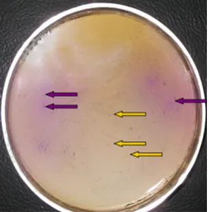
Isolation of LAB and EB. After cultivation in the lower screen medium at 37°C for 72 h, the upper colour development medium (50°C) was layered onto the lower screen medium. Positive reactions were recorded within 5 min, when upper colour development medium was applied a purple colour appearing as the amines raised the pH. A total of 154 BA-positive strains, including LAB and EB, were obtained by the screening procedure, as shown in Figures 1 and 2. Several authors have studied BA-positive bacteria isolation (Maijala & Ecrola 1993; Bover-Cid & Holzapfel 1999; Landeta et al. 2007; Moreno-Arribas & Carmen Polo 2008; Capozzi et al. 2010; Fadhlaoui-Zid et al. 2012), and their techniques are similar to that in our study. However, in those studies, the culture and colour development were carried out simultaneously, while in our study they were performed separately. The bacterial flora in the traditional Chinese sausage is diverse and complex. If the culture and colour development were carried out simultaneously, dif-ferent bacterial species would interact, as there are
some acid-forming bacteria and alkaline-forming bacteria in the sausage. In addition, different strains require different lengths of time to grow. It would be difficult to obtain optimalobservation time. In our study, the culture and colour development were carried out separately, which resulted in a dry film forming on top of the flora and ions produced by bacteria being fixed after 72 h of culture. When the upper colour development medium was applied, a clear result developed in a short time. Therefore, this method reduced the possible interactions between different bacteria and improved the isolation accuracy. In addition, some authors isolated BAs-producting bacteria by meant of plate culture, and the occur-rence of amino acid-decarboxylase activity in a single strain was investigated in the screening broth and by RP-HPLC quantitative detection (Landeta et al. 2007; Curiel et al. 2011; Chang & Chang 2012). This method results in huge workload and high costs.
[image:4.595.99.252.554.709.2]Identification of LAB and EB. The PCR-DGGE tech-nique was introduced into food microbiology in order to evaluate microbial diversity and identify microor-ganisms. The purpose of this study was to identify the bacteria isolated from the traditional Chinese sausage. The DGGE results were obtained by amplifying the V6–V8 region of the 16S rRNA gene using the U968 (GC)-L1401 primer set. The microbial ecosystem of 154 strains was compared by PCR-DGGE and five different bacteria species were identified as shown in Figure 3. The identification results are given in Table 1. The observed sequences exhibited an identity greater than 96% with the sequences from the GenBank database. A number of authors have studied Entero-coccus faecalis in fermented food. Some authors used
Figure 2. Screening of biogenic amines-producing Ente- robacteria (on figure is lower screen agar and upper colour development agar;the purple is positive and the yellow is negative)
[image:4.595.339.488.554.700.2]some Enterococcus faecalis strains as starters that were unable to generate BAs (Centeno et al. 1999; Sparo et al. 2008; Bhardwaj et al. 2010), while other authors maintained that some Enterococcus faecalis strains do produce BAs (Gardini et al. 2001; Pérez-Pulido et al. 2006; Capozzi et al. 2010; Komprda et al. 2010). Enterobacter aerogenes, Enterobacter cloacae, and Escherichia coli isolated from food are
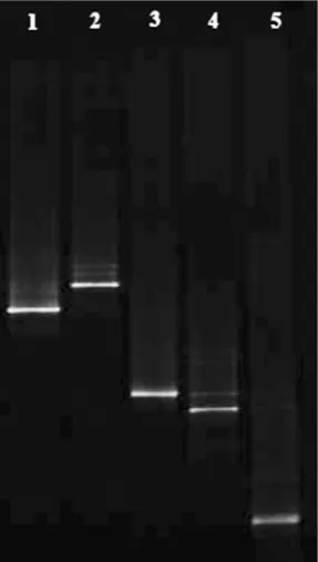
generally considered as microorganisms with a high decarboxylase activity, particularly in relation to the production of CAD and PUT (Durlu-Özkaya et al. 2001; Suzzi & Gardini 2003; Lorenzo et al. 2010). In addition, some species of Enterobacter aerogenes, Enterobacter cloacae, and Escherichia coli are pro-ducers of HIS (Hsu et al. 2009; Huang et al. 2010). Agarose electrophoresis photos of PCR amplifica-tion products by TDC and LDC-specific primers are shown in Figure 4. Bands 1 and 2 were the amplifica-tion products of TDC of 1100 bp length, while bands 3, 4, and 5 were the amplification products of LDC of 1098 bp length.
Quantification of BAs. With none of the strains as-sayed and confirmed by HPLC analysis, any false-positive or false negative reactions were observed. However, three tyramine-producing false negative strains were found by other researchers (Bover-Cid & Holzapfel 1999). May be a more appropriate pH of medium and optimalobservation time have been used in our study. Besides, it appears necessary that the culture and colour development be carried out separately.
[image:5.595.64.290.139.233.2]In this study, Enterococcus faecium and Enterococ-cus faecalis were found to produce PHE and TYR, while Enterobacter aerogenes, Enterobacter cloacae, and Escherichia coli were PUT- and CAD-producing species. Several authors have demonstrated simulta-neous production of TYR and PHE (Bover-Cid et al. 2001; Moreno-Arribas et al. 2001), and of PUT and CAD (Ansorena et al. 2002; Bunková et al. 2010). Table 1. Biogenic amines-productive strains identified by
means of 16S rDNA sequencing fragment Isolate
bands Closest relatives ID (%) Accession No. 1 Enterobacter aerogenes 98 GQ890355 2 Enterobacter cloacae 98 GQ890353 3 Escherichia coli 98 GQ890359 4 Enterococcus faecium 99 GQ890354 5 Enterococcus faecalis 9 GQ890356
bands 1, 2, 3 – CAD and PUT-producing bacteria; bands 4 and 5 – PHE and TYR-producing bacteria; ID – bands are numbered as indicated on the DGGE gels shown in Figure 3; accession number of the sequence of the closest relative found by BLAST search
Figure 4. Agarose electrophoresis photo of PCR amplifi-cation products with specific primers of biogenc amine- productive bacteria
[image:5.595.100.244.433.687.2]M –marker; line: 1 – Enterobacter aerogenes; 2 – Enterobacter cloacae; 3 – Escherichia coli; 4 – Enterococcus faecium; 5 – Enterococcus faecalis; N – negative control
Figure 3. DGGE fringerprints of biogenic amines-produ-cing bacteria classified
Line: 1 – Enterobacter aerogenes; 2 – Enterobacter cloacae; 3 – Escherichia coli; 4 – Enterococcus faecium; 5 – Enterococ-cus faecalis (Table 1)
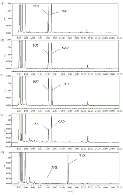
[image:5.595.308.533.506.680.2]Figure 5. HPLC chromatographic profiles of biogenic amines in bacterial culture fluid produced by (A) Enterobacter aerogenes, (B) Enterobacter cloacae, (C) Escherichia coli, (D) Enterococcus faecium, and (E) Enterococcus faecalis respectively
(A)
(B)
(C)
(D)
(E)
2.00 4.00 6.00 8.00 10.00 12.00 14.00 16.00 18.00 20.00 22.00 24.00 26.00 28.00 30.00 (min)
2.00
1.50
1.00
0.50
0.00
2.00
1.50
1.00
0.50
0.00
2.00
1.50
1.00
0.50
0.00
1.20 1.00 0.80 0.60 0.40 0.20 0.00 –0.20 1.20 1.00 0.80 0.60 0.40 0.20 0.00
AU
AU
AU
AU
AU
2.00 4.00 6.00 8.00 10.00 12.00 14.00 16.00 18.00 20.00 22.00 24.00 26.00 28.00 30.00
2.00 4.00 6.00 8.00 10.00 12.00 14.00 16.00 18.00 20.00 22.00 24.00 26.00 28.00 30.00
2.00 4.00 6.00 8.00 10.00 12.00 14.00 16.00 18.00 20.00 22.00 24.00 26.00 28.00 30.00
No other BAs were produced by the five species in our study. The chromatography profiles of the BAs produced by Enterobacter aerogenes, Enterobacter cloacae, Escherichia coli, Enterococcus faecium, and Enterococcus faecalis are shown in Figure 5.
Enterobacter aerogenes, Enterobacter cloacae, and Escherichia coli produced 5.02~3987.37 µg/ml of CAD and 3.15~3592.64 µg/ml of PUT, while 0.48~585.41 µg/ml of PHE and 0.19~3516.7 μg/ml of TYR were produced by Enterococcus faecium and Enterococcus faecalis, respectively. The BAs positive bacteria were classified on the basis of BAs productive ability by cluster analysis. Group A included bacteria showing low amine production (5.02 to 35.87μg/ml of CAD, 3.15~39.65 µg/ml of PUT, 0.48~25.41 µg/ml of PHE, and 0.19~36.7μg/ml of TYR) and accounted for 18.5, 27.3, 35.9, and 13.6% of BAs positive bac-teria, respectively. Group B included bacteria with moderate amine production (35.87~08.3 μg/ml of CAD, 39.65~189.5 µg/ml of PUT, 25.41~98.4 μg/ml of PHE, and 36.7~126.8 μg/ml of TYR) and account-ed for 15.2, 24.7, 36.1 and 27.5% of BAs positive bacteria respectively. Group C included 45.8, 37.9, 16.9, and 29.7% the positive bacteria showing high amines production (108.3–2353.05 μg/ml of CAD, 189.5~808.13 µg/ml of PUT, 98.4~296.28 µg/ml of PHE, and 126.8~2826.71μg/ml of TYR, respectively), and group D contained 20.5, 10.1, 11.1, and 29.2% of the positive bacteria showing very high levels of amines production (2353.05~3987.37 µg/ml of CAD, 808.13~3592.64 µg/ml of PUT, 296.28~585.41 µg/ml of PHE, and 2826.71~3516.7 µg/ml of TYR, respec-tively). Therefore, these five bacterial species have an important influence on the accumulation of PHE, TYR, CAD, and PUT in the traditional Chinese sausage.
CONCLUSIONS
In this study, a decarboxylase screening technique for LAB and EB was modified. It enabled an improved detection of BA-positive strains from mixed bacteria samples. Five BA-positive dominant bacteria were obtained from the traditional Chinese sausage. In addition, systematic methods for the isolation and identification of BA-positive bacteria were established.
References
Ansorena D., Montel M.C., Rokka M., Talon R., Eerola S., Rizzo A., Raemaekers M., Demeyer D. (2002): Analysis of
biogenic amines in northern and southern European sau-sages and role of flora in amine production. Meat Science, 61: 141–147.
Ayhan K., Kolsarici N., Özkan G.A. (1999): The effects of a starter culture on the formation of biogenic amines in Turk-ish soudjoucks. Meat Science, 53: 183–188.
Bhardwaj A., Gupta H., Kapila S., Kaur G., Malik R.K. (2010): Safety assessment and evaluation of probiotic potential of bacteriocinogenic Enterococcus faecium KH 24 strain under in vitro and in vivo conditions. International Journal of Food Microbiology, 141: 156–164.
Bover-Cid S., Holzapfel W.H. (1999): Improved screening procedure for biogenic amine production by lactic acid bac-teria. International Journal of Food Microbiology, 53: 33–41. Bover-Cid S., Hugas M., Izquierdo-Pulido M., Vidal-Carou
M.C. (2001): Amino acid-decarboxylase activity of bacteria isolated from fermented pork sausages. International Jour-nal of Food Microbiology, 66: 185–189.
Buňková L., Buňka F., Klčovská P., Mrkvička V., Doležalová M., Kráčmar S. (2010): Formation of biogenic amines by Gram-negative bacteria isolated from poultry skin. Food Chemistry, 121: 203–206.
Capozzi V., Ladero V., Beneduce L., Fernández M., Alvarez M.A., Benoit B., Laurent B., Grieco F., Spano G. (2010): Isolation and characterization of tyramine-producing Ente-rococcus faecium strains from red wine. Food Microbiology, 28: 434–439.
Carelli D., Centonze D., Palermo C., Quinto M., Rotunno T. (2007): An interference free amperometric biosensor for the detection of biogenic amines in food products. Biosensors and Bioelectronics, 23: 640–647.
Centeno J.A., Menendez S., Hermida M., Rodríguez-Otero J.L. (1999): Effects of the addition of Enterococcus faecalis in Cebreiro cheese manufacture. International Journal of Food Microbiology, 48: 97–111.
Chang M., Chang H.C. (2012): Development of a screening method for biogenic amine producing Bacillus spp. Inter-national Journal of Food Microbiology, 153: 269–274. Coïsson J.D., Cerutti C., Travaglia F., Arlorio M. (2004):
Pro-duction of biogenic amines in “Salamini italiani alla cac-ciatora PDO”. Meat Science, 67: 343–349.
Curiel J.A., Ruiz-Capillas C., Rivas B.D.L., Carrascosa A.V., Jiménez-Colmenero F., Muñoz R. (2011): Production of biogenic amines by lactic acid bacteria and enterobacteria isolated from fresh pork sausages packaged in different atmospheres and kept under refrigeration. Meat Science, 88: 368–373.
Durlu-Özkaya F., Ayhan K., Vural N. (2001): Biogenic amines produced by Enterobacteriaceae isolated from meat prod-ucts. Meat Science, 58: 163–166.
Fadhlaoui-Zid K., Curiel J. A., Landeta G., Fattouch S., Reverón I., de las Rivas B., Muñoz R. (2012): Biogenic amine produc-tion by bacteria isolated from ice-preserved sardine and mackerel. Food Control, 25: 89–95
Fernández M., Linares D.M., Alvarez M.A. (2004): Sequencing of the tyrosine decarboxylase cluster of Lactococcus lactis IPLA 655 and the development of a PCR method for detect-ing tyrosine decarboxylatdetect-ing lactic acid bacteria. Journal of Food Protection, 69: 2521–2529.
Gardini F., Martuscelli M., Caruso M.C., Galgano F., Crudele M.A., Favati F., Guerzoni M.E., Suzzi G. (2001): Effects of pH, temperature and NaCl concentration on the growth kinetics, proteolytic activity and biogenic amine produc-tion of Enterococcus faecalis. International Journal of Food Microbiology, 64: 105–117.
Gençcelep H., Kaban G.., Aksu M.I., Öz F., Kaya M. (2008): Determination of biogenic amines in sucuk. Food Control, 19: 868–872.
Halász A., Baráth Á., Simon-Sarkadi L., Holzapfel W. (1994): Biogenic amines and their production by microorganisms in food. Trends in Food Science and Technology, 5: 42–49. Hsu H.H., Chuang T.C., Lin H.C., Huang Y.R., Lin C.M., Kung H.F., Tsai Y.H. (2009): Histamine content and histamine-forming bacteria in dried milkfish (Chanos chanos) prod-ucts. Food Chemistry, 114: 33–938.
Hu P., Xu X.L., Zhou G.H., Han Y.Q., Xu B.C., Liu J.C. (2008): Study of the Lactobacillus sakei protective effect towards spoilage bacteria in vacuum packed cooked ham analyzed by PCR-DGGE. Meat Science, 80: 462–469.
Huang Y.R., Liu K.J., Hsieh H.S., Hsieh C.H., Hwang D.F., Tsai Y.H. (2010): Histamine level and histamine-forming bacteria in dried fish products sold in Penghu Island of Taiwan. Food Control, 21: 1234–1239.
Komprda T., Sládková P., Petirová E., Dohnal V., Burdychová R. (2010): Tyrosine- and histidine-decarboxylase positive lactic acid bacteria and enterococci in dry fermented sausages. Meat Science, 86: 870–877.
Landete J.M., de las Rivas B., Marcobal A., Muñoz R. (2007): Molecular methods for the detection of biogenic amine-producing bacteria on foods. International Journal of Food Microbiology, 117: 258–269.
Lorenzo J.M., Cachaldora A., Fonseca S., Gómez M., Franco I., Carballo J. (2010): Production of biogenic amines “in vitro”
in relation to the growth phase by Enterobacteriaceae species isolated from traditional sausages. Meat Science, 86: 684–691. Lu S., Xu X., Shu R., Zhou G., Meng Y., Sun Y., Chen Y., Wang P. (2010): Characterization of biogenic amines and factors influencing their formation in traditional Chinese sausages. Journal of Food Science, 75: M366–M372.
Maijala R., Eerola S. (1993): Contaminant lactic acid bacteria of dry sausages produce histamine and tyramine. Meat Sci-ence, 35: 387–395.
McCabe-Sellers B.J., Staggs C.G., Bogle M.L. (2006): Tyramine in foods and monoamine oxidase inhibitor drugs: a crossroad where medicine, nutrition, harmacy, and food industry con-verge. Journal of Food Composition and Analysis, 19: 58–65. Moreno-Arribas V., Torlois S., Joyeux A., Bertrand A., Lon-vaud-Funel A. (2001): Isolation, properties and behaviour of tyramine-producing lactic acid bacteria from wine. Journal of Applied Microbiology, 88: 584–593.
Moreno-Arribas M.V., Polo, M.C. (2008). Occurrence of lactic acid bacteria and biogenic amines in biologically aged wines. Food Microbiology, 25: 875–881.
Pérez-Pulido R., Abriouel H., Ben Omar N., Lucas R., Mar-tínez-Cañamero M., Gálvez A. (2006): Safety and potential risks of enterococci isolated from traditional fermented capers. Food and Chemical Toxicology, 44: 2070–2077. Roseiro C., Santos C., Sol M., Silva L., Fernandes I. (2006):
Prevalence of biogenic amines during ripening of a traditional dry fermented pork sausage and its relation to the amount of sodium chloride added. Meat Science, 74: 557–563. Şenöz B., Işikli N., Çoksöyler N. (2000): Biogenic amines in
Turk-ish sausages (Sucuks). Journal of Food Science, 65: 764–767. Sparo M., Nuñez G.G., Castro M., Calcagno M.L., García
Allende M.A., Ceci M., Najle R., Manghi M. (2008): Char-acteristics of an environmental strain, Enterococcus faecalis CECT7121, and its effects as additive on craft dry-ferment-ed sausages. Food Microbiology, 25: 607–615.
Suzzi G., Gardini F. (2003): Biogenic amines in dry fermented sausages: a review. International Journal of Food Microbiol-ogy, 88: 41–54.
Zhu W.Y., Williams B.A., Konstantinov S.R., Tamminga S., De Vos W.M., Akkermans A.D.L. (2003): Analysis of 16S rDNA reveals bacterial shift during in vitro fermentation of fermentable carbohydrate using piglet faeces as inoculum. Anaerobe. 9, 175–180.
Received: 2014–04–10 Accepted after corrections: 2014–10–08
Corresponding author.