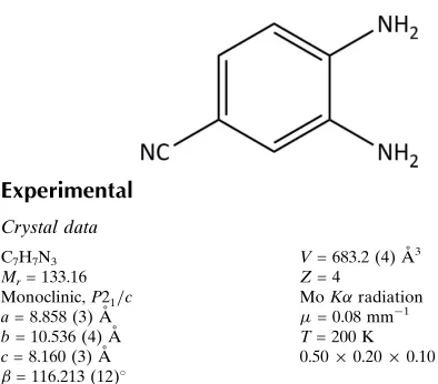
3,4-Diaminobenzonitrile
David K. Geiger* and Dylan E. Parsons
Department of Chemistry, State University of New York-College at Geneseo, 1 College Circle, Geneseo, NY 14454, USA
Correspondence e-mail: [email protected]
Received 18 February 2013; accepted 22 February 2013
Key indicators: single-crystal X-ray study;T= 200 K; mean(C–C) = 0.004 A˚;
Rfactor = 0.052;wRfactor = 0.128; data-to-parameter ratio = 11.1.
The non-H atoms in the structure of the title molecule, C7H7N3, are almost coplanar (r.m.s. deviation = 0.018 A˚ ). The two amine groups each donate two and accept one weak N— H N hydrogen bonds. N—H N hydrogen bonding between the amine and nitrile groups results in chains parallel to [101] in the crystal structure. The chains are cross-linked by N—H N hydrogen bonds between amine groups, giving rise to an infinite three-dimensional network.
Related literature
For the crystal structures of related compounds, see: Czapik & Gdaniec (2010); Sta˚lhandske (1981).
Experimental
Crystal data
C7H7N3 Mr= 133.16 Monoclinic,P21=c a= 8.858 (3) A˚
b= 10.536 (4) A˚
c= 8.160 (3) A˚ = 116.213 (12)
V= 683.2 (4) A˚3 Z= 4
MoKradiation = 0.08 mm1
T= 200 K
0.500.200.10 mm
Data collection
Bruker SMART X2S CCD diffractometer
Absorption correction: multi-scan (SADABS; Bruker, 2010)
Tmin= 0.85,Tmax= 0.99
2149 measured reflections 1188 independent reflections 662 reflections withI> 2(I)
Rint= 0.039
Refinement
R[F2> 2(F2)] = 0.052 wR(F2) = 0.128
S= 0.96 1188 reflections 107 parameters
H atoms treated by a mixture of independent and constrained refinement
max= 0.18 e A˚
3 min=0.19 e A˚
[image:1.610.45.242.449.622.2]3
Table 1
Hydrogen-bond geometry (A˚ ,).
D—H A D—H H A D A D—H A
N1—H1A N2i
0.89 (3) 2.37 (3) 3.251 (4) 168 (3)
N1—H1B N3ii 0.91 (3) 2.31 (3) 3.147 (4) 154 (3)
N2—H2A N1iii
0.90 (3) 2.36 (3) 3.246 (4) 173 (2)
N2—H2B N3ii
0.93 (3) 2.42 (3) 3.303 (4) 159 (3)
Symmetry codes: (i)xþ2;yþ1 2;zþ
3
2; (ii)xþ1;y;zþ1; (iii)x;yþ 1 2;z
1 2.
Data collection:APEX2(Bruker, 2010); cell refinement:SAINT
(Bruker, 2010); data reduction:SAINT; program(s) used to solve structure:SHELXS97(Sheldrick, 2008); program(s) used to refine structure: SHELXL97 (Sheldrick, 2008); molecular graphics:
XSHELL(Bruker, 2010) andMercury(Macraeet al., 2008); software used to prepare material for publication:publCIF(Westrip, 2010).
This work was supported by a Congressionally directed grant from the US Department of Education (grant No. P116Z100020) for the X-ray diffractometer
Supplementary data and figures for this paper are available from the IUCr electronic archives (Reference: QK2053).
References
Bruker (2010).APEX2,SAINT,SADABSandXSHELL. Bruker AXS Inc., Madison, Wisconsin, USA.
Czapik, A. & Gdaniec, M. (2010).Acta Cryst.C66, o198–o201.
Macrae, C. F., Bruno, I. J., Chisholm, J. A., Edgington, P. R., McCabe, P., Pidcock, E., Rodriguez-Monge, L., Taylor, R., van de Streek, J. & Wood, P. A. (2008).J. Appl. Cryst.41, 466–470.
Sheldrick, G. M. (2008).Acta Cryst.A64, 112–122.
Sta˚lhandske, C. (1981).Cryst. Struct. Commun.10, 1081–1086. Westrip, S. P. (2010).J. Appl. Cryst.43, 920–925.
Acta Crystallographica Section E
Structure Reports
Online
supporting information
Acta Cryst. (2013). E69, o452 [doi:10.1107/S1600536813005151]
3,4-Diaminobenzonitrile
David K. Geiger and Dylan E. Parsons
S1. Comment
Single crystals of the title compound were obtained during its purification by recrystallization for use in the synthesis of
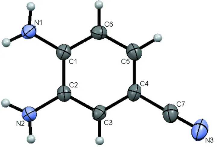
an organometallic complex. Figure 1 shows a view of the title molecule with the atom numbering scheme. The
non-hydrogen atoms are planar with a r. m. s. deviation of 0.018 Å. The maximum deviation is for C1, which is 0.034 (2) Å
out of the plane. The benzene ring is planar with a maximum deviation of 0.008 (2) Å for C4. The amine nitrogens are
decidely pyramidal with H-N-H angles of 113 (3)° and 112 (2)° for N1 and N2, respectively. N1 and N2 are 0.056 (4) and
0.073 (4) Å, respectively, below the benzene plane. All four of the hydrogen atoms of the amine groups are on the
opposite side of the benzene plane from the amine nitrogen atoms. The nitrile group carbon and nitrogen atoms are
0.046 (4) and 0.086 (5) Å, respectively, out of the benzene plane and are on the opposite side as the amine nitrogens. The
nitrile is linear (C4-C7-N = 179.2 (3)°).
There are two known crystalline forms of 1,2-diaminobenzene (Stålhandske, 1981; Czapik & Gdaniec, 2010). In both
forms, one of the N-H bonds of each amine group is coplanar with the benzene ring and an intramolecular N-H···N
interaction is exhibited. Intermolecular hydrogen bonding results in layers that are joined by additional hydrogen-bonding
interactions. The two forms are isostructural in two dimensions, but differ in the stacking of the layers (Czapik &
Gdaniec, 2010). Figure 2 shows the hydrogen-bonding network exhibited by the title compound. In contrast to
1,2-di-aminobenzene, no intramolecular hydrogen-bonding is observed. Parallel chains of molecules with an interplanar spacing
of 3.32Å are formed by hydrogen-bonds involving one hydrogen atom from each of the amines and the nitrile group on
adjacent molecules. The chains run parallel to [101] and are crosslinkeded by hydrogen bonds between amine groups.
S2. Experimental
The compound was obtained commercially (Sigma-Aldrich). Single crystals were grown by slow evaporation of an
ethanolic solution.
S3. Refinement
The H atoms bonded to carbon were refined using a riding model with C—H = 0.95 Å and Uiso = 1.2Ueq(C). The
Figure 1
Perspective view of the title molecule. Displacement ellipsoids of the nonhydrogen atoms are drawn at the 50%
probability level.
Figure 2
A view of the unit cell showing the chains parallel to [101] resulting from H-bonding between amine and nitrile groups
and the cross-linking H-bonds between amine groups. Displacement ellipsoids of the nonhydrogen atoms are drawn at the
[image:3.610.131.482.371.619.2]3,4-Diaminobenzonitrile
Crystal data
C7H7N3 Mr = 133.16 Monoclinic, P21/c Hall symbol: -P 2ybc a = 8.858 (3) Å b = 10.536 (4) Å c = 8.160 (3) Å β = 116.213 (12)° V = 683.2 (4) Å3 Z = 4
F(000) = 280 Dx = 1.294 Mg m−3
Mo Kα radiation, λ = 0.71073 Å Cell parameters from 516 reflections θ = 2.6–21.2°
µ = 0.08 mm−1 T = 200 K Prism, colourless 0.50 × 0.20 × 0.10 mm
Data collection
Bruker SMART X2S CCD diffractometer
Radiation source: fine-focus sealed tube Graphite monochromator
Detector resolution: 8.33 pixels mm-1 ω scans
Absorption correction: multi-scan (SADABS; Bruker, 2010) Tmin = 0.85, Tmax = 0.99
2149 measured reflections 1188 independent reflections 662 reflections with I > 2σ(I) Rint = 0.039
θmax = 25.2°, θmin = 3.2° h = −10→10
k = −12→11 l = −5→9
Refinement
Refinement on F2 Least-squares matrix: full R[F2 > 2σ(F2)] = 0.052 wR(F2) = 0.128 S = 0.96 1188 reflections 107 parameters 0 restraints
Primary atom site location: structure-invariant direct methods
Secondary atom site location: difference Fourier map
Hydrogen site location: inferred from neighbouring sites
H atoms treated by a mixture of independent and constrained refinement
w = 1/[σ2(F
o2) + (0.0526P)2] where P = (Fo2 + 2Fc2)/3 (Δ/σ)max < 0.001
Δρmax = 0.18 e Å−3 Δρmin = −0.19 e Å−3
Special details
Experimental. Refinement of F2 against ALL reflections. The weighted R-factor wR and goodness of fit S are based on F2, conventional R-factors R are based on F, with F set to zero for negative F2. The threshold expression of F2 >
2sigma(F2) is used only for calculating R-factors(gt) etc. and is not relevant to the choice of reflections for refinement. R -factors based on F2 are statistically about twice as large as those based on F, and R– factors based on ALL data will be even larger.
Geometry. All e.s.d.'s (except the e.s.d. in the dihedral angle between two l.s. planes) are estimated using the full covariance matrix. The cell e.s.d.'s are taken into account individually in the estimation of e.s.d.'s in distances, angles and torsion angles; correlations between e.s.d.'s in cell parameters are only used when they are defined by crystal symmetry. An approximate (isotropic) treatment of cell e.s.d.'s is used for estimating e.s.d.'s involving l.s. planes.
Fractional atomic coordinates and isotropic or equivalent isotropic displacement parameters (Å2)
x y z Uiso*/Ueq
N1 1.0289 (3) 0.4587 (3) 0.7729 (3) 0.0317 (7)
H1A 1.025 (3) 0.538 (3) 0.809 (4) 0.053 (10)*
H1B 1.135 (4) 0.433 (3) 0.798 (4) 0.063 (11)*
N3 0.3764 (3) 0.3161 (2) −0.0415 (3) 0.0495 (8)
N2 1.0370 (3) 0.2330 (2) 0.5881 (3) 0.0327 (7)
H2A 1.044 (3) 0.180 (3) 0.507 (4) 0.035 (8)*
H2B 1.142 (4) 0.264 (3) 0.670 (4) 0.068 (11)*
C1 0.8972 (3) 0.4302 (2) 0.6030 (3) 0.0259 (7)
C2 0.9008 (3) 0.3168 (2) 0.5098 (3) 0.0253 (7)
C3 0.7650 (3) 0.2879 (2) 0.3464 (3) 0.0276 (7)
H3 0.7664 0.2122 0.2838 0.033*
C4 0.6251 (3) 0.3686 (3) 0.2712 (3) 0.0298 (7)
C7 0.4877 (3) 0.3387 (3) 0.0985 (4) 0.0366 (8)
C5 0.6212 (3) 0.4783 (3) 0.3641 (3) 0.0323 (8)
H5 0.5262 0.5329 0.3150 0.039*
C6 0.7562 (3) 0.5071 (3) 0.5275 (3) 0.0305 (7)
H6 0.7525 0.5820 0.5905 0.037*
Atomic displacement parameters (Å2)
U11 U22 U33 U12 U13 U23
N1 0.0325 (16) 0.0300 (17) 0.0274 (13) −0.0019 (12) 0.0086 (13) −0.0039 (12)
N3 0.0410 (15) 0.0481 (18) 0.0418 (15) −0.0042 (13) 0.0022 (14) 0.0047 (13)
N2 0.0311 (14) 0.0309 (15) 0.0300 (13) 0.0049 (12) 0.0080 (12) −0.0032 (12)
C1 0.0271 (15) 0.0256 (16) 0.0251 (14) −0.0025 (12) 0.0115 (13) 0.0030 (12)
C2 0.0233 (14) 0.0259 (16) 0.0268 (14) −0.0006 (12) 0.0111 (13) 0.0047 (13)
C3 0.0278 (14) 0.0264 (17) 0.0258 (14) −0.0028 (12) 0.0092 (13) 0.0008 (12)
C4 0.0237 (14) 0.0338 (18) 0.0277 (15) −0.0037 (13) 0.0074 (13) 0.0011 (13)
C7 0.0305 (16) 0.036 (2) 0.0387 (17) 0.0015 (13) 0.0108 (16) 0.0062 (14)
C5 0.0272 (15) 0.0324 (19) 0.0366 (16) 0.0020 (13) 0.0134 (14) 0.0036 (13)
C6 0.0345 (16) 0.0252 (16) 0.0327 (16) 0.0016 (13) 0.0156 (14) −0.0015 (12)
Geometric parameters (Å, º)
N1—C1 1.395 (3) C2—C3 1.379 (3)
N1—H1A 0.89 (3) C3—C4 1.401 (3)
N1—H1B 0.91 (3) C3—H3 0.9500
N3—C7 1.157 (3) C4—C5 1.392 (4)
N2—C2 1.401 (3) C4—C7 1.432 (3)
N2—H2A 0.90 (3) C5—C6 1.375 (3)
N2—H2B 0.93 (3) C5—H5 0.9500
C1—C6 1.384 (3) C6—H6 0.9500
C1—N1—H1B 119 (2) C4—C3—H3 119.5
H1A—N1—H1B 113 (3) C5—C4—C3 119.7 (2)
C2—N2—H2A 112.9 (16) C5—C4—C7 120.2 (2)
C2—N2—H2B 119 (2) C3—C4—C7 120.0 (2)
H2A—N2—H2B 112 (2) N3—C7—C4 179.2 (3)
C6—C1—N1 120.7 (3) C6—C5—C4 119.4 (2)
C6—C1—C2 118.8 (2) C6—C5—H5 120.3
N1—C1—C2 120.4 (2) C4—C5—H5 120.3
C3—C2—N2 120.6 (2) C5—C6—C1 122.0 (3)
C3—C2—C1 119.1 (2) C5—C6—H6 119.0
N2—C2—C1 120.2 (2) C1—C6—H6 119.0
C2—C3—C4 121.0 (2)
Hydrogen-bond geometry (Å, º)
D—H···A D—H H···A D···A D—H···A
N1—H1A···N2i 0.89 (3) 2.37 (3) 3.251 (4) 168 (3)
N1—H1B···N3ii 0.91 (3) 2.31 (3) 3.147 (4) 154 (3)
N2—H2A···N1iii 0.90 (3) 2.36 (3) 3.246 (4) 173 (2)
N2—H2B···N3ii 0.93 (3) 2.42 (3) 3.303 (4) 159 (3)