Received 23 June 2015 Accepted 27 June 2015
Edited by J. Simpson, University of Otago, New Zealand
†
Keywords:crystal structure; dinuclear Cd complex; intramolecular C—O (ring) and N—O (ring) interactions
CCDC reference:1409269
Supporting information:this article has supporting information at journals.iucr.org/e
Structure of a dinuclear cadmium complex with
2,2
000-bipyridine, monodentate nitrate and
3-carboxy-6-methylpyridine-2-carboxylate ligands:
intramolecular carbonyl(lone pair)
p
(ring) and
nitrate(
p
)
p
(ring) interactions
Juan Granifo,a* Sebastia´n Suarezband Ricardo Baggioc*
aDepartamento de Ciencias Quı´micas y Recursos Naturales, Facultad de Ingenierı´a y Ciencias, Universidad de La
Frontera, Casilla 54-D, Temuco, Chile,bDepartamento de Quı´mica Inorga´nica, Analı´tica y Quı´mica Fı´sica,
INQUIMAE-CONICET, Facultad de Ciencias Exactas y Naturales, Universidad de Buenos Aires, Buenos Aires, Argentina, and
cGerencia de Investigacio´n y Aplicaciones, Centro Ato´mico Constituyentes, Comisio´n Nacional de Energı´a Ato´mica,
Buenos Aires, Argentina. *Correspondence e-mail: juan.granifo@ufrontera.cl, baggio@cnea.gov.ar
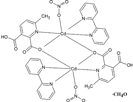
The centrosymmetric dinuclear complex bis( -3-carboxy-6-methylpyridine-2-carboxylato)-3N,O2:O2;3O2:N,O2-bis[(2,20-bipyridine-2
N,N0)(nitrato-O )-cadmium] methanol monosolvate, [Cd2(C8H6NO4)2(NO3)2(C10H8N2)2]CH3OH, was isolated as colourless crystals from the reaction of Cd(NO3)24H2O, 6-methylpyridine-2,3-dicarboxylic acid (mepydcH2) and 2,20-bipyridine in meth-anol. The asymmetric unit consists of a CdII cation bound to a -3
N,O2:O2 -mepydcH anion, an N,N0-bidentate 2,20-bipyridine group and an O -mono-dentate nitrate anion, and is completed with a methanol solvent molecule at half-occupancy. The Cd complex unit is linked to its centrosymmetric image through a bridging mepydcHcarboxylate O atom to complete the dinuclear complex molecule. Despite a significant variation in the coordination angles, indicating a considerable departure from octahedral coordination geometry about the CdII atom, the Cd—O and Cd—N distances in this complex are surprisingly similar. The crystal structure consists of O—H O hydrogen-bonded chains parallel to a, further bound by C—H O contacts alongbto form planar two-dimensional arrays parallel to (001). The juxtaposed planes form interstitial columnar voids that are filled by the methanol solvent molecules. These in turn interact with the complex molecules to further stabilize the structure. A search in the literature showed that complexes with the mepydcH ligand are rare and complexes reported previously with this ligand do not adopt the -3
coordination mode found in the title compound.
1. Chemical context
Pyridinedicarboxylate ligands derived from pyridine-2,3-di-carboxylic acid (pydcH2) have been extensively used in the
construction of a large variety of structural motifs. The two deprotonated formspydcHandpydc2have been shown to adopt a wide range of coordination modes through their carboxylate oxygen and pyridyl nitrogen atoms (Wanget al., 2009). A search in the CSD (Version 5.3; Groom & Allen, 2014) disclosed ca 200 complexes displaying diverse topolo-gies,viz. monomers (Gaoet al., 2010; Drewet al., 1971), dimers (Shankar et al., 2013), oligomers (Yu et al., 2003) as well as one-dimensional (Semerci et al., 2014), two-dimensional (C¸ olaket al., 2011) and three-dimensional (Kanooet al., 2012) polymers. In the vast majority of cases the ligand adopts an N,O-chelating mode, although there are a few exceptions to this where the binding sites attach to different metal atoms (e.g. Wang et al., 2014). By contrast, when complexes
containing similar ligands but with methyl substituents in the 6-position were sought, namely those generated from 6-methylpyridine-2,3-dicarboxylic acid (mepydcH2), only a single structure was found involving the monoanionic mepydcHligand similar to that reported here (Gurunatha & Maji, 2009). This unique structural motif appears in the form of three isostructural, monomeric MII (M = Fe, Co, Ni) complexes [M(bpee)2(mepydcH)2] (bpee= 1,2-bis(4-pyridyl)-ethylene) with octahedral geometry around MII. Both mepydcH fragments act in a simple2N,O2-chelating mode binding to a single nucleus while the two N-bound bpee ligands are trans-monodentate. The formation of these mononuclear complexes is unusual considering the obvious bridging potential of thebpeeligands. Mixed-ligand complexes based on non-methylated 2,3-pyridinedicarboxylate and 4,40 -bipyridine-like ligands usually generate stable polymeric structures with theexo-bidentate ligands adopting a bridging role (Kanooet al., 2012; Wanget al., 2009; Majiet al., 2005).
In an attempt to understand the coordination behaviour of this unusual monoanionicmepydcHligand better, we report the structure of the dinuclear complex [Cd2(2,20-bipyridine)2 -(mepydcH)2(NO3)2]MeOH (I). The uncommon bridging-chelating2-(
3
N,O2:O2) coordination behaviour and the fact that the ligand is only singly deprotonated has no counterpart in complexes of the non-methylated ligands and makes this a genuinely novel structure. The closest relatives with 2,20 -bi-pyridine as the auxiliary ligand are found with di-anionic pydc2 ligands, but these are either mononuclear (Wang & Okabe, 2005) or form coordination polymers (Li et al., 2013; Yin & Liu, 2009; Zhanget al.2013).
2. Structural commentary
The complex consists of a CdII cation to which a singly protonated 3-carboxy-6-methylpyridine-2-carboxylate ion (mepydcH) chelates through the pyridine N and carboxylate O atoms. A chelating 2,20-bipyridine that binds through both nitrogen atoms and a unidentate nitrate anion complete the
coordination sphere; the asymmetric unit also contains a non-coordinating half-occupancy methanol solvate. This five coordinate CdII unit, in turn, binds to its centrosymmetric image through the carboxylate oxygen atom of themepydcH ligand, forming a pair of Cd–O–Cd bridges. As a result, a dimeric unit forms (Fig. 1) with each CdIIatom in a six-coor-dinate N3O3 ligand environment. The Cd—X(X= N or O) distances are reasonable, spanning the range 2.304 (2)– 2.332 (3) A˚ . However, the coordination angles vary widely [X– Cd–X ranges: cis 71.15 (10)–115.79 (9); trans 142.36 (8)– 159.48 (9)]; the result is a rather distorted octahedral geometry around Cd1. Selected geometric parameters are shown in Table 1; the bridging Cd—O distances are the shortest in the coordination sphere, 2.304 (2) and 2.310 (2) A˚ , resulting in a Cd Cd separation of 3.700 (3) A˚ . This value is slightly larger than the mean for similar environments found in the CSD (3.61 A˚ for 885 cases), though well within the sample standard deviation (0.22 A˚ ).
3. Supramolecular features
[image:2.610.43.295.304.498.2] [image:2.610.316.536.497.690.2]The crystal structure, made up of isolated dimers, is sustained by three different types of non-covalent interaction, viz., hydrogen bonds (Table 2), C O and nitrate()
Figure 1
Displacement ellipsoid plot of (I) (with 40% probability ellipsoids), showing the dimeric unit with atom and ring labelling. Interactions within the dimeric unit are also shown, C—H O as dashed lines, C— O (ring) as double-dashed lines. For symmetry codes see Tables 2 and 3; additional symmetry code: (i) 1x, 1y, 1z.
Table 1
Selected bond lengths (A˚ ).
Cd1—O1Bi 2.304 (2) Cd1—N1A 2.323 (3) Cd1—O1B 2.310 (2) Cd1—O1C 2.329 (2) Cd1—N2A 2.310 (3) Cd1—N1B 2.332 (3)
contacts (Table 3). These interactions can be clearly differ-entiated according to the substructure that they support:
a) Contacts #1 (Table 2) and #9, #10 (Table 3) are internal to the dinuclear motif, as shown in Fig. 1. The first one links the bipyridine C10A—H10Agroup with the coordinating nitrate oxygen O1C. Contact #9 is a typical lone pair– interaction with a dihedral angle of 72.19 between the carboxylate and the ring plane, and a C—O Cg2 angle of 126.63. These values are close to those for the ideal geometry (90and 120,
respectively) when a lone pair provided by a carbonyl oxygen points toward the centroid of an aromatic ring (Egli & Sarkhel, 2007). By contrast, in contact #10 the orientation of the nitrate plane is more or less parallel to the ring plane (6.84), suggesting a–interaction with the-orbitals of the nitrate fragment interacting with those of the aromatic ring. A similar argument has already been applied by Fronteraet al. (2011) and Garcı´a-Raso et al. (2009) when nitrate anions interact with pyrimidinium rings. These carbonyl(lone pair) (ring) (#9) and nitrate() (ring) (#10) inter-actions in (I) fulfill a relevant function, serving to strengthen the dimeric unit (Fig. 1).
b) Strong intermolecular O—H O contacts #2 (Table 2) involving the hydrogen atom of the free carboxylic acid group of themepydcHligand with a non-bonded oxygen atom of a nitrate ligand, has the pivotal action of linking the dimers alonga, forming chains parallel to [100] (Fig. 2).
c) C—H O interactions #3, #4 and #5 (Table 2), in turn, serve to link the above chains laterally along b, to form 2D substructures parallel to (001) (Fig. 3a). These planes juxta-Table 2
Hydrogen-bonding interactions (A˚ ,) in (I).
Cg1 is the centroid of the N1A/C1A–C5Aring.
Int.# D—H A D—H H A D A D—H A #1 C10A—H10A O1C 0.93 2.52 3.143 (5) 124 #2 O4B—H4BO O3Cii 0.84 (3) 1.83 (3) 2.670 (5) 176 (6)
#3 C7A—H7A O2Biii 0.93 2.42 3.339 (4) 168 #4 C8A—H8A O2Ciii 0.93 2.59 3.51 (4) 167
#5 C9A—H9A O4Biv 0.93 2.53 3.186 (5) 127 #6 C8B—H8BC O1M 0.960 2.54 3.361 (8) 144 #7 O1M—H1M O3Biii 0.85 (5) 2.42 (9) 2.951 (9) 121 (9)
#8 C1M—H1M3 Cg1 0.96 2.78 3.640 149
[image:3.610.312.566.118.157.2]Symmetry codes: (ii) 1 +x,y,z; (iii)x, 1 +y,z; (iv)1 +x, 1 +y,z.
Figure 2
The [100] chain defined by O—H O interaction #2 (Table 2).
Table 3
X—O interactions (A˚ ,)in (I).
Cg1 is the centroid of the N1A/C1A–C5Aring andCg2 is the centroid of the N2A/C6A–C10Aring.
Int.# X—O Cg O Cg X—O Cg
#9 C6B—O2B Cg2i 3.637 (3) 126.6 (2)
#10 N1C—O2C Cg1i 3.442 (4) 104.2 (2)
[image:3.610.49.303.364.733.2]Symmetry code: (i) 1x, 1y, 1z.
Figure 3
[image:3.610.307.565.456.692.2]pose along [001] with rather weak direct interactions. In the process, however, significant columnar voids parallel to the chains are formed (with a volume 13% of the total cell volume, Fig. 3b) in which the partial occupancy methanol solvate molecules reside. These are not free, but enter instead into a number of weak C—H O, O—H O and C—H inter-actions (#6, #7 and #8 in Table 2) linking them to a framework of complex molecules, further stabilizing the structure.
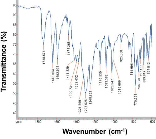
4. ATR (attenuated total reflectance) FT–IR spectroscopy
The IR spectra of mepydcH2, 2.2 0
-bipyridine and (I) were recorded on an Agilent Cary 630 FT–IR spectrometer with Varian Resolutions Pro software, using a Diamond ATR accessory. The FT–IR spectrum of (I) (Fig. 4) was recorded in the 4000–600 cm1 range, and confirms the structural data indicating the presence of the coordinating nitrate and mepydcHanions. Bands due to the unidentate NO3
group were found at 1478 and 1298 cm1 and appear due to the asym(ONO) and sym(ONO) vibrations, with a shoulder at 1010 cm1 due to the (NO) stretching modes of nitrate groups (Nakamoto, 1997). The carboxylic acid group (COOH) of themepydcHligand in complex (I) is identified by a weak band at 3083 cm1,(OH) stretching for a hydrogen-bonded system (Alisir et al., 2013), and a very strong band at 1738 cm1, (C O) stretch. The deprotonated carboxylate (COO) is characterized by the asymmetric and symmetric stretching modes asat 1593 cm1 andsat 1322 cm1. This confirms the unidentate coordination of the carboxylate O atom, with the difference between these frequencies being > 200 cm1( = as s = 271 cm
1
) (Deacon & Phillips, 1980). Finally, around 1400 cm1, a set of three bands appears (1412, 1391 and 1369 cm1) of almost equal intensity due to
the (C C) + (C N) vibrations from the coordinating 2,20-bipyridine ligand (Yanet al., 2011).
5. Synthesis and crystallization
Solid 2,20-bipyridine (0.031 g, 0.20 mmol) was added to a solution prepared by disolving Cd(NO3)4H2O (0.062 g, 0.20 mmol) and mepydcH2 (0.036 g, 0.20 mmol) in MeOH (4.0 mL). The mixture was stirred to dissolve the 2,20 -bi-pyridine and was then allowed to stand undisturbed at room temperature in an uncovered 10 mL beaker. Colourless single crystals of compound (I) suitable for X-ray diffraction were obtained within 8 h. The crystals were separated by filtration, washed with MeOH (2 x 2 mL) and diethyl ether (2 x 3 mL) (yield: 0.045 g, 44%).
6. Refinement
Relevant crystallographic data for (I) as well as pertinent experimental details are provided in Table 4. H atoms bonded to C were found in a difference Fourier map, but were then idealized and refined as riding atoms; C—Harom: 0.93 A˚ ,
Ueq(H) = 1.2Ueq(C); C—Hmethyl: 0.97 A˚ ,Ueq(H) = 1.5Ueq(C). The O—H hydrogen atom was refined with a restrained O—H distance [0.85 (1)A˚ ], and with U(H) = 1.2Ueq(O). The methanol solvate was refined at half occupancy.
Figure 4
[image:4.610.45.294.72.293.2]FT–IR spectrum of (I)
Table 4
Experimental details.
Crystal data
Chemical formula [Cd2(C8H6NO4)2(NO3)2
-(C10H8N2)2]CH4O
Mr 1053.50
Crystal system, space group Triclinic,P1 Temperature (K) 295
a,b,c(A˚ ) 8.4096 (5), 10.9626 (6), 11.5056 (4)
,,(
) 71.241 (4), 86.537 (4), 86.803 (5)
V(A˚3) 1001.79 (9)
Z 1
Radiation type MoK
(mm1) 1.14
Crystal size (mm) 0.360.140.10 Data collection
Diffractometer Oxford Diffraction Gemini CCD S Ultra
Absorption correction Multi-scan (CrysAlis PRO; Oxford Diffraction, 2009)
No. of measured, independent and observed [I> 2 (I)] reflections
21744, 4819, 4155
Rint 0.057
(sin/)max(A˚
1
) 0.684 Refinement
R[F2> 2 (F2)],wR(F2),S 0.036, 0.092, 1.01
No. of reflections 4819 No. of parameters 298 No. of restraints 4
H-atom treatment H atoms treated by a mixture of independent and constrained refinement
max,min(e A˚
3) 1.07,0.74
[image:4.610.313.564.93.393.2]Acknowledgements
The authors acknowledge the Universidad de La Frontera (Proyecto DIUFRO DI15–0027) and ANPCyT (project No. PME 2006–01113) for the purchase of the Oxford Gemini CCD diffractometer.
References
Alisir, S. H., Sariboga, B., Topcu, Y. & Yang, S.-Y. (2013).J. Inorg. Organomet. Polym.23, 1061–1067.
C¸ olak, A. T., Pamuk, G., Yes¸ilel, O. K. & Yu¨ksel, F. (2011).Solid State Sci.13, 2100–2104.
Deacon, G. B. & Phillips, R. J. (1980).Coord. Chem. Rev.33, 227–250. Drew, M. G. B., Matthews, R. W. & Walton, R. A. (1971).J. Chem.
Soc. A, pp. 2959–2962.
Egli, M. & Sarkhel, S. (2007).Acc. Chem. Res.40, 197–205. Frontera, A., Gamez, P., Mascal, M., Mooibroek, T. J. & Reedijk, J.
(2011).Angew. Chem. Int. Ed.50, 9564–9583.
Gao, E.-J., Zhu, M.-C., Huang, Y., Liu, L., Liu, H.-Y., Liu, F.-C., Ma, S. & Shi, C.-Y. (2010).Eur. J. Med. Chem.45, 1034–1041.
Garcı´a-Raso, A., Albertı´, F. M., Fiol, J. J., Tasada, A., Barcelo´-Oliver, M., Molins, E., Estarellas, C., Frontera, A., Quin˜onero, D. & Deya`, P. M. (2009).Cryst. Growth Des.9, 2363–2376.
Groom, C. R. & Allen, F. H. (2014).Angew. Chem. Int. Ed.53, 662– 671.
Gurunatha, K. L. & Maji, T. K. (2009).Inorg. Chim. Acta,362, 1541– 1545.
Kanoo, P., Matsuda, R., Kitaura, R., Kitagawa, S. & Maji, T. K. (2012).
Inorg. Chem.51, 9141–9143.
Li, W., Li, C.-H., Li, H.-F., Xu, J.-S. & Li, L. (2013).Jiegou Huaxue,32, 1567–1571.
Maji, T. K., Mostafa, G., Matsuda, R. & Kitagawa, S. (2005).J. Am. Chem. Soc.127, 17152–17153.
Nakamoto, K. (1997).Infrared and Raman Spectra of Inorganic and Coordination Compounds, 5th ed. New York: Wiley & Sons. Oxford Diffraction (2009).CrysAlis PRO. Oxford Diffraction Ltd,
Abingdon, England.
Semerci, F., Yes¸ilel, O. Z., O¨ lmez, H. & Bu¨yu¨kgu¨ngo¨r, O. (2014).
Inorg. Chim. Acta,409, 407–417.
Shankar, K., Das, B. & Baruah, J. B. (2013).Eur. J. Inorg. Chem.pp. 6147–6155.
Sheldrick, G. M. (2008).Acta Cryst.A64, 112–122. Sheldrick, G. M. (2015).Acta Cryst.C71, 3–8. Spek, A. L. (2009).Acta Cryst.D65, 148–155.
Wang, G.-H., Li, Z.-G., Jia, H.-Q., Hu, N.-H. & Xu, J.-W. (2009).
CrystEngComm,11, 292–297.
Wang, Y. & Okabe, N. (2005).Chem. Pharm. Bull.53, 366–373. Wang, D.-F., Wang, Z.-H., Zhang, T., Huang, R.-B. & Zheng, L.-S.
(2014).J. Mol. Struct.1068, 210–215.
Yan, B., Hodsdon, S. A., Li, Y.-F., Carmichael, C. N., Cao, Y. & Pan, W.-P. (2011).J. Solid State Chem.184, 3179–3184.
Yin, H. & Liu, S.-X. (2009).J. Mol. Struct.918, 165–173.
Yu, Z.-T., Li, G.-H., Jiang, Y.-S., Xu, J.-J. & Chen, J.-S. (2003).Dalton Trans.pp. 4219–4220.
sup-1
Acta Cryst. (2015). E71, 890-894
supporting information
Acta Cryst. (2015). E71, 890-894 [https://doi.org/10.1107/S2056989015012384]
Structure of a dinuclear cadmium complex with 2,2
′
-bipyridine, monodentate
nitrate and 3-carboxy-6-methylpyridine-2-carboxylate ligands: intramolecular
carbonyl(lone pair)
···
π
(ring) and nitrate(
π
)
···
π
(ring) interactions
Juan Granifo, Sebasti
á
n Suarez and Ricardo Baggio
Computing details
Data collection: CrysAlis PRO (Oxford Diffraction, 2009); cell refinement: CrysAlis PRO (Oxford Diffraction, 2009);
data reduction: CrysAlis PRO (Oxford Diffraction, 2009); program(s) used to solve structure: SHELXS97 (Sheldrick,
2008); program(s) used to refine structure: SHELXL2014 (Sheldrick, 2015); molecular graphics: SHELXTL (Sheldrick,
2008); software used to prepare material for publication: SHELXL2014 (Sheldrick, 2015) and PLATON (Spek, 2009).
Bis(µ-3-carboxy-6-methylpyridine-2-carboxylato)-κ3N,O2:O2;κ3O2:N,O2-bis[(2,2′-bipyridine-κ2N,N′ ))(nitrato-κO)cadmium] methanol monosolvate
Crystal data
[Cd2(C8H6NO4)2(NO3)2(C10H8N2)2]·CH4O
Mr = 1053.50 Triclinic, P1
a = 8.4096 (5) Å
b = 10.9626 (6) Å
c = 11.5056 (4) Å
α = 71.241 (4)°
β = 86.537 (4)°
γ = 86.803 (5)°
V = 1001.79 (9) Å3
Z = 1
F(000) = 526
Dx = 1.746 Mg m−3
Mo Kα radiation, λ = 0.71073 Å Cell parameters from 2675 reflections
θ = 3.8–28.8°
µ = 1.14 mm−1
T = 295 K Block, colourless 0.36 × 0.14 × 0.10 mm
Data collection
Oxford Diffraction Gemini CCD S Ultra diffractometer
Radiation source: fine-focus sealed tube
ω scans, thick slices
Absorption correction: multi-scan
(CrysAlis PRO; Oxford Diffraction, 2009)
21744 measured reflections
4819 independent reflections 4155 reflections with I > 2σ(I)
Rint = 0.057
θmax = 29.1°, θmin = 3.6°
h = −11→11
k = −14→14
l = −15→15
Refinement
Refinement on F2
Least-squares matrix: full
R[F2 > 2σ(F2)] = 0.036
wR(F2) = 0.092
S = 1.01 4819 reflections
298 parameters 4 restraints
Hydrogen site location: mixed
sup-2
Acta Cryst. (2015). E71, 890-894 w = 1/[σ2(F
o2) + (0.0394P)2 + 1.5425P]
where P = (Fo2 + 2Fc2)/3
(Δ/σ)max < 0.001
Δρmax = 1.07 e Å−3
Δρmin = −0.74 e Å−3
Special details
Geometry. All e.s.d.'s (except the e.s.d. in the dihedral angle between two l.s. planes) are estimated using the full covariance matrix. The cell e.s.d.'s are taken into account individually in the estimation of e.s.d.'s in distances, angles and torsion angles; correlations between e.s.d.'s in cell parameters are only used when they are defined by crystal symmetry. An approximate (isotropic) treatment of cell e.s.d.'s is used for estimating e.s.d.'s involving l.s. planes.
Fractional atomic coordinates and isotropic or equivalent isotropic displacement parameters (Å2)
x y z Uiso*/Ueq Occ. (<1)
Cd1 0.47993 (3) 0.63548 (2) 0.35527 (2) 0.01961 (9)
N1A 0.6981 (3) 0.7364 (3) 0.3922 (3) 0.0229 (6)
N2A 0.4198 (3) 0.8540 (3) 0.3047 (2) 0.0223 (6)
C1A 0.8378 (4) 0.6740 (4) 0.4272 (3) 0.0279 (7)
H1A 0.8516 0.5882 0.4306 0.033*
C2A 0.9614 (4) 0.7331 (4) 0.4581 (3) 0.0292 (8)
H2A 1.0575 0.6883 0.4821 0.035*
C3A 0.9393 (4) 0.8601 (4) 0.4526 (3) 0.0312 (8)
H3A 1.0204 0.9020 0.4740 0.037*
C4A 0.7968 (4) 0.9252 (4) 0.4152 (3) 0.0286 (7)
H4A 0.7812 1.0114 0.4104 0.034*
C5A 0.6771 (4) 0.8603 (3) 0.3851 (3) 0.0196 (6)
C6A 0.5211 (4) 0.9244 (3) 0.3409 (3) 0.0201 (6)
C7A 0.4801 (4) 1.0501 (3) 0.3369 (3) 0.0263 (7)
H7A 0.5504 1.0976 0.3629 0.032*
C8A 0.3343 (4) 1.1041 (3) 0.2940 (3) 0.0286 (8)
H8A 0.3048 1.1877 0.2920 0.034*
C9A 0.2327 (4) 1.0328 (3) 0.2540 (3) 0.0303 (8)
H9A 0.1348 1.0679 0.2230 0.036*
C10A 0.2801 (4) 0.9084 (3) 0.2614 (3) 0.0280 (7)
H10A 0.2116 0.8599 0.2351 0.034*
O1B 0.6097 (3) 0.4384 (2) 0.44192 (19) 0.0216 (5)
O2B 0.7028 (3) 0.2598 (2) 0.4031 (2) 0.0256 (5)
O3B 0.8147 (3) 0.1816 (3) 0.1644 (3) 0.0390 (6)
O4B 0.9923 (3) 0.2717 (3) 0.2400 (3) 0.0340 (6)
H4BO 1.004 (6) 0.344 (2) 0.248 (4) 0.051 (14)*
N1B 0.5964 (3) 0.5661 (3) 0.1972 (2) 0.0200 (5)
C1B 0.6745 (4) 0.4503 (3) 0.2349 (3) 0.0185 (6)
C2B 0.7605 (4) 0.3994 (3) 0.1523 (3) 0.0226 (7)
C3B 0.7607 (5) 0.4714 (4) 0.0288 (3) 0.0316 (8)
H3B 0.8164 0.4402 −0.0288 0.038*
C4B 0.6796 (5) 0.5880 (4) −0.0090 (3) 0.0343 (9)
H4B 0.6799 0.6361 −0.0919 0.041*
C5B 0.5963 (4) 0.6345 (3) 0.0780 (3) 0.0255 (7)
C6B 0.6608 (4) 0.3736 (3) 0.3714 (3) 0.0185 (6)
sup-3
Acta Cryst. (2015). E71, 890-894
C8B 0.5036 (5) 0.7606 (4) 0.0418 (3) 0.0376 (9)
H8BA 0.5131 0.7999 −0.0457 0.056*
H8BB 0.3934 0.7462 0.0669 0.056*
H8BC 0.5448 0.8166 0.0811 0.056*
N1C 0.1872 (3) 0.5118 (3) 0.2872 (2) 0.0248 (6)
O1C 0.2228 (3) 0.6168 (3) 0.3000 (3) 0.0350 (6)
O2C 0.2871 (3) 0.4227 (3) 0.2993 (3) 0.0341 (6)
O3C 0.0460 (3) 0.5009 (3) 0.2619 (3) 0.0374 (6)
O1M 0.7710 (10) 0.9624 (9) 0.0748 (8) 0.072 (2) 0.5
H1M 0.816 (4) 1.0346 (13) 0.046 (17) 0.18 (9)* 0.5
C1M 0.8993 (11) 0.8676 (10) 0.0932 (8) 0.060 (3) 0.5
H1M1 0.9936 0.9068 0.0490 0.090* 0.5
H1M2 0.8713 0.7994 0.0637 0.090* 0.5
H1M3 0.9189 0.8331 0.1792 0.090* 0.5
Atomic displacement parameters (Å2)
U11 U22 U33 U12 U13 U23
Cd1 0.02114 (13) 0.01596 (13) 0.02201 (13) 0.00148 (9) −0.00289 (9) −0.00650 (9)
N1A 0.0207 (14) 0.0214 (14) 0.0274 (14) −0.0001 (11) −0.0030 (11) −0.0087 (11)
N2A 0.0226 (14) 0.0178 (14) 0.0259 (14) 0.0016 (11) −0.0031 (11) −0.0062 (11)
C1A 0.0255 (17) 0.0252 (18) 0.0331 (18) 0.0027 (14) −0.0037 (14) −0.0095 (15)
C2A 0.0201 (16) 0.038 (2) 0.0293 (18) −0.0008 (15) −0.0038 (14) −0.0105 (16)
C3A 0.0261 (18) 0.040 (2) 0.0324 (19) −0.0088 (16) −0.0041 (15) −0.0166 (16)
C4A 0.0336 (19) 0.0228 (18) 0.0327 (18) −0.0046 (15) −0.0020 (15) −0.0129 (15)
C5A 0.0216 (15) 0.0188 (16) 0.0192 (14) −0.0028 (12) 0.0002 (12) −0.0069 (12)
C6A 0.0231 (16) 0.0182 (16) 0.0181 (14) −0.0022 (13) 0.0004 (12) −0.0048 (12)
C7A 0.0312 (18) 0.0193 (17) 0.0279 (17) −0.0039 (14) 0.0003 (14) −0.0069 (14)
C8A 0.036 (2) 0.0169 (17) 0.0293 (18) 0.0031 (14) 0.0055 (15) −0.0043 (14)
C9A 0.0277 (18) 0.0239 (18) 0.0374 (19) 0.0089 (15) −0.0044 (15) −0.0083 (15)
C10A 0.0248 (17) 0.0228 (18) 0.0363 (19) 0.0030 (14) −0.0067 (14) −0.0089 (15)
O1B 0.0278 (12) 0.0180 (11) 0.0185 (11) 0.0049 (9) 0.0004 (9) −0.0062 (9)
O2B 0.0328 (13) 0.0168 (12) 0.0271 (12) 0.0025 (10) −0.0033 (10) −0.0073 (10)
O3B 0.0471 (17) 0.0307 (15) 0.0461 (16) 0.0020 (12) −0.0037 (13) −0.0221 (13)
O4B 0.0265 (13) 0.0314 (15) 0.0481 (16) 0.0076 (11) −0.0078 (11) −0.0182 (13)
N1B 0.0196 (13) 0.0185 (14) 0.0216 (13) −0.0010 (11) −0.0033 (10) −0.0055 (11)
C1B 0.0173 (14) 0.0193 (16) 0.0208 (15) −0.0016 (12) −0.0047 (12) −0.0082 (12)
C2B 0.0197 (16) 0.0245 (17) 0.0259 (16) −0.0017 (13) −0.0012 (13) −0.0110 (14)
C3B 0.038 (2) 0.035 (2) 0.0234 (17) 0.0039 (16) 0.0038 (15) −0.0126 (15)
C4B 0.047 (2) 0.032 (2) 0.0203 (16) 0.0064 (17) −0.0004 (15) −0.0051 (15)
C5B 0.0295 (18) 0.0226 (17) 0.0234 (16) 0.0013 (14) −0.0052 (13) −0.0055 (13)
C6B 0.0156 (14) 0.0181 (16) 0.0237 (15) −0.0006 (12) −0.0029 (12) −0.0088 (12)
C7B 0.0265 (17) 0.0270 (19) 0.0283 (17) 0.0012 (14) 0.0046 (14) −0.0135 (15)
C8B 0.051 (2) 0.032 (2) 0.0256 (18) 0.0103 (18) −0.0058 (17) −0.0053 (16)
N1C 0.0250 (15) 0.0292 (16) 0.0218 (13) −0.0013 (12) −0.0019 (11) −0.0104 (12)
O1C 0.0264 (13) 0.0315 (15) 0.0561 (17) 0.0001 (11) −0.0092 (12) −0.0254 (13)
O2C 0.0268 (13) 0.0282 (14) 0.0484 (16) 0.0019 (11) −0.0004 (11) −0.0143 (12)
sup-4
Acta Cryst. (2015). E71, 890-894
O1M 0.078 (6) 0.075 (6) 0.077 (5) −0.016 (5) −0.010 (4) −0.039 (5)
C1M 0.052 (6) 0.097 (9) 0.036 (5) −0.018 (6) −0.010 (4) −0.023 (5)
Geometric parameters (Å, º)
Cd1—O1Bi 2.304 (2) O1B—C6B 1.281 (4)
Cd1—O1B 2.310 (2) O2B—C6B 1.220 (4)
Cd1—N2A 2.310 (3) O3B—C7B 1.205 (4)
Cd1—N1A 2.323 (3) O4B—C7B 1.326 (4)
Cd1—O1C 2.329 (2) O4B—H4BO 0.845 (10)
Cd1—N1B 2.332 (3) N1B—C5B 1.336 (4)
N1A—C5A 1.336 (4) N1B—C1B 1.348 (4)
N1A—C1A 1.342 (4) C1B—C2B 1.397 (4)
N2A—C10A 1.336 (4) C1B—C6B 1.525 (4)
N2A—C6A 1.349 (4) C2B—C3B 1.386 (5)
C1A—C2A 1.377 (5) C2B—C7B 1.496 (5)
C1A—H1A 0.9300 C3B—C4B 1.367 (5)
C2A—C3A 1.375 (5) C3B—H3B 0.9300
C2A—H2A 0.9300 C4B—C5B 1.398 (5)
C3A—C4A 1.378 (5) C4B—H4B 0.9300
C3A—H3A 0.9300 C5B—C8B 1.497 (5)
C4A—C5A 1.387 (5) C8B—H8BA 0.9600
C4A—H4A 0.9300 C8B—H8BB 0.9600
C5A—C6A 1.490 (4) C8B—H8BC 0.9600
C6A—C7A 1.389 (5) N1C—O2C 1.230 (4)
C7A—C8A 1.379 (5) N1C—O3C 1.260 (4)
C7A—H7A 0.9300 N1C—O1C 1.261 (4)
C8A—C9A 1.380 (5) O1M—C1M 1.431 (8)
C8A—H8A 0.9300 O1M—H1M 0.855 (10)
C9A—C10A 1.377 (5) C1M—H1M1 0.9600
C9A—H9A 0.9300 C1M—H1M2 0.9600
C10A—H10A 0.9300 C1M—H1M3 0.9600
O1Bi—Cd1—O1B 73.38 (8) N2A—C10A—C9A 123.1 (3)
O1Bi—Cd1—N2A 101.83 (9) N2A—C10A—H10A 118.4
O1B—Cd1—N2A 159.48 (9) C9A—C10A—H10A 118.4
O1Bi—Cd1—N1A 94.90 (9) C6B—O1B—Cd1i 128.9 (2)
O1B—Cd1—N1A 89.17 (9) C6B—O1B—Cd1 118.42 (19)
N2A—Cd1—N1A 71.15 (10) Cd1i—O1B—Cd1 106.62 (8)
O1Bi—Cd1—O1C 88.36 (9) C7B—O4B—H4BO 109 (3)
O1B—Cd1—O1C 112.97 (9) C5B—N1B—C1B 120.4 (3)
N2A—Cd1—O1C 86.44 (9) C5B—N1B—Cd1 124.9 (2)
N1A—Cd1—O1C 157.56 (10) C1B—N1B—Cd1 114.7 (2)
O1Bi—Cd1—N1B 142.36 (8) N1B—C1B—C2B 121.7 (3)
O1B—Cd1—N1B 71.66 (8) N1B—C1B—C6B 117.5 (3)
N2A—Cd1—N1B 115.79 (9) C2B—C1B—C6B 120.7 (3)
N1A—Cd1—N1B 98.09 (9) C3B—C2B—C1B 117.6 (3)
sup-5
Acta Cryst. (2015). E71, 890-894
C5A—N1A—C1A 119.8 (3) C1B—C2B—C7B 124.1 (3)
C5A—N1A—Cd1 117.1 (2) C4B—C3B—C2B 120.4 (3)
C1A—N1A—Cd1 123.0 (2) C4B—C3B—H3B 119.8
C10A—N2A—C6A 118.9 (3) C2B—C3B—H3B 119.8
C10A—N2A—Cd1 123.4 (2) C3B—C4B—C5B 119.6 (3)
C6A—N2A—Cd1 116.7 (2) C3B—C4B—H4B 120.2
N1A—C1A—C2A 122.1 (3) C5B—C4B—H4B 120.2
N1A—C1A—H1A 119.0 N1B—C5B—C4B 120.4 (3)
C2A—C1A—H1A 119.0 N1B—C5B—C8B 117.7 (3)
C3A—C2A—C1A 118.3 (3) C4B—C5B—C8B 121.9 (3)
C3A—C2A—H2A 120.9 O2B—C6B—O1B 126.7 (3)
C1A—C2A—H2A 120.9 O2B—C6B—C1B 118.0 (3)
C2A—C3A—C4A 119.9 (3) O1B—C6B—C1B 115.3 (3)
C2A—C3A—H3A 120.0 O3B—C7B—O4B 120.6 (3)
C4A—C3A—H3A 120.0 O3B—C7B—C2B 122.0 (3)
C3A—C4A—C5A 118.9 (3) O4B—C7B—C2B 117.1 (3)
C3A—C4A—H4A 120.5 C5B—C8B—H8BA 109.5
C5A—C4A—H4A 120.5 C5B—C8B—H8BB 109.5
N1A—C5A—C4A 121.0 (3) H8BA—C8B—H8BB 109.5
N1A—C5A—C6A 116.7 (3) C5B—C8B—H8BC 109.5
C4A—C5A—C6A 122.3 (3) H8BA—C8B—H8BC 109.5
N2A—C6A—C7A 121.0 (3) H8BB—C8B—H8BC 109.5
N2A—C6A—C5A 116.6 (3) O2C—N1C—O3C 121.1 (3)
C7A—C6A—C5A 122.4 (3) O2C—N1C—O1C 121.0 (3)
C8A—C7A—C6A 119.4 (3) O3C—N1C—O1C 117.9 (3)
C8A—C7A—H7A 120.3 N1C—O1C—Cd1 118.4 (2)
C6A—C7A—H7A 120.3 C1M—O1M—H1M 104.7 (13)
C7A—C8A—C9A 119.4 (3) O1M—C1M—H1M1 109.5
C7A—C8A—H8A 120.3 O1M—C1M—H1M2 109.5
C9A—C8A—H8A 120.3 H1M1—C1M—H1M2 109.5
C10A—C9A—C8A 118.2 (3) O1M—C1M—H1M3 109.5
C10A—C9A—H9A 120.9 H1M1—C1M—H1M3 109.5
C8A—C9A—H9A 120.9 H1M2—C1M—H1M3 109.5
![Figure 3Two projections along [100], presenting (within square brakets) views ofthe two-dimensional substructures parallel to (001), formed by the [100]columns linked along b](https://thumb-us.123doks.com/thumbv2/123dok_us/438656.541664/3.610.49.303.364.733/figure-projections-presenting-brakets-dimensional-substructures-parallel-columns.webp)