Lactic acid bacteria (LAB) are common micro-organisms in foods and also constitute the natural intestinal microbiota of humans and most animals (Rojo-Bezares et al., 2006). LAB produce a wide range of antimicrobial metabolites which include organic acids, diacetyl, hydrogen peroxide, antibi-otics and bacteriocins. Bacteriocins are ribosomally synthesized, extracellularly released bioactive pep-tides which have a bactericidal or bacteriostatic effect on other (usually closely related) species. They are generally low molecular weight proteins that gain entry into target cells by binding to cell surface receptors. Their bactericidal mechanisms vary and may include pore formation of cell wall or cytoplasmic membrane, degradation of cellu-lar DNA, disruption through specific cleavage of 16S rRNA, and inhibition of peptidoglycan
syn-thesis (Todorov et al., 2011). At first, bacteriocins received much attention mainly because of their potential application as food additives in the con-trol of food spoilage and pathogenic food-borne microorganisms (Cleveland et al., 2001; Bromberg et al., 2005). Later, the effects of bacteriocin or bacteriocin-producing probiotics on animal per-formance have been studied (Stern et al., 2006).
Probiotics have received increasing attention as an alternative to in-feed antibiotics and for the purpose of improving productivity in the poul-try induspoul-try (Shin et al., 2008). LAB, particularly
Lactobacillus spp., is one of the probiotic groups which make up a large group of microorganisms in the GIT of all humans and animals. They can be tolerant to acid and bile, adhere to the intestinal epithelium of the hosts; they show an antagonistic
Bacteriocin-producing lactic acid bacteria as a probiotic
potential from Thai indigenous chickens
H. Musikasang
1, N. Sohsomboon
1, A. Tani
2, S. Maneerat
11Department of Industrial Biotechnology, Faculty of Agro-Industry, Prince of Songkla University,
Thailand
2Institute of Plant Science and Resources, Okayama University, Japan
ABSTRACT: Bacteriocin-producing lactic acid bacteria (LAB) were isolated and screened from the gastrointesti-nal tract (GIT) of Thai indigenous chickens. The bacteriocinogenic activities and the primary probiotic properties were determined. The bacteriocins produced by 14 strains of selected LAB displayed inhibitory activity against indicator strains after the supernatants were neutralized with NaOH in the following species: Lactobacillus sakei
subsp. sakei JCM1157, Enterococcus faecalis VanB,Bacillus sp., and Listeria monocytogenes. The antagonistic acti-vity of selected LAB was inactivated or decreased after being treated with proteolytic enzymes (α-chymotrypsin and trypsin). CR5-1 strain exhibited the highest level of activity (5120 AU/ml) in the stationary phase against
L. sakei subsp. sakei JCM1157 in MRS broth at 37°C. The nine isolates of selected LAB were investigated for primary probiotic properties. The survival of the nine isolates was found to decrease approximately by 3 log CFU/ml after passing through the gastrointestinal conditions. All isolates exhibited protein digestion on agar plates but no isolates showed the ability to digest starch and lipid. Most of them showed high susceptibilities to some antibiotics (penicillin G, tetracycline and erythromycin). Thirteen LAB strains producing bacteriocin with strongly inhibitory activity were identified as Lactobacillus salivarius and only one strain was identified by 16S rDNA sequence analysis as Lactobacillus agilis.
Keywords: poultry; gastrointestinal tract; probiotic; antibiotic susceptibility
activity against pathogenic bacteria and keep their viability during processing and storage (Musikasang et al., 2009).
LAB are well known for their production of bac-teriocins. They have the potential to be used in the food and feed industry to substitute for chemical preservation (Gao et al., 2010). Bacteria intended for probiotic use should be screened for antibiotic resistance to avoid any potential carriage of un-desirable antibiotic resistance into the intestinal environment (Huyghebaert et al., 2011). To avoid a source of antibiotic resistant LAB, Thai indig-enous chickens not fed any commercial feed, were used as a source of bacteriocin-producing LAB and probiotic strains.
In general, for the production of Thai indigenous chickens backyard or village production systems by homesteads (extensive system) are preferred since the production costs are very low. The chickens are allowed to scavenge on their own for resources
around the homestead during the day and this is sup-plemented with concentrated feeds in the evening when they come back to roost and shelter for night (Wattanachant, 2008). In this rearing system, the chickens are not fed any commercial feed and antibi-otics. Native chickens are well-suited to small-farm conditions; they have a good resistance to diseases and they tolerate a large variety of locally available feeds (Haitook et al., 2003). Therefore, there is an intestine compost of a large variety of bacteria spe-cies in Thai indigenous chickens due to the kind of feeds and rearing positions. For this reason, Thai indigenous chickens were chosen for LAB screening in this study to isolate many species of LAB and to avoid a source of antibiotic-resistant LAB.
[image:2.595.67.531.383.672.2]The purpose of this study was to isolate and screen bacteriocin-producing LAB from the GIT of Thai indigenous chickens. The bacteriocinogenic activities and the primary probiotic properties were also studied.
Table 1. Microorganisms, strains and growth conditions
Microorganisms Gram stain Growth medium Growth conditions (h/°C)
Lactobacillus sakei subsp. sakei JCM1157a + MRS 24/37
Pediococcus pentosaceus DMST 18752b + MRS 24/37
Enterococcus faecalis VanBc + MRS 24/37
Listeria monocytogenesd + TSB 24/37
Listeria monocytogenes DMST 17303b + TSB 24/37
Bacillus sp.d + TSB 24/30
Bacilluscereus DMST 5040b + TSB 24/30
Staphylococcus aureusd + TSB 24/37
Staphylococcus aureus DMST 8840b + TSB 24/37
Salmonella Stanley 42c – TSB 24/37
Salmonella Typhimurium DMST 16809b – TSB 24/37
Salmonella Enteritidis DMST 15676b – TSB 24/37
Escherichia colid – TSB 24/37
Escherichia coli DMST 4212b – TSB 24/37
Vibrio parahaemolyticusd – TSB 24/37
Pasteurella multocida DMST 16357b – TSB 24/37
aobtained from the Japan Collection of Microorganisms, Japan
bobtained from Culture Collection for Medical Microorganism, Department of Medical Sciences, Thailand
cobtained from The WHO Global Salm-Surv Regional Centre of Excellence, South-East Asia and Western Pacific, Faculty
of Veterinary Science, Chulalongkorn University, Thailand
dobtained from Songklanagarind Hospital, Prince of Songkla University, Thailand
MATERIAL AND METHODS
Bacterial strains and culture conditions Reference strains of pathogenic and spoilage mi-croorganisms were chosen to test antimicrobial activity of the strains presumed to produce bac-teriocin or bacbac-teriocin-like substances (Table 1). All strains were stored at –20°C in their respective media with added glycerol (25%).
Isolation of LAB from chicken intestinal tract
The gastrointestinal tracts (crop, small intestine, large intestine and cecum) of six Thai indigenous chickens were used as LAB sources. 25 g of each section of the gut was homogenized in 225 ml phosphate-buffered saline (PBS: 50mM KH2HPO4, 50mM K2HPO4×3H2O, 0.85% NaCl, pH 7.0) for 5 min using the Stomacher® 400 Circulator (Seward Ltd., Worthing, UK). Appropriate serial dilutions were plated onto MRS agar (HiMedia Laboratories, Pvt. Ltd., India) supplemented with 0.02% bromo-cresol purple (Ajax Finechem, Australia) and incu-bated anaerobically at 37°C for 24 h. Colonies which exhibited a clear halo were randomly selected from the highest dilutions of each MRS agar plate. Bacterial colonies were then purified 2–3 times by restreaking on MRS agar. The pure cultures were characterized using Gram stain, cell morphology and catalase reaction tests. Gram-positive and catalase-negative isolates were stored at –20°C in MRS broth supplemented with 25% (v/v) glycerol. For routine analysis, the strains were subcultured twice in MRS broth at 37°C for 24 h (Musikasang et al., 2009).
Antimicrobial assay and estimation of bacteriocinogenic activity
Primary screening. Antimicrobial activity was de-termined by the agar-spot test method using L. sakei
subsp. sakei JCM1157 as the indicator strain. Briefly, each isolate of LAB was spotted on MRS agar and incubated under anaerobic conditions at 37°C for 24 h. Then, the plate was overlaid with 10 ml of soft MRS agar (1% agar, Fluka, USA) seeded with 250 µl overnight cultureof L. sakei subsp. sakei JCM1157 strain. This was incubated under the same
condi-tions at 37°C for 24 h. Inhibition was detected as a clear zone around the tested organism.
Secondary screening. The selected strains from primary screening were grown in MRS broth at 37°C for 24 h. Cell-free supernatants were collected by cen-trifugation (8500 rpm, 10 min, 4°C) of overnight MRS broth cultures. The pH value of the culture superna-tants was adjusted to 6.5 with NaOH (6M) to elimi-nate the effect of organic acids. After adding catalase (300 U/ml) (Fluka, USA), the cell-free supernatants (pH 6.5) were incubated at 37°C for 1 h to eliminate the effect of hydrogen peroxide. Then they were heat-ed at 90°C for 10 min to stop the enzyme reaction. The same supernatants without catalase were used as control and all culture supernatants were filter-sterilized to eliminate the possible presence of viable cells (Gurban Oglu Gulahmadov et al., 2006).
The inhibition activity was examined by means of the diameters of inhibition zones using the agar well diffusion method (Batdorj et al., 2006). Briefly, 50 μl of cell-free supernatants were placed into wells (6.0 mm in diameter) on the appropriate media agar plates (Table 1) seeded with indicator strains (final concentration 106 CFU/ml). After
in-cubation for 24 h, the diameters of inhibitory zones were measured.
The proteinaceous character of the bacteriocin substance was confirmed by treating neutralized cell free supernatant (NCFS) with a-chymotrypsin
(Fluka, USA) and trypsin (Fluka, Switzerland) at a final concentration of 1 mg/ml at 37°C for 3 h. After stopping the enzyme activity by heat, the agar well diffusion method was used to estimate the bacte-riocinogenic activity.
Production of bacteriocin from selected LAB
An overnight culture of selected bacteriocin-pro-ducing LAB was inoculated (OD600 = 0.5, 1%) into 1 l of MRS broth and cultivated with moderate stirring (50 rev/min) at 37°C for 24 h. Samples were taken every 3 hours for 24 h and changes in optical density (600 nm), pH value, viable count and bacteriocin tivity were measured. To quantify the bacteriocin ac-tivity, the agar well diffusion method was used. NCFS was serially diluted twofold in sterile deionized water and 50 µl of each dilution was added into the wells. The titre was defined as 2n, where n is the
of antibacterial activity per ml was defined as (2n ×
1000 µl)/50 µl (Cheikhyoussef et al., 2009). L. sakei
subsp. sakei JCM1157 was used as a bacteriocin sensitive indicator strain to determine bacteriocin activity levels.
Resistance to simulated intestinal juice after sequential incubation in simulated gastric juice of isolated LAB
A simulated gastric juice was prepared by suspend-ing 3 mg/ml pepsin (Fluka, Japan) in sterile saline (0.85% NaCl, w/v) and the pH was adjusted to 3.0 with 1.0M HCl. Twenty-four hour 1.0 ml cultures of the strains (Table 2) were subjected to centrifu-gation in Eppendorf Centrifuge 5415R (Eppendorf AG, Hamburg, Germany) at 8500 rpm for 10 min. They were then washed twice with sterile saline before being re-suspended in simulated gastric juice. Resistance was assessed in terms of the vi-able colony count and enumerated after incubation at 42°C for 2 h. After 120 min of gastric digestion, cells were harvested and suspended in simulated in-testinal fluid which contained 1 mg/ml pancreatin (Sigma-Aldrich Chemie GmbH, Germany) and 0.5% freeze-dried chicken bile at pH 8.0. The suspension was incubated at 42°C for 6 h and the viable count was determined by drop plating (modified from Musikasang et al., 2009).
Starch, protein and lipid digesting capabilities
Three modified MRS agars were prepared as follows: MRS agar containing 2% skimmed milk (HiMedia Laboratories Pvt. Ltd., India), MRS agar without dextrose containing 1% palm oil (Fluka, USA), and MRS agar without dextrose contain-ing 2% soluble starch (Labchem, Ajax Finechem, Australia). These media were used for detecting the protein, lipid and starch digesting capabilities of selected LAB strains, respectively. The overnight cultures of LAB (10 µl) were dropped on the modi-fied MRS agar and incubated at 42°C for 24 h. Then the diameters of the halo zone on the agar plate were measured. The digesting capability of the tested strains was classified as positive when the diameters of the clear zone were more than 1 mm. Each assay was performed in triplicate (modified from Musikasang et al., 2009).
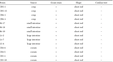
Antibiotic susceptibility
Nine isolates of selected LAB (S6-10, CE5-3, CE5-10, CR5-15, CR5-1, CR6-1, CR6-3, L3-7, and L5-4) (Table 2) were tested for resistance to four antimicrobial agents. These were: penicillin G (Fluka, Austria) (β-lactam group, inhibitors of cell wall synthesis), tetracycline (Fluka, China)
(inhibi-Table 2. Source and some characteristics of selected bacteriocin-producing lactic acid bacteria
Strain Source Gram-stain Shape Catalase test
CR5-1 crop + short rod –
CR5-15 crop + short rod –
CR6-1 crop + short rod –
CR6-3 crop + short rod –
S4-17 small intestine + short rod –
S4-18 small intestine + short rod –
S6-10 small intestine + short rod –
L3-2 large intestine + short rod –
L3-7 large intestine + short rod –
L5-4 large intestine + short rod –
CE4-4 cecum + short rod –
CE4-5 cecum + short rod –
CE5-3 cecum + short rod –
[image:4.595.65.533.499.756.2]tors of protein synthesis), chloramphenicol (Sigma-Aldrich Chemie GmbH, Germany) (inhibitors of protein or mRNA synthesis, broad spectrum) and erythromycin (Fluka, Italy) (inhibitors of protein synthesis – Gram positive spectrum). Each of the antibiotic powders was weighed, dissolved, diluted in appropriate diluents and filter sterilized prior to the addition to MRS medium. Serial dilutions of antibiotics ranging from 256 to 0.25 µg/ml for penicillin G, tetracycline, and chloramphenicol and 320 to 2.5 µg/ml for erythromycin were prepared.
Minimal inhibitory concentration (MIC) values for all bacterial isolates were determined by the broth microdilution procedure. The MRS broth containing antibiotics was added to micro-well plate and serial twofold dilutions were prepared. The final concen-tration of inoculum was adjusted to 105 CFU/ml. The
inoculum was derived from a broth culture which was incubated at 42°C for 24 h and 20 μl of the inocu-lum was used to inoculate each well. The trays were covered and incubated at 42°C for 24 h. Then mini-mal bactericidal concentration (MBC) values were measured by streaking on MRS agar and incubation at 42°C for 24 h. The experiments were replicated at least three times to verify the reproducibility of the methodology when using the above-mentioned conditions (D’Aimmo et al., 2007).
Strain identification
To identify bacteriocin-producing strains, the selected isolates were finally identified along the full length of 16S rDNA sequence analysis. Some isolates were identified by 16S rDNA V3 regions of bacterial sequence. PCR amplifications were performed in a total reaction volume of 50 µl containing 0.2µM of each of the primers, 25 µl of EmeralAmp Mastermix (Takara, Japan), Dnase and Rnase free water 14 µl (Takara, Japan), and 1 µl of template DNA. All amplification reactions were performed in a PCR Sprint temperature cycling system (Techne, UK) using the following amplification conditions: the first cycle was preceded by incubation at 95°C for 5 min, followed by 30 cycles at 95°C for 1 min, 50°C for 30 s and at 72°C for 1.5 min. The reactions were terminated with 5 min of incubation at 72°C, and then chilled to 4°C. 10 µl of the PCR products were subjected to electrophoresis in a 1% agarose gel (ISC Bio Express®, USA) and were subsequently visual-ized by UV illumination after staining with SYBR Safe (InvitrogenTM, USA). The oligonucleotide
primers (AITbiotech Pte Ltd., Singapore) used in this study are as follows: Forward primer 8F: 5’ AGA GTT TGA TCC TGG CTC AG 3’, Reverse primer 1492R: 5’ GGC TAC CTT GTT ACG ACT T 3’, yielding a product of approximately 1500 base pairs (bp). Forward primer 341F: 5’ CCT ACG GGA GGC AGC AG 3’ and Reverseprimer 518R: 5’ ATT ACC GCG GCT GCT GG 3’ giving a prod-uct of 192 bp. The PCR fragments of the 16S rDNA were purified and then subsequently sequenced (1st BASE Pte Ltd., Singapore). Sequences were
ana-lysed with the sequencing software Chromaspro from Technelysium that opens chromatogram files produced by the Applied Biosystems equipment. The sequences obtained were sent to the National Centre for Biotechnology Information (NCBI) to be analysed for the nucleotide-nucleotide BLAST database in FASTA format (http://blast.ncbi.nlm.nih.gov/Blast. cgi) (modified from González etal., 2007).
RESULTS AND DISCUSSION
Isolation of LAB from chicken intestinal tract
In total, 307 LAB strains were isolated from the GIT of six Thai indigenous chickens. All of them were isolated from four gut parts: 66 isolates were isolated from crop, 72 from small intestine, 71 from large intestine, and 98 isolates were isolated from caecum. They were Gram-positive, catalase negative, mostly short rod-shaped and facultatively anaerobic bacteria. The results were in accordance with previ-ous studies establishing that the major LAB in native chicken or poultry intestinal tract were rod shaped (Lima et al., 2007; Musikasang et al., 2009).
Antimicrobial activity
of bacteriocin-producing LAB
Only 14 isolates (L3-2, L3-7, S4-17, S4-18, CE4-4, CE4-5, CR5-1, CR5-15, L5-4, CE5-3, CE5-10, CR6-1, CR6-3, and S6-10) (Table 2) out of the 307 isolates of selected LAB could inhibit L. sakei subsp. sakei
JCM1157. This was used as an indicator strain for the primary bacteriocin screening step.
For the secondary screening, the neutralized su-pernatants from 14 strains of selected LAB exhibited inhibitory activity against indicator strains including
18752, Ent. faecalis VanB,Lis. monocytogenes, Lis. monocytogenes DMST 17303, Bacillus sp. and B. ce-reus DMST 5040 (Table 3). However, the NCFS of selected LAB could not inhibit S. aureus, S. aureus
DMST 8840, Sal. Stanley 42, Sal. Typhimurium DMST 16809, Sal. Enteritidis DMST 15676, E. coli,
E. coli DMST 4212, V. parahaemolyticus,and P. mul-tocida DMST 16357.
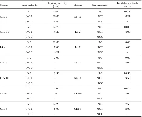
The proteinaceous character of the bacteriocin substance was confirmed. After treating NCFS with α-chymotrypsin, the inhibitory activity toward
L. sakei subsp. sakei JCM1157 of 12 selected LAB strains disappeared (Table 4). The destruction of the antimicrobial activity by proteases suggested that the compound in the supernatant could be a peptide or bacteriocin (Bromberg et al., 2005). In addition,
the inhibitory activities of NCFS after being treated with trypsin were decreased 0.5 fold (Table 3).
The LAB isolated from chickens could produce many kinds of bacteriocins such as acidocin A which is secreted by Lactobacillus acidophilus
[image:6.595.66.531.112.515.2]TK9201 (Kanatani et al., 1995). Bacteriocin OR7 is produced by L. salivarius (NRRL B-30514) (Cole et al., 2006; Stern et al., 2006). Ent. faecium EF55 isolated from the gastrointestinal tract of chick-ens produced enterocins A and P (Strompfová and Lauková, 2007). In addition, bacteriocins produced from LAB strains were also reported to inhibit the growth of pathogenic bacteria in many studies (Ogunbanwo et al., 2004; De Kwaadsteniet et al., 2005; Cole et al., 2006; Pilasombut et al., 2006; Stern et al., 2006; Lima et al., 2007; Gao et al., 2010).
Table 3. Inhibitory activity of selected lactic acid bacteria strains against bacterial indicator strains after eliminat-ing effect of organic acid and H2O2 by using agar well diffusion method
Indicator strains Antibacterial activity
L3-2 L3-7 S4-17 S4-18 CE4-4 CE4-5 CR5-1 CR5-15 L5-4 CE5-3 CE5-10 CR6-1 CR6-3 S6-10
Lactobacillus sakei subsp.
sakei JCM1157 + + + + + + + + + + + + + +
Pediococcus pentosaceus
DMST 18752 + + + + + + + + + – – + + +
Enterococcus
faecalis VanB + + + + + + + + + – – + + +
Listeria monocytogenes + + + + + + + + + – + – + +
Listeria monocytogenes
DMST 17303 + + + + + + + + + – – + + +
Bacillus sp. – – – – – – + + + – – – + +
Bacilluscereus
DMST 5040 – – – – – – + + – – – – – +
Staphylococcus aureus – – – – – – – – – – – – – –
Staphylococcus aureus
DMST 8840 – – – – – – – – – – – – – –
Salmonella Stanley 42 – – – – – – – – – – – – – –
Salmonella Typhimurium
DMST 16809 – – – – – – – – – – – – – –
Salmonella Enteritidis
DMST 15676 – – – – – – – – – – – – – –
Escherichia coli – – – – – – – – – – – – – –
Escherichia coli
DMST 4212 – – – – – – – – – – – – – –
Vibrio parahaemolyticus – – – – – – – – – – – – – –
Pasteurella multocida
DMST 16357 – – – – – – – – – – – – – –
In our study, the bacteriocins produced from selected LAB showed strongly inhibitory activity toward many Gram-positive bacteria. However, all of the Gram-negative indicators (Table 1) used were not inhibited by any of the bacteriocins produced (Table 3). Bacteriocins produced by LAB have at-tracted great interest in terms of safety, but most of them only inhibit some Gram-positive pathogenic bacteria. However, some bacteriocins are effective against Gram-negative spoilage and pathogenic bac-teria such as sakacin C2 produced by Lactobacillus sake C2 strongly inhibited E. coli ATCC 25922. This activity against many Gram-negative bacteria was not frequently seen in bacteriocins from LAB (Gao et al., 2010). In addition, the combination of
bacte-riocin with some natural antimicrobial compounds is able to enhance the inhibition of Gram-negative pathogen such as interactions of nisin, lysozyme, and EDTA had an effect on the growth of Sal. ty-phimurium (Gill and Holley, 2000).
[image:7.595.64.533.126.513.2]Gram-negative spoilage organisms and patho-gens are especially problematic due to their in-herent resistance to some antimicrobial agents such as bacteriocins. This inability is due to the protective outer membrane which covers the cy-toplasmic membrane and peptidoglycan layer of the cells. This asymmetrical membrane contains glycerophospholipids and lipopolysaccharide (LPS) molecules in which the anchoring of divalent cati-ons is involved. Treatment with metal-chelating
Table 4. Inhibitory activity of selected lactic acid bacteria strains against Lactobacillus sakei subsp. sakei JCM1157 after eliminating effect of organic acid, H2O2 and effect of protease enzyme on antagonistic activity by using agar well diffusion method
Strains Supernatants Inhibitory activity (mm) Strains Supernatants Inhibitory activity (mm)
CR5-1
NC 14.50
S6-10
NC 10.75
NCT 10.50 NCT 5.25
NCC 5.50 NCC –
CR5-15
NC 12.75
L3-2
NC 10.00
NCT 4.25 NCT 4.00
NCC – NCC –
L5-4
NC 11.50
L3-7
NC 9.00
NCT 7.00 NCT 3.00
NCC 4.25 NCC –
CE5-3
NC 7.00
S4-17
NC 9.00
NCT – NCT 4.00
NCC – NCC –
CE5-10
NC 1.50
S4-18
NC 10.50
NCT – NCT 3.50
NCC – NCC –
CR6-1
NC 3.00
CE4-4
NC 10.50
NCT – NCT 3.00
NCC – NCC –
CR6-3
NC 12.25
CE4-5
NC 7.50
NCT 6.00 NCT 3.00
NCC – NCC –
Figure 1. Bacteriocin production during the growth of CR5-1 (a), CE4-4 (b), CR6-3 (c), and CR5-15 (d) strains in MRS broth at 37°C. The viable counts (bar), optical density, absorbance at 600 nm (®) and pH (¡) of the culture were measured at the time intervals as indicated. The antibacterial activity () was also assayed and expressed as AU/ml
7.0 6.5 6.0 5.5 5.0 4.5 4.0 3.5 3.0 7.0 6.5 6.0 5.5 5.0 4.5 4.0 3.5 3.0 10 9 8 7 6 5 4 3 2 1 0 700 600 500 400 300 200 100 0 3000 2500 2000 1500 1000 500 0 6000 5000 4000 3000 2000 1000 0 3000 2500 2000 1500 1000 500 0 1.0 0.9 0.8 0.7 0.6 0.5 0.4 0.3 0.2 0.1 0.0 1.0 0.9 0.8 0.7 0.6 0.5 0.4 0.3 0.2 0.1 0.0 1.0 0.9 0.8 0.7 0.6 0.5 0.4 0.3 0.2 0.1 0.0 1.0 0.9 0.8 0.7 0.6 0.5 0.4 0.3 0.2 0.1 0.0 10 9 8 7 6 5 4 3 2 1 0 10 9 8 7 6 5 4 3 2 1 0 10 9 8 7 6 5 4 3 2 1 0 7.0 6.5 6.0 5.5 5.0 4.5 4.0 3.5 3.0 7.0 6.5 6.0 5.5 5.0 4.5 4.0 3.5 3.0 pH pH pH pH
0 3 6 9 12 15 18 21 24
0 3 6 9 12 15 18 21 24
0 3 6 9 12 15 18 21 24
0 3 6 9 12 15 18 21 24
OD
. (600 mm)
OD
. (600 mm)
OD
. (600 mm)
OD
. (600 mm)
agents such as EDTA generally results in removal by chelation of divalent cations with a consequent dis-ruption of the outer membrane. Lactic acid and its salts are also potent outer membrane disintegrating agents as evidenced by their ability to cause LPS re-lease. These permeabilizers increase susceptibility to hydrophobic substances such as bacteriocins by enabling them to penetrate the cell wall (Alakomi et al., 2003; Castellano et al., 2008).
Production kinetics of bacteriocin from selected LAB
Out of the 14 selected LAB tested, four strains (CR5-1, CE4-4, CR6-3, and CR5-15) showed rela-tively high levels of antimicrobial activity against
L. sakei subsp. sakei JCM1157 (Figure 1).
Bacteriocin produced from strain CR5-1 gave the highest level of activity against L. sakei subsp.
sakei JCM1157 when cultured in MRS broth at 37°C. This strain started to produce bacteriocin of about 40 AU/ml at 6 h during the middle logarithmic growth phase at a cell count of 8.19 log CFU/ml. Bacteriocin concentration reached the maximum (5120 AU/ml) after 12 h of incubation at a cell count of 9.26 log CFU/ml. This was at the beginning of the stationary phase of growth, the level of inhibi-tion thereafter remained constant (Figure 1a). Both the CE4-4 and CR6-3 strains showed inhibitory activity against L. sakei subsp. sakei JCM1157 at 2560 AU/ml after 9 and 12 h of incubation, respec-tively. However, the production started to decrease after 15 and 12 h, respectively, which may be due to proteolytic degradation or changes in the
envi-ronmental conditions (Huang et al., 2009). During bacterial growth, the pH of the medium dropped from pH 6.57 (0 h) to pH 3.85 (24 h) (Figure 1b, c).
Resistance to simulated intestinal juice after sequential incubation in a simulated gastric juice of isolated LAB
Tolerance to low pH, bile, and pancreatic fluid in vitro is expected to predict the survival of a strain in the conditions present in the gut. All of the nine strains (CE5-3, CR6-3, CR5-1, S6-10, CE5-10, CR5-15, L5-4, CR6-1, and L3-7) (Table 2) could survive after the sequential method was studied. The viable LAB cell numbers initially decreased approximately 3 log CFU/ml for most strains (Figure 2). However, the lab cells could survive over 106 CFU/ml even after 2 h of
exposure to the simulated gastric juice (pH 2.5) and their number slightly decreased after 4 h incubation in the simulated intestinal juice. It corresponds with previous studies reporting that LAB isolated from chickens were less stable in the gastric juice condi-tions (pH 2.6) (Lin et al., 2007; Musikasang et al., 2009). However, most of the isolated LAB were re-duced to some extent at pH 3.0 after 2 h of incubation (data not shown). The pH levels of the gastric juice may vary from 2.0 to 3.5 depending on the feeding time, the growing stage or the kind of animal (Yu and Tsen, 1993).
Intestinal origin has been reported to be a relevant criterion for selecting probiotic strains. This is be-cause probiotic strains may be expected to function better in an environment similar to that from which they were originally isolated. However,
CR5-1 CR5-15 CR6-1 CR6-3 S6-10 L3-7 L5-4 CE5-3 CE5-10 Lactic acid bacteria strains
0 h 2 h 6 h
10 9 8 7 6 5 4 3 2 1 0
Vi
able c
oun
ts (lo
g C
[image:9.595.65.351.561.745.2]FU/ml)
cal aspects such as the viability in the product have to be considered in the probiotic selection (Saarela et al., 2000; Ruiz-Moyano et al., 2008).
Starch, protein, and lipid digestive capabilities
The agar plate assays were used to study the di-gestive capability of nine isolates (L3-7, CR5-1, CR5-15, L5-4, CE5-3, CE5-10, CR6-1, CR6-3, and S6-10) of the selected bacteriocin-producing LAB. Sterilized skimmed milk, palm oil, and soluble starch were used for detecting protein, lipid and starch digestive capabilities, respectively. All the nine isolates exhibited protein digestion. In par-ticular, the L3-7 strain showed the highest activity observed from the size of the clear halo. However, neither starch nor lipid digestions were detected (data not shown).
The beneficial effects of enzymes in the Lacto- bacillus show the importance of enzymatic ac-tivities in bacterial strains of chicken probiotics. Probiotics exhibiting enzymatic activities can im-prove digestion especially in newly hatched chicks (Taheri et al., 2001). In addition, Lactobacillus
producing high-active amylase could improve the feed conversion ratio in pigs (Lee et al., 2001). Utilization of some LAB strains in nutrition is due to their production of enzymes such as α-amylase, phytase, lecitinase, lipase, and/or protease.
Antibiotic susceptibility
Table 5 shows MIC and MBC values obtained from different antibiotics tested in our selected LAB strains. Penicillin G MIC values detected in all LAB strains were low (MIC < 0.25 μg/ml) and their MBC values were also low (MIC range from < 0.25 to 4 μg/ml). Similarly to penicillin G, all LAB strains showed high susceptibility to erythromycin (MIC < 0.25 μg/ml) and tetracycline (MIC range was from < 0.25 to 4 μg/ml). L3-7, L5-4, CR5-1, CR5-15, CE5-10, and S6-10 strains showed high MBC values of erythromycin (MBC range from 20 to 320 μg/ml). The other tested strains (CE5-3, CR6-1, and CR6-3) exhibited a lower range of MBC values (< 0.25 to 5 μg/ml).Tetracycline MBC values detected in most of our strains were generally very high. However, the CR5-15 and CR6-3 strains showing tetracycline MBC values < 0.25 μg/ml were the exception. All selected LAB strains showed a moderate resistance to chloramphenicol (MBC 8 μg/ml) and most of them exhibited high MBC values (64–128 μg/ml).
In this study, MIC values of the selected LAB were mostly lower than 0.25–8 μg/ml, so the bacteria may be considered moderately resistant (Walsh, 2003; D’Aimmo et al., 2007). Therefore, the prob-ability of resistances of antibiotic genes to environ-mental changes may be low.
Bacteria can develop numerous strategies to counteract the action of antibiotics, although most of them can be included in three types of
resist-Table 5. Minimum inhibitory concentration (MIC) and minimum bactericidal concentration (MBC) of antibiotics toward selected lactic acid bacteria (in µm/ml)
Strains
Antibiotic susceptibility
penicillin G tetracycline chloramphenicol erythromycin
MIC MBC MIC MBC MIC MBC MIC MBC
L3-7 < 0.25 < 0.25 1.0 64 8 64 < 0.25 40
L5-4 < 0.25 < 0.25 4.0 64 8 128 < 0.25 40
CR5-1 < 0.25 < 0.25 4.0 128 8 256 < 0.25 20
CR5-15 < 0.25 1.0 < 0.25 < 0.25 8 > 256 < 0.25 40
CE5-3 < 0.25 < 0.25 1.0 16 8 16 < 0.25 5
CE5-10 < 0.25 4.0 0.5 128 8 256 < 0.25 40
CR6-1 < 0.25 0.5 1.0 64 8 64 < 0.25 < 2.5
CR6-3 < 0.25 < 0.25 < 0.25 < 0.25 4 8 < 0.25 < 2.5
[image:10.595.65.533.539.760.2]ance mechanisms. These are: (1) inactivation of the antibiotics by some enzyme(s), (2) active efflux pumps that extrude the molecule outside the cell, and (3) modification of the subcellular target where the antibiotics bind (Walsh, 2003; Rojo-Bezares et al., 2006).
Strain identification
The 14 bacteriocin-producing LAB strains were all Gram-positive, catalase-negative and facultatively anaerobic short rods. They were identified by a com-parison of the full length of 16S rDNA sequence or 16S rDNA V3 region sequence. Strains CE4-4, CE4-5, CE5-3, CR5-1, CR5-15, L3-2, L3-7, S4-17, and S4-18 were similar to Lactobacillus salivarius at 100%. The CE5-10, CR6-3,L5-4, and S6-10 strains were identi-fied as L. salivarius with 97, 97, 99 and 99% similar-ity, respectively. Strain CR6-1 was similar to L. agilis
at 99%. The rDNA sequences were deposited in DDBJ/ EMBL/GenBank under accession numbers AB612958, AB612959, AB612960, AB612962, AB612963, AB612966, AB612967, AB612969, AB612970, AB612961, AB612965, AB612968, AB612971, and AB612964, respectively.
Lactobacillus salivarius is able to produce many kinds of bacteriocins such as Abp 118 alpha and beta from L. salivarius UCC118 (Flynn et al., 2002). Salivaricin CRL 1328 was produced by L. salivarius
CRL 1328 which was isolated from a healthy human vagina (Pingitore et al., 2009). Bacteriocin OR7 was also produced by L. salivarius (NRRL B-30514) (Cole et al., 2006; Stern et al., 2006). In addition, L. salivarius
K7 isolated from the chicken intestinal tract could produce bacteriocin FK22 which was homologous to Abp 118 beta produced by L. salivarius UCC118 (Pilasombut et al., 2006).
CONCLUSION
The results indicate that the 14 LAB strains of 307 isolates showed bacteriocin activity. Thirteen isolates were identified as L. salivarius. Only one strain was identified as L. agilis. The bacte-riocins produced from the selected LAB showed many strongly inhibited Gram-positive bacteria. However, all the Gram-negative indicators used were not inhibited by any of the bacteriocins pro-duced. In this research, it has been demonstrated
that bacteriocin-producing LAB have some good primary probiotic properties. Based on the physi-ological characteristics of L. salivarius and biologi-cal properties of bacteriocin, the strain may be used as probiotics for poultry. Future investigation is warranted on in vivo probiotic properties on poul-try production of L. salivarius CR5-1. Additional experiments proving the safety of the strain and its bacteriocin need to be undertaken.
Acknowledgement
The work was granted by the office of the Higher Education Commission. Miss Hatairat Musikasang was supported by CHE Ph.D. scholarship.
REFERENCES
Alakomi H., Saarela M., Helander J. (2003): Effect of EDTA on Salmonella enterica serovar Typhimurium involves a component not assignable to lipopolysac-charide release. Microbiology, 149, 2015–2021. Batdorj B., Dalgalarrondo M., Choiset Y., Pedroche J.,
Métro F., Prévost H. (2006): Purification and charac-terization of two bacteriocins produced by lactic acid bacteria isolated from Mongolian airag. Journal of Ap-plied Microbiology, 101, 837–848.
Bromberg R., Moreno I., Delboni R.R., Cintra H.C., Oli-veira P.T.V. (2005): Characteristics of the bacteriocin produced by Lactococcus lactis subsp. cremoris CTC 204 and the effect of this compound on the mesophilic bacteria associated with raw beef. World Journal of Microbiology and Biotechnology, 21, 351–358. Castellano P., Belfiore C., Fadda S., Vignolo G. (2008): A
review of bacteriocinogenic lactic acid bacteria used as bioprotective cultures in fresh meat produced in Argentina. Meat Science, 79, 483–499.
Cheikhyoussef A., Pogori N., Chen H., Tian F., Chen W., Tang J., Zhang H. (2009): Antimicrobial activity and partial characterization of bacteriocin-like inhibitory substances (BLIS) produced by Bifidobacterium infan-tis BCRC 14602. Food Control, 20, 553–559.
Cleveland J., Montville T.J., Nes I.F., Chikindas M.L. (2001): Bacteriocins: safe, natural antimicrobials for food preservation. International Journal of Food Mi-crobiology, 71, 1–20.
Levchuk V.P., Pokhilenko V.D., Borzenkov V.N., Svetoch O.E., Kudryavtseva T.Y., Reyes-Herrera I., Blore P.J., Solis de los Santos F., Donoghue D.J. (2006): Bacteriocins reduce Campylobacter colonization and alter gut morphology in Turkey poults. Poultry Sci-ence, 85, 1570–1575.
D’Aimmo M.R., Modesto M., Biavati B. (2007): Antibiotic resistance of lactic acid bacteria and Bifidobacterium
spp. isolated from dairy and pharmaceutical products. International Journal of Food Microbiology, 115, 35–42. De Kwaadsteniet M., Todorov S.D., Knoetze H., Dicks
L.M.T. (2005): Characterization of a 3944 Da bacte- riocin, produced by Enterococcus mundtii ST15, with activity against Gram-positive and Gram-negative bacteria. International Journal of Food Microbiology, 105, 433–444.
Flynn S., Sinderen D.V., Thornton G.M., Holo H., Nes I.F., Collins J.K. (2002): Characterization of the genetic locus responsible for the production of ABP-118, a novel bacteriocin produced by the probiotic bacterium
Lactobacillus salivarius subsp. salivarius UCC118. Microbiology, 148, 973–984.
Gao Y., Jia S., Gao Q., Tan Z. (2010): A novel bacteriocin with a broad inhibitory spectrum produced by Lacto-bacillus sake C2, isolated from traditional Chinese fermented cabbage. Food Control, 21, 76–81.
Gill A.O., Holley R.A. (2000): Surface application of lysozyme, nisin, and EDTA to inhibit spoilage and pathogenic bacteria on ham and bologna. Journal of Food Protection, 63, 1338–1346.
González L., Sandoval H., Sacristán N., Castro J.M., Fresno J.M., Tornadijo M.E.(2007): Identification of lactic acid bacteria isolated from Genestoso cheese throughout ripening and study of their antimicrobial activity. Food Control, 18, 716–722.
Gurban Oglu Gulahmadov S., Batdorj B., Dalgalarrondo M., Chobert J., Alekper Oglu Kuliev A., Haertlé T. (2006): Characterization of bacteriocin-like inhibitory substances (BLIS) from lactic acid bacteria isolated from traditional Azerbaijani cheeses. European Food Research and Technology, 224, 229–235.
Haitook T., Tawfik E., Zöbisch M. (2003): Options for native chicken (Gallus domesticus) production in Northeastern Thailand. In: Conf. on Int. Agricultural Research for Development. Deutscher Tropentag, Oc-tober 8–10, Göttingen, Germany.
Huang Y., Luo Y., Zhai Z., Zhang H., Yang C., Tian H., Li Z., Feng J., Liu H., Hao Y. (2009): Characterization and application of an anti-Listeria bacteriocin produced by
Pediococcus pentosaceus 05-10 isolated from Sichuan Pickle, a traditionally fermented vegetable product from China. Food Control, 20, 1030–1035.
Huyghebaert G., Ducatelle R., Immerseel F.V. (2011): An update on alternatives to antimicrobial growth promot-ers for broilpromot-ers. Veterinary Journal, 187, 182–188. Kanatani K., Oshimura M., Sano K. (1995): Isolation and
characterization of acidocin A and cloning of the bac-teriocin gene from Lactobacillus acidophilus. Applied and Environmental Microbiology, 61, 1061–1067. Lee H.S., Gilliland S.E., Carter S. (2001): Amylolytic
cul-tures of Lactobacillus acidophilus: potential probiotics to improve dietary starch utilization. Journal of Food Science, 66, 338–344.
Lima E.T., Filho R.L.A., Okamoto A.S., Noujaim J.C., Bar-ros M.R., Crocci A.J. (2007): Evaluation in vitro of the antagonistic substances produced Lactobacillus spp. isolated from chickens. Canadian Journal of Veterinary Research, 71, 103–107.
Lin W.H., Yu B., Jang S.H., Tsen H.Y. (2007): Different probiotic properties for Lactobacillus fermentum
strains isolated from swine and poultry. Anaerobe, 13, 107–113.
Musikasang H., Tani A., H-kittikun A., Maneerat S. (2009): Probiotic potential of lactic acid bacteria iso-lated from chicken gastrointestinal digestive tract. World Journal of Microbiology and Biotechnology, 25, 1337–1345.
Ogunbanwo S.T., Sanni A.I., Onilude A.A. (2004): Influ-ence of bacteriocin in the control of Escherichia coli
infection of broiler chickens in Nigeria. World Journal of Microbiology and Biotechnology, 20, 51–56. Pilasombut K., Sakpuaram T., Wajjwalku W.,
Nitisinpra-sert S., Swetwiwathana A., Zendo T., Fujita K., Nakaya- ma J., Sonomoto K. (2006): Purification and amino acid sequence of a bacteriocin produced by Lactobacillus salivarius K7 isolated from chicken intestine. Songkla-nakarin Journal of Science and Technology, 28, 121–131. Pingitore E.V., Hébert E.M., Nader-Macías M.E., Sesma
F. (2009): Characterization of salivaricin CRL 1328, a two-peptide bacteriocin produced by Lactobacillus salivarius CRL 1328 isolated from the human vagina. Research in Microbiology, 160, 401–408.
Rojo-Bezares B., Sáenz Y., Poeta P., Zarazaga M., Ruiz-Larrea F., Torres C. (2006): Assessment of antibiotic susceptibility within lactic acid bacteria strains isolated from wine. International Journal of Food Microbiology, 111, 234–240.
Ruiz-Moyano S., Martín A., Benito M.J., Nevado F.P., de Guía Córdoba M. (2008): Screening of lactic acid bacteria and bifidobacteria for potential probiotic use in Iberian dry fermented sausages. Meat Science, 80, 715–721.
func-tional and technological properties. Journal of Biotechnology, 84, 197–215.
Shin M.S., Han S.K., Ji A.R., Kim K.S., Lee W.K. (2008): Isolation and characterization of bacteriocin-produc-ing bacteria from the gastrointestinal tract of broiler chickens for probiotic use. Journal of Applied Micro-biology, 105, 2203–2212.
Stern N.J., Svetoch E.A., Eruslanov B.V., Perelygin V.V., Mitsevich E.V., Mitsevich I.P., Pokhilenko V.D., Lev-chuk V.P., Svetoch O.E., Seal B.S. (2006): Isolation of a
Lactobacillus salivarius strain and purification of its bacteriocin, which is inhibitory to Campylobacter je-juni in the chicken gastrointestinal system. Antimicro-bial Agents and Chemotherapy, 50, 3111–3116. Strompfová V., Lauková A. (2007): Microbial host
inter-actions in vitro study on bacteriocin production of Enterococci associated with chickens. Anaerobe, 13, 228–237.
Taheri H.R., Moravej H., Tabandeh F., Zaghari M., Shivazad M. (2009): Screening of lactic acid bacteria
toward their selection as a source of chicken probiotic. Poultry Science, 88, 1586–1593.
Todorov S.D., Rachman C., Fourrier A., Dicks L.M.T., van Reenen C.A., Prévost H., Dousset X. (2011): Char-acterization of a bacteriocin produced by Lactobacillus sakei R1333 isolated from smoked salmon. Anaerobe, 17, 23–31.
Walsh C. (2003): Antibiotics resistance. In Walsh C. (ed.): Antibiotics, Actions, Origins, Resistance. ASM, Wash-ington, USA, 89–143.
Wattanachant S. (2008): Factors affecting the quality characteristics of Thai indigenous chicken meat. Suranaree Journal of Science and Technology, 15, 317–322.
Yu B., Tsen H.Y. (1993): Lactobacillus cells in the rabbit digestive tract and the factors affecting their distribu-tion. Journal of Applied Bacteriology, 75, 269–275.
Received: 2011–08–03 Accepted after corrections: 2011–11–30
Corresponding Author
Dr. Suppasil Maneerat, Prince of Songkla University, Faculty of Agro-Industry, Department of Industrial Biotechnology, Hat Yai 90112, Thailand