F. Zerrin Yetkin, Victor M. Haughton, Robert W. Cox, James Hyde, Rasmus M. Birn, Eric C. Wong, and Robert Prost
PURPOSE: To evaluate the effect of objects moving outside the field of view on functional MR
imaging. METHODS: Echo-planar image sequences were acquired in the sagittal plane of a stationary phantom or of the head of a volunteer subject while a second phantom was moved periodically outside the field of view. The signal intensity changes in each pixel within the field of view were measured. RESULTS: Movement of the phantom outside the field of view produced signal intensity changes in the field of view that equaled or exceeded typical functional activation without the latency that characterizes activation. The greatest changes occurred at the bottom and top edges in the phantom and at the interfaces in the head. CONCLUSION: If temporally correlated with the performance of a task, movement of objects or tissues outside the field of view may produce artifactual changes in signal intensity. The artifactual signal intensity changes were characterized by their location, greater magnitude, and more rapid rise to maximum than seen with typical “activation.”
Index terms: Magnetic resonance, functional; Magnetic resonance, phantoms
AJNR Am J Neuroradiol17:1005–1009, June 1996
Functional magnetic resonance (MR) imag-ing shows changes in signal intensity (“activa-tion”) linked to changes in neuronal activity. Activation correlated with auditory, visual, and tactile stimuli and with motor tasks has been described (1–16). Functional MR images are processed from echo-planar or other rapidly ac-quired images with and without the subject per-forming a specific task in order to select pixels with signal intensity changes that correspond temporally to the performance of the tasks. Movement of the subject’s head synchronously with the performance of a task may produce signal intensity changes in pixels in which the proportions of higher and lower signal intensity tissues change as a result of movement. These stimulus-related motion artifacts may simulate
(17) or obscure true activation on functional MR images. Motion of objects outside the field of view that cause changes in the bulk magnetic field is another potential source of artifacts on functional MR images. These artifacts, like those caused by stimulus-correlated motion, can mimic or obscure the functional activation. Movement of the chest wall, for example has been suggested as the cause of artifacts on functional MR images (Noll DC, Schneider W, Cohen JD, “Artifacts in Functional MRI Using Conventional Scanning,” Book of Abstracts, New York, NY: Society of Magnetic Resonance in Medicine, 1993). The movement of the jaw, lips, or even the eyes or hand during a func-tional MR imaging acquisition theoretically may result in artifacts. The purpose of this study was to model signal intensity changes produced by movement of tissues outside the field of view and to compare these signal intensity changes with the activation on functional MR images.
Materials and Methods
We used a 1.5-T clinical scanner equipped with an experimental end-capped bird cage radio frequency coil and a three-axis gradient coil (3, 4, 9, 12). Planes for functional imaging in the phantoms or in the human sub-jects were identified by means of conventional spin-echo
Received May 31, 1995; accepted after revision December 19. Supported by grants AR33667, CA41464, and MH51358 from the National Institutes of Health.
From the Departments of Radiology (F.Z.Y., V.M.H., R.P.) and Biophys-ics (R.W.C., J.H., R.M.B., E.C.W.), The Medical College of Wisconsin, Milwaukee.
Address reprint requests to Victor M. Haughton, MD, Department of Radiology, 151, Medical College of Wisconsin, Doyne Hospital, 8700 W Wisconsin Ave, Milwaukee, WI 53226.
AJNR 17:1005–1009, Jun 1996 0195-6108/96/1706 –1005
qAmerican Society of Neuroradiology
images with parameters of 600/20/2 (repetition time/echo time/excitations), 24-cm field of view, and a 10-mm sec-tion thickness. A series of 140 consecutive blipped, single-shot echo-planar images was acquired in the sagittal plane at the rate of one per second. The acquisition time was divided into four 20-second baseline periods alternating with three activity periods. Studies were performed with either a phantom or a volunteer’s head in the coil and an additional phantom outside the coil. For phantom studies, both phantoms were motionless during the baseline peri-ods and the phantom outside the field of view was moved periodically (see below). Technical factors for the echo-planar images in the sequence included imaging parame-ters of 1000/40, section thickness of 10 mm, field of view of 24 cm, and a matrix of 64 3 64. The images were processed by plotting the signal intensity in each pixel as a function of time. The acquired images were analyzed by computing a difference image and a cross-correlation im-age, methods that are both commonly used in functional MR image analysis. The cross-correlation image was ob-tained by comparing the time course of the signal intensity of each pixel with an ideal square wave response function, as described by Bandettini et al (18). The first 5 seconds of acquisition were disregarded in this cross correlation. A thresholded cross-correlation image was also generated by accepting only those pixels whose correlation coeffi-cient exceeded a given threshold, in this case 0.7, setting all other pixel values to zero. This threshold corresponds to a Pvalue of .0043, meaning that there is only a 0.43% chance (per pixel) that the observed correlation could have occurred by chance. Signal intensity of the pixels in the center column of the phantom image were plotted along with thresholded correlation coefficient values of correlation images of the corresponding pixels.
Difference images were calculated for each pixel that passed the threshold by subtracting an average of all im-ages in the time course from an average of imim-ages corre-sponding to the activated states. In computing these av-erages, images acquired during the transition from a nonactivated to an activated state, or vice versa, were disregarded. The percentage of change in the signal inten-sity of the pixels in regions of interest at the top edge, the center, and the bottom edge of the phantom was mea-sured.
For the phantom studies, a cylindrical polyethylene bottle (first phantom) 15 cm in diameter and 20 cm in height, filled with aqueous 0.005 mol/L copper sulfate and 0.05 mol/L sodium chloride solution, was placed in the coil with its long axis parallel to the z-axis. For the second phantom, a plastic syringe with 30 mL of the same aque-ous solution was used. The second phantom was placed 5 cm outside the coil and aligned with the axis of the coil. The second phantom was moved manually 3 cm toward the coil at the beginning of each activity period and 3 cm back to its initial position at the end of the period by an investigator standing 1 meter from the coil, by means of a wooden dowel 0.63 cm (0.25 in) in diameter and 1 m in length attached to the second phantom. These
experi-ments with movement of the second phantom were re-peated with the syringe in various positions in front of the coil. For controls, the same series of echo-planar images were acquired without motion of the phantoms. The time course plots of each experiment were inspected for changes in signal intensity that corresponded temporally to the movement of the syringe. Positive and negative correlations and mean signal intensity change were calcu-lated. The location of pixels with signal intensity changes was determined by reference to the images.
For the head studies, echo-planar images were ac-quired in two human subjects. The anatomic reference images and several series of 140 echo-planar images were acquired as control studies with the subject not moving or performing any task. The acquisitions were repeated with the second phantom positioned 15 cm from the axis of the coil and 5 cm anterior to the edge of the coil, as in the phantom experiments. The acquisitions were repeated with the second phantom moved 3 cm toward the coil at the beginning of each activity period and returned to its original position at the conclusion of each activity period, as in the phantom studies. Signal intensity changes in the time course plots obtained in control studies were com-pared with those obtained in volunteer studies with sub-jects holding their heads still while the second phantom was moved outside the field of view.
Results
In the images acquired without motion of the first or second phantom, signal intensity fluctu-ated less than 1%. In the images acquired with intermittent motion of the second phantom, sig-nal intensity fluctuated synchronously with ac-tivity periods by 4% to 25% of the baseline sig-nal intensity (Fig 1). The sigsig-nal intensity changed from baseline to its maximum or vice versa with no lag time.
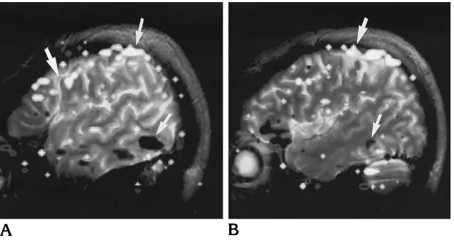
In the echo-planar imaging acquisitions in volunteers, both positive and negative signal intensity changes synchronous with motion of the second phantom were evident. Control stud-ies with no motion of the phantom showed no such synchronous signal intensity changes. As in the phantom experiments, there were more positive (in-phase) signal intensity changes at the superior edge and more negative (out-of-phase) signal intensity changes at the inferior edge of the brain (Fig 3).
Discussion
The study was designed to evaluate the effect on functional MR images of tissues or objects
moving in the magnetic field outside the field of view. Others have investigated the possible ef-fects of head motion temporally correlated with the performance of a task (stimulus-correlated motion) on functional MR images obtained with a surface coil (17). Stimulus-correlated motion during a visual task in that study was identified as a source of artifacts with a “striking” similar-ity to functional activation. Artifactual signal in-tensity changes caused by stimulus-correlated head motion occurred where signal intensity changed over a short distance, such as the boundaries between brain and cerebrospinal fluid.
In our study, significant signal intensity changes were produced by the movement of a phantom outside the field of view without move-ment of the head or of the scanned phantom. Quantitative analyses of the results of the phan-tom experiment showed positively and nega-Fig 1. Time course plots for nine contiguous pixels at the
periphery of the phantom for a series of echo-planar images acquired with acquired with intermittent movement of a second phantom located within the magnetic field but not within the field of view. The plots illustrate increases and decreases in signal intensity (vertical axis) temporally correlated (horizontal axis) with the movement of the second phantom, the timing of which is indicated in two of the pixels by the horizontal lines to which arrowspoint.
Analysis of pixels significantly correlated with the motion of the syringe phantom
Positively Correlated Negatively Correlated
Fraction of Pixels That Showed Correlation, %
Mean Signal
Change, % Fraction of Pixels, %
Mean Signal Change, %
Interior 33 10.4 29 20.5
Top edge 77 110.5 12 23.7
[image:3.612.59.304.80.325.2]Inferior edge 15 134 70 23.3
[image:3.612.57.556.651.729.2]tively correlated signal intensity changes most conspicuously at the edges of the phantom. Ac-quisitions obtained with external phantom mo-tion while the volunteers were not performing any motor, sensory, or cognitive task also showed a distribution of activated pixels similar to that observed in phantom experiments, with more and positively correlated pixels at the su-perior edge. Motion of tissues in or outside the field of view may therefore result in signal inten-sity changes that simulate activation.
In future experiments, more precisely con-trolled experimental conditions may be pro-duced by designing a mechanical device to reg-ulate the motion of phantoms. The magnitude of the motion in our second phantom was only approximately controlled. The phantoms did not exactly reproduce the structure, magnetic susceptibility, and signal intensities normally encountered in head imaging.
Either motion of the object being imaged or motion of objects with magnetic susceptibility outside the field of view may produce artifacts that simulate or obscure functional activation. The physical mechanisms by which the motion produces artifacts in the two cases are different. In the one case, the object’s registration to the magnetic field changes; in the other, the spatial configuration of the B0 magnetic field is changed. That latter change also alters the reg-istration of the magnetic field to the object being studied. In regions of the head, with signal in-tensity changes over short distances, the changes in registration that result from a change in the longitudinal magnetic field may increase or decrease signal intensity. If these magnetic field changes are temporally corre-lated with the sequence of task and rest, these
changes are falsely registered as activation. Ad-ditional effects caused by changes in B0include changes in the effective volume of voxels and through-section dephasing of the transverse magnetization. The signal intensity changes prominent at interfaces may be modified by truncation artifacts, Nyquist ghosting, and time-dependent intravoxel phase changes within a single echo-planar imaging shot. The latter causes the signal intensity to fluctuate even at a great distance from the susceptibility gradients because the effective complex point-spread function changes within each shot. Because of the short acquisition times, the blurring and ghosting seen on spin-echo echo-planar images are not seen on gradient-echo echo-planar im-ages.
Movement of the object in the field of view or distortion of the homogeneity of the magnetic field due to external motion that occurs during the acquisition of a functional MR imaging se-quence may produce changes in signal inten-sity and artifacts on functional MR. The artifac-tual signal intensity changes from either of these effects differ from the activations seen on functional MR images and may be distinguished by means of postprocessing or field maps. The magnitude of the signal intensity change in ac-tivation is typically between 4% and 6%, but in artifacts it can be much higher. The artifactual signal intensity changes are instantaneous with the motion or the magnetic field changes while the activations are characterized by a lag time of several seconds. Finally, the artifacts typi-cally include both in-phase and out-of-phase signal intensity changes whereas the activations are predominantly in-phase only. On the basis of the time course plots, artifactual signal inten-Fig 3. Two contiguous functional
[image:4.612.231.557.86.259.2]sity changes and activation can often be distin-guished by the magnitude, rise time, and pro-portion of in-phase and out-of-phase changes. The shorter repetition time possible with echo-planar imaging has advantages over the longer repetition time of fast low-angle shot imaging for the determination of rise times. The ob-served rise time of the signal magnitude, how-ever, will also depend on the speed of the ex-ternal motion and the steepness of the intensity change. Thus, a fast rise time indicates a pos-sible external motion artifact, but a slow rise time does not exclude this possibility.
Motion artifacts in a clinical functional MR imaging study may result from motion of body parts as well as from motion of the head. Move-ment of the patient’s chest, throat, jaw, or eyes or movement of a device within the scanner room may produce artifacts. Motion artifacts may explain some of the difference in results obtained with a silent versus an audible word-generation task (19). Movement of paramag-netic gaseous oxygen in the magparamag-netic field near or in the field of view may also produce artifacts as a result of its effect on the bulk magnetization (20). These results suggest that functional MR imaging paradigms should be designed with minimal movement in the magnetic field or with suitable controls for movement.
Acknowledgments
We gratefully acknowledge the help of Debbie Bauer, Jane Harmeyer, and Sue Madden.
References
1. Frahm J, Bruhn H, Merboldt K, Hanicke W. Dynamic MR Imaging of human brain oxygenation during rest and photic stimulation.J Magn Reson Imaging1992;2:501–505
2. Belliveau JW, Kennedy DN, McKinstry RC, et al. Functional map-ping of the human visual cortex by magnetic resonance imaging. Science1991;254:716 –719
3. Binder JR, Rao SM, Hammeke TA, et al. Functional magnetic resonance imaging of human auditory cortex.Ann Neurol1994; 35:662– 672
4. Hammeke TA, Yetkin FZ, Mueller WM, et al. Functional magnetic resonance imaging of somatosensory stimulation.Neurosurgery 1994;35:677– 681
5. Yetkin FZ, Mueller WM, Hammeke TA, Morris GL, Haughton VM. Functional magnetic resonance imaging mapping of the sensori-motor cortex with tactile stimulation.Neurosurgery1995;36:921– 925
6. Kim S-G, Georgopolis AP, Merkle H, et al. Functional imaging of human motor cortex at high magnetic field.J Neurophysiol1993; 69:297–301
7. Kwong KK, Belliveau JW, Chesler DA, et al. Dynamic magnetic resonance imaging of human brain activity during primary sen-sory stimulation.Proc Nat Acad Sci U S A1991;89:5675–5679 8. Yetkin FZ, Papke AR, Mark LP, Daniels DL, Mueller WM, Haughton
VM. Location of the sensorimotor cortex: functional and conven-tional MR compared.AJNR Am J Neuroradiol 1995;16:2109 – 2113
9. Morris GL, Mueller WM, Yetkin FZ, et al. Functional magnetic resonance imaging in partial epilepsy.Epilepsia1994;35:1194 – 1198
10. Ogawa S, Menon RS, Tank DW, et al. Functional brain mapping by blood oxygen level-dependent contrast magnetic resonance imaging: a comparison of signal characteristics with a biophysical model.J Biophys Soc1993;64:803– 812
11. Turner R, Jezzard P, Wen H, Kwong KK, Le Bihan D, Balaban RS. Functional mapping of the human visual cortex at 4 T and 1.5 T using deoxygenation contrast EPI. Magn Reson Med 1993;29: 277–279
12. Rao SM, Binder JR, Bandettini PA, et al. Functional magnetic resonance imaging of complex human movements.Neurology 1993;43:2311–2318
13. Ogawa S, Tank DW, Menon R, et al. Intrinsic signal changes accompanying sensory stimulation: functional brain mapping with magnetic resonance imaging. Proc Natl Acad Sci U S A 1992;89:5951–5999
14. Cao Y, Towle VL, Levin DN, Balter J. Functional mapping of human cortical activation by conventional MRI at 1.5T.J Magn Reson Imaging1993;3:869 – 875
15. Blamire AM, Ogawa S, Ugurbil K, et al. Dynamic mapping of the human visual cortex by high speed magnetic resonance imaging. Proc Natl Acad Sci U S A1992;89:11069 –11073
16. Jack CR Jr, Thompson RM, Butts RK, et al. Sensory motor cortex: correlation of presurgical mapping with functional MR imaging and invasive cortical mapping.Radiology1994;190:85–92 17. Hajnal JV, Myers R, Oatridge A, Schwieso JE, Young IR, Bydder
GM. Artifacts due to stimulus related motion in functional imaging of the brain.Magn Reson Med1994;31:283–291
18. Bandettini PA, Jesmanowicz A, Hyde JS, et al. Processing strat-egies for time-course data sets in functional MRI of the human brain.Magn Reson Med 1993;30:161–173
19. Yetkin FZ, Hammeke TA, Swanson SJ, et al. A comparison of functional MR activation patterns during silent and audible lan-guage tasks.AJNR Am J Neuroradiol1995;16:1087–1092 20. Bates S, Yetkin FZ, Jesmanowicz A, et al. Artifacts in functional
magnetic resonance imaging from gaseous oxygen.J Magn Re-son Imaging1195;4:443– 445