Open Access
Vol 10 No 2
Research article
Evaluation of the current knowledge limitations in breast cancer
research: a gap analysis
Alastair Thompson
1, Keith Brennan
2, Angela Cox
3, Julia Gee
4, Diana Harcourt
5, Adrian Harris
6,
Michelle Harvie
7, Ingunn Holen
8, Anthony Howell
9, Robert Nicholson
4, Michael Steel
10,
Charles Streuli
2for Breast Cancer Campaign Gap Analysis Meeting (2 November 2006, London,
UK)
1Department of Surgery and Molecular Oncology, University of Dundee, Ninewells Avenue, Dundee DD1 9SY, UK
2Wellcome Trust Centre for Cell Matrix Research, Faculty of Life Sciences, University of Manchester, Oxford Road, Manchester M13 9PT, UK 3Institute for Cancer Studies, University of Sheffield Medical School, Beech Hill Road, Sheffield S10 2RX, UK
4Tenovus Centre for Cancer Research, Welsh School of Pharmacy, Cardiff University, Redwood Building, King Edward VII Avenue, Cardiff CF10
3NB, UK
5The Centre for Appearance Research, School of Psychology University of the West of England, Frenchay Campus, Coldharbour Lane, Bristol BS16
1QY, UK
6Cancer Research UK Molecular Oncology Laboratories, Weatherall Institute of Molecular Medicine, John Radcliffe Hospital, University of Oxford,
Headley Way, Headington, Oxford OX3 9DS, UK
7Family History Clinic, Nightingale & Genesis Prevention Centre, Wythenshawe Hospital, Southmoor Road, Manchester M23 9LT, UK
8Academic Unit of Clinical Oncology, School of Medicine and Biomedical Sciences, University of Sheffield, Beech Hill Road, Sheffield S10 2RX, UK 9Breast Cancer Prevention Centre, South Manchester University Hospitals NHS Trust, Wilmslow Road, Manchester M20 4BX, UK
10University of St Andrews, Bute Medical School, University of St Andrews, Fife KT16 9TS, UK
Corresponding author: Alastair Thompson, [email protected]
Received: 30 Aug 2007 Revisions requested: 24 Jan 2008 Revisions received: 13 Mar 2008 Accepted: 27 Mar 2008 Published: 27 Mar 2008
Breast Cancer Research 2008, 10:R26 (doi:10.1186/bcr1983)
This article is online at: http://breast-cancer-research.com/content/10/2/R26 © 2008 Thompson et al.; licensee BioMed Central Ltd.
This is an open access article distributed under the terms of the Creative Commons Attribution License (http://creativecommons.org/licenses/by/2.0), which permits unrestricted use, distribution, and reproduction in any medium, provided the original work is properly cited.
Abstract
Background A gap analysis was conducted to determine which areas of breast cancer research, if targeted by researchers and funding bodies, could produce the greatest impact on patients.
Methods Fifty-six Breast Cancer Campaign grant holders and prominent UK breast cancer researchers participated in a gap analysis of current breast cancer research. Before, during and following the meeting, groups in seven key research areas participated in cycles of presentation, literature review and discussion. Summary papers were prepared by each group and collated into this position paper highlighting the research gaps, with recommendations for action.
Results Gaps were identified in all seven themes. General barriers to progress were lack of financial and practical resources, and poor collaboration between disciplines. Critical gaps in each theme included: (1) genetics (knowledge of genetic changes, their effects and interactions); (2) initiation of breast cancer (how developmental signalling pathways cause ductal elongation and branching at the cellular level and
influence stem cell dynamics, and how their disruption initiates tumour formation); (3) progression of breast cancer (deciphering the intracellular and extracellular regulators of early progression, tumour growth, angiogenesis and metastasis); (4) therapies and targets (understanding who develops advanced disease); (5) disease markers (incorporating intelligent trial design into all studies to ensure new treatments are tested in patient groups stratified using biomarkers); (6) prevention (strategies to prevent oestrogen-receptor negative tumours and the long-term effects of chemoprevention for oestrogen-receptor positive tumours); (7) psychosocial aspects of cancer (the use of appropriate psychosocial interventions, and the personal impact of all stages of the disease among patients from a range of ethnic and demographic backgrounds).
Conclusion Through recommendations to address these gaps with future research, the long-term benefits to patients will include: better estimation of risk in families with breast cancer and strategies to reduce risk; better prediction of drug response and patient prognosis; improved tailoring of treatments to patient subgroups and development of new therapeutic
approaches; earlier initiation of treatment; more effective use of resources for screening populations; and an enhanced experience for people with or at risk of breast cancer and their
families. The challenge to funding bodies and researchers in all disciplines is to focus on these gaps and to drive advances in knowledge into improvements in patient care.
Introduction
Significant advances in the prevention, diagnosis and manage-ment of breast cancer have been made in recent years based on the clinical application of scientific discoveries. However, breast cancer remains a complex disease process affecting millions worldwide, and further advances in scientific knowl-edge and clinical care could improve many lives. It is timely to review the current position of breast cancer research because funding bodies, researchers and clinicians work in an exciting age of discovery but have limited resources.
In November 2006, the research charity Breast Cancer Cam-paign convened a panel of leading breast cancer researchers, as an initial event, to debate and identify the limitations of cur-rent research into the pathophysiology, detection, treatment, prevention and psychosocial aspects of breast cancer. The aims of this analysis were as follows: To determine the gaps in our knowledge of breast cancer that, if resolved, could result in benefits to patients; To encourage breast cancer research-ers and funding bodies worldwide to focus their resources on the highlighted areas of research to achieve a substantive impact for patients; To make recommendations for priority action.
This gap analysis represents a unique insight into breast can-cer research in the UK and the challenges involved in directing efforts to areas in need of further investigation likely to result in advances in the management of breast cancer.
Materials and methods
Current and former members of the Breast Cancer Campaign Scientific Advisory Board leading scientists and clinicians res-ident in the UK were invited to participate in the gap analysis meeting. The choice of participants was based on publication record, research activity and clinical stature, and selected using a database of researchers developed since the incep-tion of the Breast Cancer Campaign in 1988.
Seven key research areas were selected for review in the gap analysis by the Breast Cancer Campaign and the Scientific Advisory Board, taking into account UK, European and USA themes in scientific meetings focused on breast cancer and UK Government analyses of research funding streams: Genet-ics of breast cancer; Initiation of breast cancer; Progression of breast cancer; Therapies and targets in breast cancer; Dis-ease markers in breast cancer; Prevention of breast cancer; Psychosocial aspects of breast cancer.
Prior to the event, participants were asked to review relevant literature and construct short presentations summarising their areas of expertise and identifying potential research gaps. Key participants had already conducted, published and/or reviewed systematic evidence, literature reviews and evi-dence-based guidelines. As a result they were considered to be opinion leaders in their field. Further, additional, systematic literature reviews for each of the seven areas under consider-ation was not performed.
For each theme, 6 to 10 UK breast cancer researchers of national or international standing in their fields of research were invited and accepted participation in the event. Twenty-three invitees declined to contribute to the gap analysis.
On 2 November 2006 a one-day meeting was convened in London, UK. In the initial subgroup sessions, each participant gave a presentation to their group on pre-agreed topics rele-vant to the gap analysis for their assigned breast cancer research area on which they were experts. There were con-structive debates of the content and the issues raised by the presentations were explored further. Breast Cancer Campaign staff members acted as facilitators throughout the analysis process.
Issues explored during the gap analysis were structured around the following questions: What do we already know; What are the gaps in our knowledge; What are the problems that need to be overcome to fill these gaps; What are the translational implications?
After collating the information resulting from these discussions for each of the seven themes, this four-point structure was used to present the content of each theme to the other partic-ipants and to discuss their findings in an open forum.
This iterative process continued as evidence-based expert opinion from the one-day meeting was cross-referenced, shaped and developed during subsequent weeks. Each group formulated a summary paper for their research area, incorpo-rating key references, which was then circulated to the partic-ipants of the respective groups for further refinement. These seven themes were collated into a unified position paper, which is what we present here.
Results and discussion
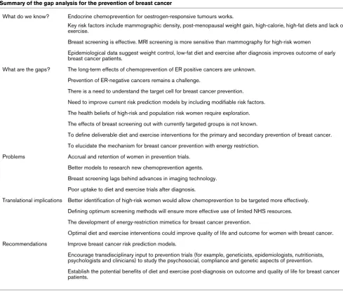
1. Genetics of breast cancer
What do we know?
Several genes bearing high-penetrance mutations have been implicated in inherited predisposition to breast cancer, the most important of these being in the BRCA1 and BRCA2
genes. However, BRCA1 and BRCA2 account for less than 5% of all breast cancer and, in recent years, breast cancer susceptibility genes other than BRCA1 and BRCA2 have been identified. These genes fall into two broad categories: Those containing rare moderate-penetrance alleles such as
CHEK2, ATM and BRIP1 [1-3]; Those carrying more common
low-penetrance alleles [4].
Large genome-wide association and candidate gene studies to identify the latter are just beginning to bear fruit [5-8].
Intermediate phenotypes such as radiosensitivity and mammo-graphic density have quite strong genetic components and further study of these may provide some insight into novel breast cancer susceptibility genes.
Since the breast cancer genes BRCA1 and BRCA2 were cloned in 1994 and 1995, respectively, research efforts have concentrated on developing an understanding of the cellular functions of the large multidomain proteins encoded by
BRCA1 and BRCA2 and the mechanisms by which loss of
their functions causes breast cancer. An understanding of these mechanisms is relevant not only to families with BRCA1
and BRCA2 mutation carriers but also in sporadic breast
can-cer, in which the same or related genetic pathways may also be aberrant.
Cells deficient in BRCA1 and BRCA2 are extremely sensitive to DNA-damaging agents and are defective in repairing DNA double-strand breaks by homologous recombination, being impaired in the recruitment and filament formation of the recombination protein RAD51 [9,10]. More recently, new functions have been identified for both proteins: BRCA1 and its partner BARD1 form an E3-ubiquitin ligase that is recruited to sites of DNA damage and activated by the DNA damage checkpoint, promoting ubiquitylation [11]. The first in vivo sub-strate for such ubiquitylation events has been identified as CtIP [12].
An important development in recent years has been the iden-tification of the links between the BRCA1 and BRCA2 path-ways and proteins involved in Fanconi anaemia. Several genes cause Fanconi anaemia, and most of these encode proteins that are involved in a complex that ubiquitylates FANCD2 at Table 1
Summary of the gap analysis for the genetics of breast cancer
What do we know? Multiple genes of different penetrance are involved in the predisposition to breast cancer.
Genome wide screens and somatic genetic approaches are identifying further genes involved in breast cancer.
What are the gaps? Detailed understanding of the actions of BRCA1 and BRCA2.
Knowledge of large-scale genetic rearrangements in tumour cells.
The important variants, effects and interactions of low-penetrance genes.
Further identification of point mutations and epigenetic changes.
Problems The quality, quantity and accessibility of materials.
Funding for large-scale experiments (such as sequencing) using expensive equipment.
Bioinformatic analysis skills.
Translational implications Classifying breast tumours according to the signalling pathways that are disrupted to predict prognosis and response to therapy.
Determining the relevance of somatic events to prognosis and response to therapy.
Generate new, targeted therapies based on target discovery.
Better genetic risk estimation.
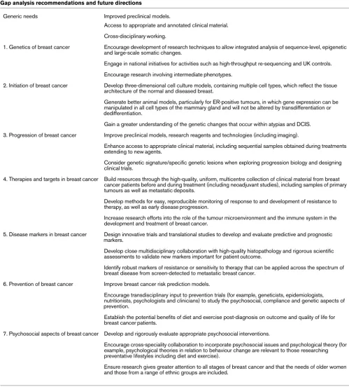
Recommendations Encourage development of research techniques to allow integrated
analysis of sequence level, epigenetic and large-scale somatic changes.
Engage in national initiatives for activities such as high-throughput re-sequencing and UK controls.
K561. The FANCD1, FANCJ and FANCN gene products act downstream of this step. FANCD1 was found to be the
BRCA2 gene [13]. FANCJ encodes BRIP1, which interacts
with the BRCT domain of BRCA1 and, of note, mutations in this gene can also cause breast cancer [3]. Lastly, FANCN
encodes PALB2, which interacts with BRCA2, and is also a breast cancer susceptibility gene [14].
What are the gaps?
There remain gaps in our knowledge of cancer predisposition genes, both in identifying genes responsible for low-pene-trance disease and the interactions with environmental factors. Increasing knowledge of BRCA1 and BRCA2 acts as an exemplar resulting in improved patient care. However, defi-ciencies remain in our understanding of how BRCA1 or BRCA2 dysfunction causes breast cancer, for example, it is unclear why BRCA1 deficiency is associated with triple-nega-tive (basal-like) breast cancer. We also need to find other pro-teins that interact with BRCA1 and BRCA2 and elucidate the post-translational modifications that occur as a result of these interactions. For example, the functional consequences of CtIP ubiquitylation and its implications for RAD51 recruitment are not yet known; furthermore, it is likely that there are other substrates for BRCA1/BARD1.
Only a relatively small proportion of breast cancers are caused by the loss of BRCA1 or BRCA2 function; most arise as a result of somatic mutation or changes in expression of a number of other genes. The list of genes showing somatic point mutations in breast cancers is beginning to be identified using genome-wide sequencing approaches [15]. Epigenetic changes such as DNA methylation and histone modification can cause loss or gain of gene expression, and genome-wide screens for these are being actively pursued. More work is needed in both of these areas.
To date, little progress has been made towards cataloguing larger-scale genetic rearrangements, such as translocations, deletions and amplifications, which occur frequently and are the hallmarks of tumour cells and have been particularly useful in the haematological malignancies. These changes also need to be related to tumour subtype. Reciprocal translocations seem to be common; for example, one translocation causes abnormal expression of the NRG1 gene which encodes lig-ands of the epidermal growth factor receptor (EGFR) family [16]. Work has begun to characterise and catalogue these events using high-resolution DNA microarrays. In the future it will be important to devise approaches to examine sequence-level, epigenetic and large-scale changes together and relate these to clinical features to form a complete and integrated picture.
As common, low-penetrance alleles continue to be identified, future challenges lie in identifying the causative variants within the haplotype blocks containing the associated marker single
nucleotide polymorphism (SNP). First, this requires high-throughput sequencing capacity to detect all common variants in haplotype blocks spanning typically 30 to 150 kb, in at least 48 people. Second, case and control DNA collections from non-European populations are needed to separate alleles that are completely correlated in European populations. Third, sen-sitive biological assays need to be developed to determine the differences in function of the potentially causative alleles. It will be important to encourage a high degree of collaboration between research groups, in particular between epidemiology and basic biology. Furthermore, these studies would benefit from shared resources including first-class sample collections, such as a UK national control set.
A potentially useful but currently under-developed approach is to examine other phenotypes linked with breast cancer that are themselves determined genetically. These include mammo-graphic density, radiation sensitivity, cell migration and circu-lating levels of hormones and growth factors.
What are the problems?
The number of patients required for gene searches requires large national and international consortia. The quality, quantity and form of the clinical material (blood derivatives, frozen tis-sue, formalin fixed tissues), and the handling or products from these present a significant challenge. The increasing sophisti-cation of equipment and the level of technical expertise are reflected in the need to integrate the data in a meaningful way presenting a substantial bioinformatics challenge. Techniques for high-throughput re-sequencing are being developed, but funding is needed to make these accessible. Thus, this type of research is particularly resource intensive and requires a high level of collaboration.
Translational implications
Therapies based on developing an understanding of the role of BRCA1 and BRCA2 in DNA repair are already in clinical tri-als (including cisplatin and PARP inhibitors), and improving our understanding of the many functions of BRCA1 and BRCA2 will no doubt generate further targets for therapy. Increasing efforts to understand genetic events should allow us to perform the following functions: Classify breast tumours according to the signalling pathways that are disrupted and to predict prognosis and response to therapy; Determine the rel-evance of somatic events to prognosis and response to ther-apy; Generate new, targeted therapies based on target discovery.
complimentary data, international exchanges of such informa-tion should enhance future patient management.
2. Initiation of Breast Cancer
A summary of the gap analysis for the initiation of breast can-cer is given in Table 2.
To decipher the molecular basis of the initiation and progres-sion of breast cancer, it is critical that we fully understand the key features and genes involved in normal mammary develop-ment. Changes to developmental processes may lead to tumour initiation and the influences of endocrine agents, growth factors and environmental carcinogens on normal and developing breast components is largely unknown,
What do we know?
Significant progress has been made in determining the local factors that control all stages of mammary development, largely through the generation of an extensive array of trans-genic and knockout mouse strains [17]. Such animal models are complemented by classic embryological approaches that enable the transplantation of a complete mammary gland, duc-tal rudiments or, more recently, stem cells into cleared fat pads [18-20]. These approaches have imparted considerable knowledge about a wide array of developmental signalling
pathways that are now known to be dysregulated in tumours. For example, amphiregulin/EGFR signalling is required for the branching and outgrowth of the ductal epithelial tree during pubertal development [21,22], while overexpression of both the ligand and receptor, and the related receptor ErbB2, is associated with poor prognosis in breast cancer [23]. Other examples include the IGF, integrin, Notch, NF-κB, STAT,
TGF-β and Wnt pathways [24-29].
What are the gaps? Tissue architecture
Many of the signalling pathways controlling normal mammary development have been identified and the genetic circuit dia-grams that link the different pathways are emerging [17]. How-ever, it is not clear how these signals cause ductal elongation or branching at the cellular level, or even how they maintain normal ductal or acinar architecture. Although analysis has focused on epithelial cells, the stromal, endothelial and immune components are also crucial for development [30]. For example, fibroblasts and macrophages are required for ductal growth [31]. However, many of the stroma-derived sig-nals are poorly understood, as is the reciprocal communica-tion between the epithelium and stroma and the signalling pathways controlled by the interaction between luminal and myoepithelial cells.
In addition, the importance of cell adhesion and the extracellu-lar matrix has been underestimated, although it is becoming
[image:5.612.56.543.422.700.2]increasingly clear that both adhesion and matrix-derived sig-nals modify many signalling pathways and provide a spatial Table 2
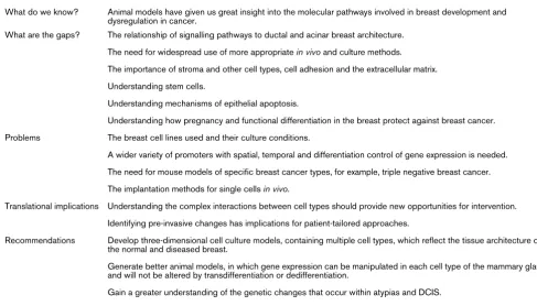
Summary of the gap analysis for the initiation of breast cancer
What do we know? Animal models have given us great insight into the molecular pathways involved in breast development and dysregulation in cancer.
What are the gaps? The relationship of signalling pathways to ductal and acinar breast architecture.
The need for widespread use of more appropriate in vivo and culture methods. The importance of stroma and other cell types, cell adhesion and the extracellular matrix.
Understanding stem cells.
Understanding mechanisms of epithelial apoptosis.
Understanding how pregnancy and functional differentiation in the breast protect against breast cancer.
Problems The breast cell lines used and their culture conditions.
A wider variety of promoters with spatial, temporal and differentiation control of gene expression is needed.
The need for mouse models of specific breast cancer types, for example, triple negative breast cancer.
The implantation methods for single cells in vivo.
Translational implications Understanding the complex interactions between cell types should provide new opportunities for intervention.
Identifying pre-invasive changes has implications for patient-tailored approaches.
Recommendations Develop three-dimensional cell culture models, containing multiple cell types, which reflect the tissue architecture of the normal and diseased breast.
Generate better animal models, in which gene expression can be manipulated in each cell type of the mammary gland and will not be altered by transdifferentiation or dedifferentiation.
checkpoint for developmental decisions [32]. Critically, changes in cell adhesion, through matrix remodelling or altered adhesion receptors, underpin both tissue disorganisation in early breast cancer and progression to malignancy [33,34], but their involvement in tumour initiation is not well understood.
Stem cells
The recent development of technologies allowing enrichment of mammary gland stem cells has been a significant step for-ward [19,20,35]. However, it is still not possible to purify these cells to homogeneity, and we do not fully understand their location within the ducts or the mechanisms involved in their differentiation into luminal and myoepithelial cells [36,37]. Moreover, it is not certain whether cancer stem cells are derived from normal stem cells or an intermediate progenitor cell, or how they are influenced by stromal factors [38]. In fact, it is far from clear that tumours are derived from stem/progen-itor cells at all, as opposed to reprogrammed differentiated cells, let alone whether differences between cancer stem cells might be responsible for the development of different tumour subtypes.
Although considerable progress has been made towards understanding the mechanisms controlling epithelial apopto-sis in the mammary gland, we know little about the sensitivity of stem/progenitor cells to apoptosis signals [39,40]. Cells of the main ducts survive involution, while alveoli and terminal ducts are lost, although the reason for this difference is not clear.
We also have little understanding of how early pregnancy and functional differentiation of the breast protects against cancer, and whether this is related to stem cell dynamics [41].
Consequently, we urgently need more appropriate in vivo and culture models to resolve the mechanisms of both normal breast development and tumour initiation. Furthermore, use of these sophisticated models needs to become more widespread.
What are the problems? Culture models
Breast cancer has traditionally been studied by cell and molec-ular biologists using long-lived cell lines that are derived from late stage tumours and which display few of the cellular prop-erties of normal breast epithelial cells. In addition, we now appreciate that breast cancer involves growth in three dimen-sions and the contribution of various breast cell types. Further-more, techniques for studying the initiation and progression of cancer are largely restricted to analysing the luminal epithe-lium. Recently there has been a shift from two-dimensional to three-dimensional culture models, which better reflect the tis-sue environment found in vivo [42,43]. However, most of these models still contain only luminal epithelial cells. In the
future, culture models containing all mammary cell types, as well as those amenable to examining ductal branching, model-ling the stem cell niche and even assessing whether an iso-lated cell is a stem cell, will provide fertile avenues for analysis [44].
Excellent imaging technologies are emerging to explore the dynamic nature of mammary gland development and neoplasia both in culture and in vivo; applying them to human cancer cells and ensuring their widespread use will be of enormous value [45,46].
Genetic analyses
Expression of transgenes or gene ablation specific to the mammary gland is usually achieved by placing the transgene or Cre recombinase under the control of a milk gene promoter or the MMTV promoter [47]. However, these promoters are active only in the luminal epithelium and, in the case of the milk protein genes, limited to a particular developmental stage. More recently, the keratin 5 promoter has been used to target myoepithelial and basal cells [48]. Despite this, a wider variety of promoters would improve the spatial and/or temporal con-trol gene of expression. This will be helped considerably by generating an atlas of mammary gland development. Further refining markers to identify stem and progenitor cells, and fibroblasts, will pinpoint other valuable promoters. Greater use of inducible transgene systems, such as the Tet-On system, will allow transgene expression to be restricted to specific time intervals [49].
Another significant problem with existing promoters is that their expression in the luminal epithelium depends on its differ-entiation; activity is therefore lost if a transgene causes transdifferentiation or dedifferentiation within a tumour. The problem can be overcome through the development of 'hit and run' transgenics. These include lines where the expression of a transgene is under the control of a housekeeping gene but is prevented by a lox-stop-lox cassette. Excision of the stop codon by crossing with mice carrying a gland-specific Cre recombinase then leads to continuous and consistent trans-gene expression. These and similar sophisticated strategies will allow the more realistic activation of breast cancer onco-genes, and will provide better opportunities to understand how they cause disease within the correct tissue environment. It would also be valuable to produce a series of transgenic reporter gene mice equivalent to the TOPGAL strain, to moni-tor changes in developmental signalling pathways [50].
mice are required. Significant progress has been made in gen-erating such humanised models, but it is still not possible to implant individual cells [51].
Translational implications
Continuing to study normal breast development will provide many useful insights into the earliest stages of breast cancer initiation. Tumour development depends on signals between the stroma, myoepithelial cells and luminal cells, and therefore gaining a better understanding of how these cells communi-cate in normal mammary gland and early breast disease will provide new opportunities for intervention.
In addition, we know little about the genetic changes that occur in atypias, lobular or ductal carcinoma in situ (DCIS) [52], although they are most likely to be within components of the signalling pathways that control normal development.
Pro-ducing timelines of these mutations for different breast cancer subtypes, similar to those generated for colon cancer [53], would be a major step forward. Molecular profiling of breast cancers has begun to classify tumours [54], and relating them to the TNM classification will be clinically valuable for biomar-ker analysis and therapy. Differences between profiles may represent alterations in specific signalling pathways, so map-ping this information onto a timeline of when mutations occur in breast cancer will enable clinicians to tailor specific treat-ments to individuals.
3. Progression of Breast Cancer
A summary of the gap analysis for the progression of breast cancer is given in Table 3.
Intracellular inputs What do we know?
The oestrogen receptor, receptor tyrosine kinase (RTK) and DNA repair pathways are key research areas in understanding
[image:7.612.58.555.313.701.2]the intracellular inputs for growth and progression of invasive breast cancer.
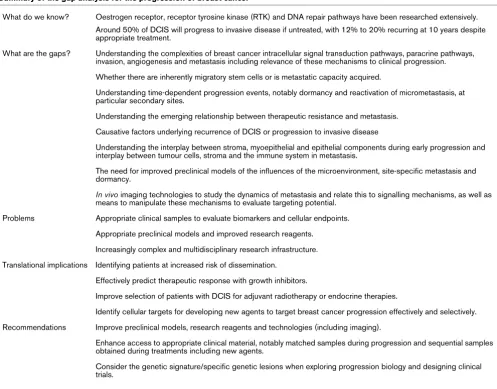
Table 3
Summary of the gap analysis for the progression of breast cancer
What do we know? Oestrogen receptor, receptor tyrosine kinase (RTK) and DNA repair pathways have been researched extensively.
Around 50% of DCIS will progress to invasive disease if untreated, with 12% to 20% recurring at 10 years despite appropriate treatment.
What are the gaps? Understanding the complexities of breast cancer intracellular signal transduction pathways, paracrine pathways, invasion, angiogenesis and metastasis including relevance of these mechanisms to clinical progression.
Whether there are inherently migratory stem cells or is metastatic capacity acquired.
Understanding time-dependent progression events, notably dormancy and reactivation of micrometastasis, at particular secondary sites.
Understanding the emerging relationship between therapeutic resistance and metastasis.
Causative factors underlying recurrence of DCIS or progression to invasive disease
Understanding the interplay between stroma, myoepithelial and epithelial components during early progression and interplay between tumour cells, stroma and the immune system in metastasis.
The need for improved preclinical models of the influences of the microenvironment, site-specific metastasis and dormancy.
In vivo imaging technologies to study the dynamics of metastasis and relate this to signalling mechanisms, as well as means to manipulate these mechanisms to evaluate targeting potential.
Problems Appropriate clinical samples to evaluate biomarkers and cellular endpoints.
Appropriate preclinical models and improved research reagents.
Increasingly complex and multidisciplinary research infrastructure.
Translational implications Identifying patients at increased risk of dissemination.
Effectively predict therapeutic response with growth inhibitors.
Improve selection of patients with DCIS for adjuvant radiotherapy or endocrine therapies.
Identify cellular targets for developing new agents to target breast cancer progression effectively and selectively.
Recommendations Improve preclinical models, research reagents and technologies (including imaging).
Enhance access to appropriate clinical material, notably matched samples during progression and sequential samples obtained during treatments including new agents.
It is now established that steroid hormone or oestrogen recep-tor alpha (ERα) input, a fundamental driver for the growth of many breast cancers, should not be considered independent of additional signalling networks. This nuclear ERα may inter-play with ERβ and there is emerging evidence that membrane-localised ERα may also have a role. A web of RTK signalling also contributes to breast cancer growth and progression, and these pathways can interact with ER when present [55-57]. Previous experimental deciphering of ER and RTK pathways has provided proof of principal that useful biomarkers and ther-apies (for example, endocrine therther-apies, erbB and kinase inhibitors) can stem from concerted research in breast cancer growth signalling biology [58].
DNA damage response (DDR) pathways and mechanisms for mitotic chromosome segregation are also of interest when considering breast cancer growth, progression and selectivity. They can be subject to genetic alteration, associating with tumourigenesis for inherited (for example, BRCA proteins) and for sporadic (for example, Aurora A: 30% to 60% overexpres-sion) breast cancer because they cause genetic instability that may permit secondary alteration, accelerating the develop-ment of cancer. Equally, there are major implications for thera-peutic efficacy, for example, for poly(ADP-ribose) polymerase inhibitors and BRCA mutations, and also resistance to taxanes when Aurora A is amplified [59].
What are the gaps?
Research into ER and RTK signalling is intense. Further deci-phering of the complexities of breast cancer signal transduc-tion pathways, their regulatransduc-tion and interplay, including elucidating downstream gene effects, is important, not least because therapeutic resistance is a persistent problem for all therapies targeting known pathways.
There are also limitations in our ability to subsequently evalu-ate RTK and ER signalling aspects emerging from experimen-tal studies in clinical disease and to rapidly translate findings into clinically useful biomarkers and targets for new drug development.
Furthermore, the breadth of DDR and mitotic regulator altera-tions underlying the pathogenesis of breast cancer and its subtypes, or the point at which DDR alterations might contrib-ute to disease progression is unknown.
Extracellular inputs What do we know?
Local paracrine pathways (for example, those involving TGFβs), cell invasion into the extracellular matrix and angio-genesis all contribute to cancer growth and metastasis. Selec-tive inhibitors of anti-Src and anti-HGF/Met, for some of the identified pathways involved in metastasis and inhibition of angiogenesis (for example, by targeting vascular endothelial growth factor signalling) are already in clinical practice [60].
What are the gaps?
Substantial questions remain about the biology of angiogen-esis and metastasis. While studies of aggressive breast can-cer cells in vitro have allowed significant progress in understanding adhesion and migration through matrix, matrix degradation (including mechanisms of matrix metalloproteinases) and in vitro invasiveness, subsequent translational applications have proved limited.
We have yet to fully explore the breadth of potential positive and negative regulators of invasion and metastasis, their mechanisms and interplay, and the role of the interaction of tumour cells with the stromal microenvironment and immune system during metastasis. The propensity for cancer to metas-tasise, apparently selectively, to certain end organs is poorly understood. Whether human breast cancers contain inher-ently migratory stem cells or whether metastatic capacity is acquired, and also time-dependent progression events, nota-bly dormancy and reactivation of micrometastasis at particular secondary sites, remain poorly defined.
Equally, little is known about the emerging relationship between resistance to conventional therapies (for example, endocrine therapies) and metastasis [61] or the degree of redundancy of invasive elements or angiogenic pathways that may contribute to therapeutic resistance to anti-invasive and anti-angiogenic approaches.
Ductal carcinoma in situ and very early progression in breast cancer
What do we know?
Around 50% of DCIS will progress to invasive disease if untreated, with 12% to 20% recurring locally at 10 years despite breast conserving surgery and radiotherapy. Links have been suggested between early progression and mecha-nistic alterations both within the epithelial cells and in the inter-play with associated basement membrane, myoepithelial and stromal cells, and mechanical constraints [62,63]. HER2 pos-itivity and HER4 negativity, and high levels of cyclo-oxygenase-2 (COX-cyclo-oxygenase-2) may have some relevance to invasive recurrence [64].
What are the gaps?
Issues regarding the very early progression of breast cancer reflect those for invasive breast cancer. There are substantial gaps in our ability to perform biological investigation of the intracellular and extracellular factors (notably interplay among stroma, myoepithelial and epithelial components) underlying growth and progression of DCIS, to dynamically monitor early progression, and to subsequently manipulate implicated path-ways to address their therapeutic potential.
What are the problems? Improved preclinical models
We need to develop better preclinical models in order to more accurately reveal the mechanisms involved in growth and progression of breast cancer and to evaluate potential targets. This is particularly important when trying to interpret the mech-anisms of metastasis because dissemination commonly occurs at inaccessible sites, making clinical research material extremely difficult to obtain. However, it is equally relevant if we are to build on findings made using in vitro monolayer culture studies for growth signalling mechanisms.
Models are needed to allow researchers to investigate more accurately the influence of the microenvironment, the impact of the immune system and site-specific metastasis, and time-dependent progression including the phenomenon of dor-mancy. In vivo models will be essential to study these areas, and researchers may also benefit from in vitro three-dimen-sional assays encompassing stromal components, matrix and tumour epithelial cells [65], as well as studies over extended culture time in vitro and associated animal models.
Increased use of genetically modified animal models will be valuable, as will genetic manipulation of individual targets (for example, using RNAi) alone or in combination with existing agents in model systems to address how mechanisms of growth and spread might be exploited to provide new targeted therapies. Studies of very early progression will also require improved models, notably use of in vitro cultures of primary or mammosphere DCIS (again extending to longer-term culture), and of human DCIS xenograft and transgenic (for example, MMTV/HER2) models.
Alongside targeting individual candidate elements, more spec-ulative approaches are warranted to broadly screen for syn-thetic lethality, for example, using RNAi libraries. We also need to embrace fully in vivo imaging technologies to decipher the dynamics of metastasis (and equally of very early progression) at a cellular level and relate this to signalling mechanisms (for example, using in vivo fluorescent reporter assays).
Powerful real-time imaging studies in animal models are emerging which indicate that the motile phenotype may be transient and confined to a subpopulation of cells at the tumour periphery in disease metastasising in vivo. Such heter-ogeneity is not easily modelled with in vitro studies. These
observations illustrate the power of imaging systems for deci-phering breast cancer biology, where the data have implica-tions for interpreting whole tumour microarray/proteomic profiles and clearly confirm that time-dependent study of metastasis at a cellular level is essential.
Appropriate clinical samples
Appropriate clinical samples are needed to translate experi-mental findings into useful predictive biomarkers, and to con-firm that therapeutic strategies stemming from basic research are relevant in patients. Meaningful study will require, where possible, improved access to clinical material with parallel therapeutic response and survival data, encompassing tissue microarray (TMA) and full-section resources. Studies will ben-efit from improved access to clinical samples of primary, local recurrent, lymph node and distant metastatic (where accessi-ble) breast cancer, ideally comparing matched samples from patients to track potential biomarkers of progression. Studies of very early progression will also need increased access to clinical DCIS material to verify the relevance of experimental associations with invasive recurrence, considering gene expression signatures both for DCIS and invasive disease, preferably from the same patient.
It is also important that researchers obtain material from sequential samples (with parallel outcome data), taken before and during treatment with conventional therapies, and from biologically directed innovative trials of targeted therapies. Clinical trial design should increasingly incorporate improved tissue collection and pathology support (which has often been considered as an afterthought). Experience indicates that neo-adjuvant studies and, where possible, access to patient sam-ples treated longer term will be particularly valuable.
Studying samples obtained during treatment with anti-invasive agents (or, indeed, any new agents) will allow researchers to evaluate the ability of biomarkers and cellular endpoints stem-ming from experimental studies to provide surrogates for drug response, an important aim in the absence of long-term out-come data. This could be achieved by examining circulating tumour cells and sampling lymph nodes during treatment. In addition, evaluation of anti-invasive agents could benefit signif-icantly from improvements in tumour tissue imaging of func-tional reporters.
capacity, and so enhanced trial design and better recruitment of DCIS patients is also essential.
Consideration of gene profiles
Greater attention needs to be given to gene profiles and the impact of genetic lesions in malignant epithelial cells and in the stromal background in clinical trial design and during the selection of patients for therapy. Expression profiling at the mRNA and protein level has revealed several subtypes of breast cancer, and it is important that intracellular and extracel-lular processes of growth and progression are further explored in relation to these, both through models that aim to recapitu-late each class and through representative clinical material.
The non-tumour content of biopsies should also be consid-ered since evidence is emerging to suggest that this has a sig-nificant effect on gene expression profiles [66]. Equally, several studies have indicated the importance of considering specific lesions in relation to therapeutic response, for exam-ple, Aurora kinase overexpression and taxane resistance, BRCA2 loss and response to platinum-based therapy [67], topoisomerase II alpha amplification and epirubicin response [68]. Considering these parameters may not only improve patient stratification for more effective trial design, but may also allow smaller patient cohorts to be studied, if these are selected rationally for therapy according to the status of the drug target or molecular lesion.
Improved research reagents
Better research reagents are needed to continue to accurately define new pathways in experimental material and to study clinical samples to verify these pathways as biomarkers in rela-tion to pathology and outcome.
Signalling mechanisms in clinical samples can be detected using immunohistochemistry, including the use of phospho-specific antibodies [69,70]. However, if associations are to be accurate and meaningful, this urgently requires assays that have a reproducible, sensitive and specific performance in such material, incorporating improved quality control as has been achieved for ERα and HER2 assays [71-73]. Technolo-gies such as fluorescence resonance energy transfer (FRET) have the potential to measure interplay between elements (for example, receptor dimerisation) in such material. However, in all instances these methodologies will require consensus regarding evaluation which should also aim for quantitative analysis, for example, through image analysis. More accurate quantification may potentially incorporate luminescent quan-tum dots as an alternative approach to visualise quan-tumour markers.
Research to reveal new biomarkers and drug targets in exper-imental and clinical material will also increasingly need to incorporate high-throughput gene profiling with microarrays and TMA validation, as well as proteomics. In these latter
areas, quality control urgently needs to be addressed and bio-informatic capabilities must be significantly enhanced on a national level if we are to manage and meaningfully interpret the increasing volume of signalling data that will emerge in relation to cellular endpoints and clinical outcome.
Research infrastructure
Overcoming these various barriers has obvious implications for research infrastructure. Studies will need to be increasingly multidisciplinary if we are to identify relevant determinants of breast cancer growth and progression; for example, requiring the integration of multiple expression/signalling studies, bioin-formatics, imaging technologies, improved in vitro and in vivo
models, genetic manipulation and clinical examination. This will depend on a backbone of realistic supportive funding, not only to maintain core strategies and associated quality control, but to ensure access to new technologies to pursue innovative research avenues (for example, in vivo imaging, genetically engineered models and high-throughput genomic screening).
A critical mass of expert staffing is essential, including expand-ing the breast cancer research talent pool through improved research training and more clearly structured career develop-ment. The need for carefully collected and documented clinical tissue with serial biopsies taken during therapy with defined treatments, TMAs, samples from distant sites and local recur-rences made available to investigators is key. Recent legisla-tive changes have made this more difficult and both surgical and pathology support are needed at a senior level. Both spe-cialities have suffered severe cutbacks; collaborative contribu-tions by academics in these areas are important and deserve funding, in addition to supporting the highest quality peer-reviewed independent research.
Pathology training will be increasingly important as we expand our technical capabilities for investigating clinical material. Standardisation of antibodies and other reagents is needed to compare results between investigators. Research would also benefit from increased sharing of experimental and clinical resources. Finally, increased investigator-driven studies and collaboration with industry will be essential if we are to improve patient recruitment for clinical studies aimed at understanding biological factors driving selective response.
Translational implications
If research can be tailored to the diverse questions regarding the intracellular and extracellular processes underlying breast cancer growth and progression, this should link tumour mech-anisms to disease classification and prognosis.
How-ever, we need to build on these studies: first, so we can iden-tify earlier patients at increased risk of dissemination and therefore allow selection for anti-invasive therapy and, second, to provide robust biomarkers to effectively predict therapeutic response with growth inhibitors. Filling the gaps outlined for very early progression may improve selection of patients with DCIS for adjuvant radiotherapy or endocrine therapies, while avoiding unnecessary treatment in others.
Equally, it is likely that we will reveal cellular targets for devel-oping new agents to target breast cancer progression effec-tively and seleceffec-tively (as well as being able to measure the target pathways dynamically during therapy to monitor clinical efficacy of novel inhibitors and improve trial design). Together, successful research should allow therapy to be increasingly individualised and include combination strategies aiming at maximally subverting tumour resistance and disease progres-sion, improving the outlook for patients.
4. Therapies and targets in breast cancer
A summary of the gap analysis for therapies and targets in breast cancer is given in Table 4.
The treatment of breast cancer has improved over recent years and has led to an increased survival rate for patients with tumours confined to the breast. This is partly due to breast screening resulting in early diagnosis but also the appropriate selection for patients of the surgical approach, radiotherapy, chemotherapy regimen and more recent therapies.
What do we know?
The introduction of new therapeutic strategies, including newer adjuvant endocrine treatments, radiotherapy schedul-ing, chemotherapy combinations and novel agents such as trastuzumab, has contributed to the increase in disease-free and, in some cases, overall survival. However, breast cancer recurs, sometimes many years after diagnosis, and the treat-ment of metastatic disease remains palliative. Thus, not all therapies used are effective and a proportion of patients (per-haps a majority for some therapies) receive one or more treat-ments which either are not required or fail to stem the disease.
Selection of multimodality therapy for an individual patient by a multidisciplinary team is based on the extensive evidence
[image:11.612.60.558.379.689.2]base for individual and combination therapies summarised elsewhere.
Table 4
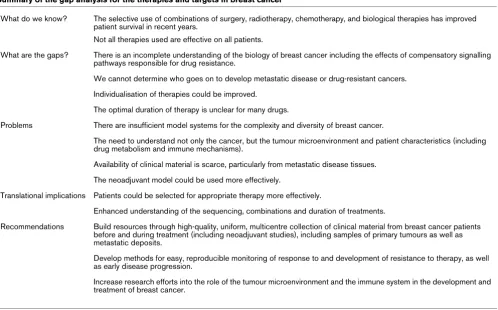
Summary of the gap analysis for the therapies and targets in breast cancer
What do we know? The selective use of combinations of surgery, radiotherapy, chemotherapy, and biological therapies has improved patient survival in recent years.
Not all therapies used are effective on all patients.
What are the gaps? There is an incomplete understanding of the biology of breast cancer including the effects of compensatory signalling pathways responsible for drug resistance.
We cannot determine who goes on to develop metastatic disease or drug-resistant cancers.
Individualisation of therapies could be improved.
The optimal duration of therapy is unclear for many drugs.
Problems There are insufficient model systems for the complexity and diversity of breast cancer.
The need to understand not only the cancer, but the tumour microenvironment and patient characteristics (including drug metabolism and immune mechanisms).
Availability of clinical material is scarce, particularly from metastatic disease tissues.
The neoadjuvant model could be used more effectively.
Translational implications Patients could be selected for appropriate therapy more effectively.
Enhanced understanding of the sequencing, combinations and duration of treatments.
Recommendations Build resources through high-quality, uniform, multicentre collection of clinical material from breast cancer patients before and during treatment (including neoadjuvant studies), including samples of primary tumours as well as metastatic deposits.
Develop methods for easy, reproducible monitoring of response to and development of resistance to therapy, as well as early disease progression.
What are the gaps?
Incomplete understanding of the biology of breast cancer Our understanding of the many cellular and molecular proc-esses involved in the development of breast cancer is still incomplete. This hampers the identification of new therapeutic targets as well as the optimal use of the targets we know about. We have limited knowledge of which signals drive breast cancer cell growth, and how they promote the invasive nature of the disease. In addition, the role of the surrounding healthy tissues in tumour development, both at the primary and metastatic sites, needs to be clarified.
Current thinking is that to eradicate cancer cells we may need a combination of therapies targeting the tumour cells, their microenvironment and, potentially, their blood supply.
We cannot determine who goes on to develop advanced disease
Despite our best efforts a proportion of patients will develop advanced disease, and we do not currently have reliable tools to predict who these patients are. By using tumour grade, pathological node status, tumour size and other pathology fea-tures, a number of models have been designed to assess the risk of patients developing metastatic disease including the Nottingham Prognostic Index and Adjuvant Online. However, there are no established molecular markers used in clinical practice to determine with certainty whether a breast tumour is likely to metastasise to other sites, and therefore no easy way of selecting patients at early stages of the disease that will require more intensive treatment to prevent tumour progression.
In addition, there are no simple, non-invasive methods availa-ble for detecting the early stages of tumour progression, and patients often present with relatively advanced (symptomatic) disease.
Insufficient knowledge to provide precise, individualised therapies
One of the main problems when treating breast cancer is to determine which patients will benefit from particular therapeu-tic strategies, ensuring optimal results for each individual patient. Not only is this key to achieving the best possible outcome for patients who are likely to respond to any given treatment, but also to avoid treating those who will not benefit.
A few targeted treatments are available (for example, endo-crine treatments and trastuzumab) that rely on identifying a receptor present on the tumour cells. Understanding the biol-ogy of breast cancer better is likely to help us develop new anti-cancer agents that effectively target specific receptors present in only a subset of patients.
In addition to the biological characteristics of the tumours, each patient has an individual capacity to metabolise drugs.
This leads to variations in drug half-life that may partly explain why the response rate varies between patients receiving iden-tical treatments. For example, many commonly used anti-can-cer agents (including cisplatin, doxorubicin, tamoxifen and etoposide) are metabolised in the liver by enzymes of the p450 group, and there are documented variations in the activity of these enzymes between individuals.
Finally, the optimisation and combination of the current thera-pies to fit individual patients is often based on a trial and error approach, rather than a clear understanding of the biology of the tumour.
How to decide when to stop treatment?
We lack suitable methods for the early determination of recur-rence and treatment failure.
For most current therapies, there is little long-term data to sup-port when it is safe to stop treatment. If patients experience no side-effects and are free from cancer they are likely to want to continue their therapies even in the absence of any proven benefit. There are no easy, non-invasive, reproducible methods available for routinely monitoring subclinical disease progres-sion and response to adjuvant treatment, and we rely on patients to present with symptoms in order to establish whether the tumour has recurred.
Who will develop drug-resistant tumours?
The basis of drug resistance is not well understood, and as a consequence we have no reliable methods of predicting who will go on to develop resistance to the commonly used thera-pies. We do not know how to avoid a resistant phenotype developing, and is it not clear whether changes in the fre-quency of drug administration and/or length of treatment con-tribute to this process.
Incomplete understanding of the role of the immune system We do not fully understand how best to use the immune sys-tem to our advantage in breast cancer treatment, either as a vaccine or in the form of immunotherapy. The lack of suitable model systems for studying the immune response, as well as the many fundamental differences between the species most commonly used (rodents) and humans have hampered progress in this area. In addition we also lack understanding of how tumour cells suppress the immune response to ensure their survival and growth.
How do anti-cancer treatments adversely affect cancer cells?
various hormone-responsive breast cancer models that main-tain residual downstream kinase activity, proliferation and cell survival [76]. Interestingly, 'compensatory' induction of alterna-tive signal transduction is a phenomenon shared by other types of anti-cancer therapy, including anti-growth factors, chemotherapy and radiotherapy [77-79].
The full breadth and cellular impact of such compensatory sig-nalling is largely unknown in breast cancer. To fill this gap we need to use high-throughput discovery tools to profile multiple signalling pathways and to explore the concept in a broader panel of models reflective of the various breast cancer sub-types. In addition, although changes in some signalling elements (for example, HER2, activity of various kinases including MAPK, JNK and p38) have been reported to be in place by the time of relapse, the drug-induced concept is largely unexplored in patients. We need increased access to 'on therapy' clinical samples to rectify this.
What are the problems?
There are many reasons for these gaps in our knowledge, including the following: A lack of suitable model systems reflecting the complexity and diversity of breast cancer; Lim-ited access to clinical material from patients before and, in par-ticular, during treatment; A severe lack of material from metastatic deposits made available for studies of target hits and biological response to therapies.
The neoadjuvant model provides a window of opportunity where therapy can be tested in vivo in humans to assess the effects of an intervention. It allows biological evaluation of tumour markers and normal tissue responses by histological, biochemical, molecular, imaging or clinical techniques. How-ever, neoadjuvant studies have not usually involved adequate numbers of patients for what can be intensive study and indi-vidual centres often recruit too few patients in specific groups to ensure meaningful analysis; a multicentre approach is required to ensure progress.
Identifying new therapeutic targets is hampered by our limited understanding of the role of the tumour microenvironment and interactions with cancer stem cells in the development and progression of breast cancer. In addition, we do not understand the mechanisms underlying the acquired resist-ance to anti-cresist-ancer therapies. There are too few studies across disciplines to increase our understanding of the role of the immune system, an area where there is a lack of appropri-ate model systems and insufficient high-quality studies carried out in humans. Not enough attention has been paid to how drug metabolism by individuals affects response to treatment. This important point is not considered in drug trials on an indi-vidual basis, or linked to measurements of response, but may partly explain why there is such variation between patients receiving identical treatments. As assays for drug metabolising
cytochrome p450s become available (for example, 2D6 for tamoxifen metabolism), this may move into clinical practice.
These gaps may be filled by developing improved model sys-tems that seek to reflect the complexity of the human disease, combined with increased efforts to design multicentre studies using clinical material collected and processed in a uniform way. Particular areas that we need to strengthen are the increased use of neoadjuvant studies, providing researchers with valuable clinical material from breast cancer patients dur-ing treatment in the form of repeated biopsies.
Few studies have involved investigations of metastatic depos-its, so our understanding of the biological changes of the tumour cells as they adapt to new environments is limited. Not many patients will undergo procedures that allow the collec-tion of material from metastatic sites, and no single centre is likely to be able to collect significant numbers of quality spec-imens for research. As building these types of 'biorepositories' for future research is likely to take many years to accumulate numbers that allow meaningful data analysis, this requires a collaborative, long-term approach for which funding may be difficult to obtain (most funding bodies operate on a three- to five-year timescale before results are expected).
We therefore see an urgent need for high-quality, comprehen-sive, longitudinal sample collections (tumour, DNA, serum, plasma, urine) from breast cancer patients, coupled with extensive clinical information. Where appropriate, collabora-tions with researchers in other fields (for example, immunolo-gists working in rheumatology or auto-immune diseases) are needed.
Translational implications
5. Disease Markers in Breast Cancer
A summary of the gap analysis for disease markers in breast cancer is given in Table 5.
Developing new treatments for breast cancer and refining existing regimens are clearly important and exciting areas of research. The challenge is to ensure that new therapies reach the patients who will benefit most, and to identify patients for whom the harms outweigh the benefits or for whom the treatment will be ineffective [80]. To achieve these aims we need validated predictive and prognostic markers.
What do we know?
The gold standard for comparing new markers is testing against high-quality pathological assessment of tumour type, size, grade and lymph node stage.
Only two markers have been established so far in the routine assessment of breast cancer: ER (for predicting response to endocrine therapies) [80]; HER2 (for predicting response to trastuzumab) [80,81].
Although theses markers for predicting the response to endo-crine and biological therapies are already available, even ER and HER2 are far from perfect; for example, assessment of HER2 status will still include some non-responding patients [81].
Intelligent trial design involving multidisciplinary teams is essential to ensure new treatments are tested in patient groups stratified using biomarkers. Large-scale studies of invasive breast cancer that have and are using these principles successfully include the trials of trastuzumab after adjuvant chemotherapy in HER2-positive breast cancer [81]. Biomar-ker-based trials may also be used to assess treatments for advanced breast cancer, but may be confounded by prior exposure of such cancers to multiple therapies.
Ideally, however, pre-operative (neoadjuvant) studies are required, using clinically relevant models and crossover designs to provide early evidence of a therapeutic effect and to differentiate responsive from non-responsive groups of patients [82].
What are the gaps?
Innovative trial and study design
[image:14.612.57.552.448.725.2]Disease marker concepts should be applied to trials of treat-ments for pre-invasive disease including DCIS and to models of sentinel lymph node assessment, where funding is limited and where long-term follow-up is required to obtain robust clinical data, but where we need a better understanding of the pathophysiological processes involved. Two areas we need to address are as follows: The level of sentinel lymph node involvement that has a clinical impact; Optimum protocols for
Table 5
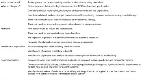
Summary of the gap analysis for disease markers in breast cancer
What do we know? Patient groups can be successfully stratified in clinical trials using biomarkers.
What are the gaps? Optimum protocols for pathological assessment of DCIS and sentinel lymph nodes.
Combining clinical, radiological, pathological and genomic data in trial populations.
No robust validated markers have yet been developed for predicting response to chemotherapy or radiotherapy.
There is no consensus for markers indicative of resistance to therapy.
There is a need for improved prognostic indices based on disease markers.
Problems New assays must be robust and reproducible.
There is a need for standardisation of tissue handling.
The impact of legislation, industrial involvement and academic pressures.
Networks of collaboration employing systems biology are required.
Translational implications Accurate recognition of the diversity of breast cancer.
Identification of patients most likely to benefit.
Identification of patients least likely to benefit from therapy and hence able to avoid toxicity.
Recommendations Design innovative trials and translational studies to develop and evaluate predictive and prognostic markers.
Develop close multidisciplinary collaboration with high-quality histopathology and rigorous scientific assessments to validate new markers important for patient outcome.
pathological assessment and systems for reproducibly cate-gorising clinically relevant pathological metastatic disease [83].
Additional challenges include recognising the differences between laboratory studies and in vivo studies in humans where interactions between tumour and stroma, three-dimen-sional effects and vascularisation become relevant. Further-more, combining clinical, radiological, pathological and genomic data in trial populations with innovative trial designs (such as in the MINDACT trial [84]) will allow us to relate, com-pare and combine established markers to, and with, new tech-nologies in a range of settings. Success depends on close multidisciplinary collaboration at an early stage, as well as the highest quality histopathological and scientific evaluation.
Validating new markers
New markers may be best validated in trials of neoadjuvant or adjuvant therapies and in advanced disease, where clinical data and outcomes are robust and statistically significant. However, large numbers of patients may be required for small incremental differences in outcome.
The key question is: does introducing a new marker change clinical practice and therefore patient outcome? In the past, researchers have not paid enough attention to experimental design when assessing this; consequently the results have not always been standardised, reproducible or robust enough to apply to clinical practice. Developing rigorously controlled rea-gents, technical methods and appropriate interpretation requires adequate resources and to date this has rarely been forthcoming.
While ER-negative patients rarely respond to hormone thera-pies, a proportion of those classified as having ER-positive dis-ease will also not respond [85]. No robust validated markers have yet been developed for predicting response to chemo-therapy or radiochemo-therapy. Many markers are favoured by local enthusiasts (for example, progesterone receptor [PgR], PS2 and cathepsin D) but we need high-quality clinical evidence to support their more widespread use.
Markers indicative of resistance to therapy (for example, ER-positive, PgR-negative tamoxifen-resistant cancers) have been proposed, but there is little agreement about methodology or cut-offs of scores for clinical application, or indeed their overall value. In addition, some markers may not be useful once regi-mens or therapies are superseded. We therefore need to com-pare, and potentially combine, markers such as the ER and PgR with pathological markers (such as histological type, grade and node metastasis), which have prognostic impor-tance. Funding for robust studies evaluating these markers is crucial, but is rarely achieved without financial support from the pharmaceutical industry.
Researchers have identified validated intermediate endpoints, such as the effects on apoptosis, proliferation and ER down-regulation (for example, with fulvestrant therapy). However, fur-ther work is urgently needed using tumour pathology (for example, lymphovascular invasion and microstaging) and molecular biology, and exploring the potential of disease response markers, such as serum proteomic markers and cir-culating tumour cells. Subclass-specific markers based on microarray approaches have been identified and validated by immunohistochemistry [86,87], but have yet to be applied in clinical trials and clinical practice.
Therefore, although breast cancer can be classified according to histology and expression of RNA and protein, for example, into basal, HER2, luminal A and luminal B and normal breast-like subtypes [86,87], we need to develop this further to pre-dict the prognosis of each subtype and the likelihood of response to therapies.
A further challenge is in understanding the complex factors influencing prognosis and in improving the quality control of reagents used in new technologies (such as proteomics, phosphoproteomics, epigenetics and assays of microRNA) so that they can be effectively applied to routinely available clini-cal material. In particular, integrating old and new methods and combining techniques (such as TMA, immunohistochemistry, fluorescence in situ hybridisation, data storage and analytical methods) may allow us to develop new composite prognostic indices. A significant challenge to the development and valida-tion of predictive markers is counteracting the effects of stor-ing tumours and blood derivatives on proteins (includstor-ing phosphorylated proteins) and in proteomic studies. We clearly need careful research into these processes and how to take them into account.
What are the problems?
Any new assays that are developed must stand up to the day-to-day challenges of clinical practice. The quality of service delivered in routine clinical practice varies, even for basic markers such as ER and HER2 [88]. RNA-dependent assays have been considered less robust than protein- or DNA-based assays in the breast cancer setting. However, RNA-based polymerase chain reaction (PCR) technology may well become a standard of care as an intra-operative detection method for tumour in sentinel lymph nodes. Techniques must undergo regular quality assurance both during development (in the research setting) and in subsequent laboratory and clin-ical use; marker validation requires time and resources and subsequently convincing the professions to apply them.