ISSN Online: 2165-3364 ISSN Print: 2165-3356
DOI: 10.4236/ojvm.2018.84006 Apr. 30, 2018 47 Open Journal of Veterinary Medicine
Excessive Moderator Band in a Female Cat’s
Left Ventricle
Eduarda Aléxia Nunes Louzada Dias Cavalcanti
1*, Stefânia dos Santos Gazzinelli
2,
Mary Suzan Varaschin
3, Guilherme Albuquerque de Oliveira Cavalcanti
11Department of Veterinary Medicine, Universidade Federal de Pelotas, Pelotas, Brazil 2Faculdade de Estudos Administrativos de Minas Gerais (FEAD), Belo Horizonte, Brazil 3Department of Veterinary Medicine, Universidade Federal de Lavras, Lavras, Brazil
Abstract
This report describes a case of cardiomyopathy associated with excessive moderator bands in a 7 years old cat. In the cat echocardiography examina-tion was observed cardiomegaly, decreased systolic funcexamina-tion and thinning of the left ventricle wall. The electrocardiogram showed the occurrence of su-praventricular tachycardia. The treatment was initiated with digoxin, furose-mide and taurine supplementation, with an improvement of the clinical signs, however the animal died in a fall. The diagnosis of cardiomyopathy associated with excessive moderator band, was based on the multiple cardiac bunds which connect the septum to the free wall of the left ventricle observed in the cat necropsy.
Keywords
Cardiomyopathy, Feline, Cardiac Muscle Bundles
1. Introduction
The excessive moderator bands in the left ventricle (MBLV) are a rare congenital anomaly in domestic cats, whose clinical signs are similar to those of most heart diseases [1] [2].
Moderator bands are defined as tendon muscle bundles which connect the septum to the free wall ventricle [3] and in mammals are often found in the right ventricle, and are rarer in the left ventricle [1] [3]. It’s believed that the function of these structures is to prevent a great strain during ventricular diastole [2].
While moderator bands aren’t related to heart disease in most species, when presented in excess in the left ventricle in cats, they are associated with heart How to cite this paper: Cavalcanti,
E.A.N.L.D., dos Santos Gazzinelli, S., Vara-schin, M.S. and de Oliveira Cavalcanti, G.A. (2018) Excessive Moderator Band in a Female Cat’s Left Ventricle. Open Journal of Veterinary Medicine, 8, 47-53.
https://doi.org/10.4236/ojvm.2018.84006
Received: February 2, 2018 Accepted: April 27, 2018 Published: April 30, 2018
Copyright © 2018 by authors and Scientific Research Publishing Inc. This work is licensed under the Creative Commons Attribution International License (CC BY 4.0).
DOI: 10.4236/ojvm.2018.84006 48 Open Journal of Veterinary Medicine The objective of this report is to present and discuss the clinical, hematologi-cal, radiologihematologi-cal, echocardiography, electrocardiographic aspect, the specific treatment and the necropsy findings in a cat that developed congestive heart failure (CHF) due to MBLV.
2. Case Report
A female mongrel cat (7 years-old, 3.3 kg), was treated at a veterinary hospital located in Belo Horizonte/MG. According to the owner, the animal has had dif-ficulty breathing and decreased appetite for about 30 days, and has been treated for 7 days with enrofloxacin (2.5 mg/kg twice daily), furosemide (2 mg/kg twice daily) and prednisolone (1 mg/kg twice daily), with the symptoms worsening in the days prior to the visit.
On a physical examination, the patient had severe dyspnea, with wheezing and abdominal breathing pattern, and chest expansion restriction. The auscultation revealed muffled sounds in the broncho-alveolar ventral thoracic region, and crackles in the lung’s fields of the dorsal region. The chest percussion sound has showed massive in its ventral portion. At thoracentesis, a 200 mL of serosan-guinous fluid were obtained, and there was improvement in breathing pattern. Hypothermia, tachycardia and dehydration were also observed. Hydration, oxy-gen and antibiotics therapy was initiated.
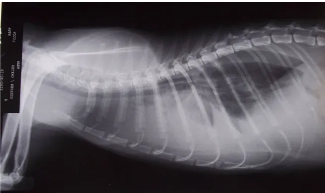
The Radiography performed a few hours after the initial treatment already in-dicated new accumulation of fluid in the chest (Figure 1). However, radio-graphic studies performed shortly after the second thoracentesis, in which 250 mL of fluid was obtained, demonstrated the occurrence of cardiomegaly, with a large increase of 11 VHS (VHS reference value 7.5 ± 0.3) [6].
On the third day of the hospital internment, we opted for the insertion of a chest tube with the purpose of draining the effusion, because daily thoracenteses was necessarily, in which an average of 250 mL fluid was obtained.
DOI: 10.4236/ojvm.2018.84006 49 Open Journal of Veterinary Medicine
Figure 1. Increased of thoracic ventral radiopacity in lateral-lateral radiographic view, a
typical finding of pleural effusion.
Echocardiography examination performed on the fifth day of hospitalization showed large bi-ventricular increase (Left ventricular internal diameter in dias-tole-19 mm; reference 9 mm), left atrial enlargement (Ae/AoB-3.2), right atrial subjectively enlargement (Figure 2), decreased systolic function (EF 27% AD% and 11%) and thinning of the left ventricle wall (LV free wall in diastole = 3 mm) [7]. The electrocardiogram showed the occurrence of supraventricular ta-chycardia.
After the echocardiogram, the treatment was initiated with digoxin (0.005 mg/kg twice daily), furosemide (4.0 mg/kg twice daily) and taurine supplemen-tation (250 mg twice daily), with an improvement of the clinical picture, with progressive reduction in pleural effusion, reaching 8 ml in four days. The elec-trocardiogram was normalized and the animal received hospital discharge four days after the initiation of the treatment mentioned above.
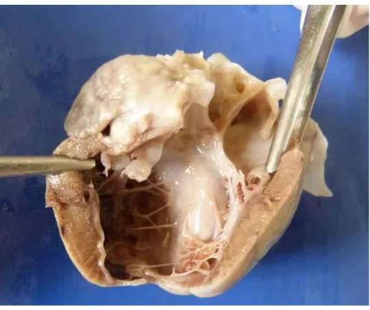
In a return 10 days after hospital discharge, the owner reported mild tachyp-nea and 220 mL of liquid were obtained at thoracentesis. However, the patient was stable for three months, when she fell from the fourth floor of the building where she lived and became deceased. The autopsy showed abdominal effusion, and thoracic and cardiac dilation and a discrete steeper widespread quantity in LV. It was observed that the endocardium still presented itself diffusely thick-ened and whitthick-ened and the presence of moderator bands in both loads between the septum and the ventricular papillary muscle and between the walls of LV (Figure 3).
DOI: 10.4236/ojvm.2018.84006 50 Open Journal of Veterinary Medicine
Figure 2. Cardiac enlargement evidenced in the right parasternal view of the
[image:4.595.208.537.70.270.2]echocardiogram. LA-left atrium; RA-right atrium; LV-left ventricle; RV-right ventricle.
Figure 3. The image shows the endocardium presenting diffusely
thick-ened and white and the presence of moderator bands in both loads be-tween the septum and the ventricular Papillary muscle and bebe-tween the walls of left ventricle.
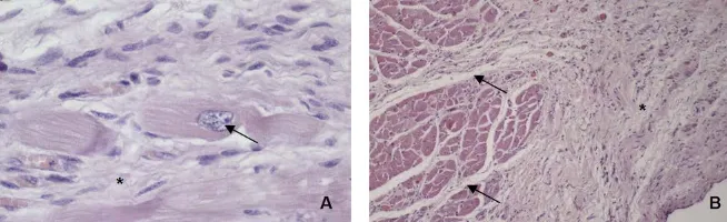
volume, and there was a rare focus of lymphocytic infiltration (Figure 4).
3. Discussion
[image:4.595.239.509.310.536.2]DOI: 10.4236/ojvm.2018.84006 51 Open Journal of Veterinary Medicine
Figure 4. Histopathology slides stained with hematoxylin and eosin shows fibrosis (*)
and increased myocyte nucleus (arrow) in (A) (×200), and the endocardium with marked proliferation of collagen fibers (*) and fibrous connective tissue (arrow) in (B) (×40).
they can cause congestive heart failure (CHF), diastolic deficiency or arrhythmia [8] [9].
The excessive moderator bands in the cat’s left ventricle (MBLV) represent a congenital anomaly, and if there isn’t a confirmed racial predisposition, cats of any age can be affected [10].
The left ventricular cardiomegaly is the most common abnormality (58%) among patients with MBLV and it’s also more common in older animals suffer-ing from this condition [5]. In this report we observed LV dilation with tapersuffer-ing chamber, matching the most common form of the disease’s symptom, besides being the one that occurs the most in older animals carrying these diseases.
It is believed, based on the observed etiology, pathogenesis and histopatholo-gy, that a diastolic disability has occurred in this case, with compensatory con-centric hypertrophy due to the low cardiac output, leading to the death of the cardiomyocytes and replacement by fibrous tissue with subsequent cardiac dila-tion, thinning of ventricular wall and a decrease in systolic function observed in echocardiography, as already alerted [2] [11] [12]. However, some authors [1] [13] don’t believe in the possibility of MBLV causing heart disease in cats.
There are no significant differences in humans in the prevalence of moderat-ing bands among normal hearts and those with congenital abnormalities [8], and there have also been similar findings in dogs [4]. In felines, however, 90% of animals that has MBLV have heart disease [5], which leads us to believe that in this species the excessive MBLV constitute a cause of heart disease.
[image:5.595.210.537.73.173.2]cardiomyo-DOI: 10.4236/ojvm.2018.84006 52 Open Journal of Veterinary Medicine miting, paraplegia due to thrombi, syncope and gallop rhythm [2] [5]. The cat in this report showed the age and the most common clinical signs described in the literature: dyspnea, hypothermia, dehydration, anorexia and pleural effusion.
The ECG changes occur due to cardiac dilatation or the ability of MBLV in conducting erratic electrical stimuli and include right bundle branch block, atrioventricular block, left anterior fascicular, sinus bradycardia [5] and supra-ventricular tachycardia [14]. In this report suprasupra-ventricular tachycardia was at-tributed to severe atrial dilation, which was resolved with the use of dioxin.
It is reported in the scientific community that the levels of ALT, AST, ALP, bi-lirubin serum, phosphorus and nitrogen may increase due to the hepatic conges-tion or due to a reducconges-tion in renal blood flow which occurs at ICC [15]. Hypo-kalemia may also occur in anorexic animals [1], so changes in the biochemical evaluation found in this animal are compatible with the CHF.
In cases of MBLV the therapy is applied according to the developed cardi-omyopathy. When dilated cardiomyopathy occurs, the indicated treatment is based on the use of digitalis, diuretics, and vasodilators [16], while cases of hypertrophic cardiomyopathy are treated with beta blockers such as atenolol, calcium channel blockers and diuretics [10].
4. Conclusions
The animal presented in this report was treated with dioxin and furosemide, with an appropriate framework control during the three follow-up months. The administration of taurine was instituted due to the evidence of dilated cardi-omyopathy, which in cats could be related to the deficiency of the amino acid taurine, but the authors believe that the supply of the amino acid did not influ-ence the clinical outcome.
We have concluded, therefore, that treatment with digoxin and furosemide in cats with MBLV CMD may be effective in the short term, however the scientific community needs further studies to evaluate whether the MBLV is actually causing heart disease in domestic cats.
References
[1] Fox, P.R. (1999) Textbook of Canine and Feline Cardiology, Principle and Clinical Practice. WB Saunders, Philadelphia, 621-678.
[2] Wray, J.D., Gajanayake, J. and Smith, S.H. (2007) Congestive Heart Failure Asso-ciated with a Large Transverse Left Ventricular Moderator Band in Cats. Journal Feline Medicine Surgery, 9, 56-60. https://doi.org/10.1016/j.jfms.2006.03.006
DOI: 10.4236/ojvm.2018.84006 53 Open Journal of Veterinary Medicine
on Moderator Bands in the Heart of Ostrich (Stuthio camelus). Global Veterinária, 4, 374-379.
[4] Koie, H., Hara, A., Sakai, M., Takiyama, N. and Uechi, M. (2007) Clinical Evalua-tion of Left Ventricular Moderator Band in 12 Dogs. Journal Veterinary Medicine Science, 69, 965-967. https://doi.org/10.1292/jvms.69.965
[5] Liu, S., Fox, P.R. and Tilley, L.P. (1982) Excessive Moderator Bands in the Left Ven-tricle of 21 Cats. JAVMA, 180, 1215-1219.
[6] Litster, A.L. and Buchanan, J.W. (2000) Vertebral Scale System to Measure Heart Size in Radiographs of Cats. Journal of the American Veterinary Medical Associa-tion, 216, 210-214. https://doi.org/10.2460/javma.2000.216.210
[7] Boon, J.A. (1998) Manual of Veterinary Echocardiography. Willians & Wilknis, Baltmore, 478.
[8] Gerlis, L.M., Wright, H.M., Wilson, N., Erzengin, F. and Dickison, D.F. (1984) Left Ventricular Bands: A Normal Anatomical Features. British Heart Journal, 52, 641-647. https://doi.org/10.1136/hrt.52.6.641
[9] Schober, K. and Todd, A. (2010) Echocardiographic Assessment of Left Ventricular Geometry and the Mitral Valve Apparatus in Cats with Hypertrophic Cardiomyopathy. Journal Veterinary Cardiology, 12, 1-16.
https://doi.org/10.1016/j.jvc.2009.09.004
[10] Ferasin, L. (2009) Feline Myocardial Disease 2: Diagnosis, Prognosis and Clinical Management. Journal Feline Medice Surgery, 11, 183-194.
https://doi.org/10.1016/j.jfms.2009.01.002
[11] Baty, C.J., Malarkey, D.E., Atkins, C.E., Defrancesco, T.C., Sidley, J. and Keene, B.W. (2001) Natural History of Hypertrophic Cardiomyopathy and Aortic Throm-boembolism in a Family of Domestic Shorthair Cats. Journal Veterinary Internal Medicine, 15, 595-599. https://doi.org/10.1111/j.1939-1676.2001.tb01598.x
[12] Wolf, O.A., Imgrund, M. and Wess, G. (2017) Echocardiographic Assessment of Feline False Tendons and Their Relationship with Focal Thickening of the Left Ventricle. Journal of Veterinary Cardiology, 19, 14-23.
https://doi.org/10.1016/j.jvc.2016.08.008
[13] Kimura, Y., Karakama, S., Kobayashi, M. and Machida, N. (2015) Incidence, Dis-tribution and Morphology of Left Ventricular False Tendons in a Cat’s Hearts.
Journal of Veterinary Medicine, 45, 490-493..
[14] Ferasin, L., Sturgess, C.P., Cannon, M.J., Caney, S.M., Gruffydd-Jones, T.J. and Wotton, P.R. (2003) Feline Idiopathic Cardiomyopathy: A Retrospective Study of 106 Cats (1994-2001). Journal Feline Medicine Surgery, 5, 151-159.
https://doi.org/10.1016/S1098-612X(02)00133-X
[15] Cavalcanti, G.A.O., Tatibana, L.S., Varaschin, M.S., Araújo, R.B. and Costa Val, A.P. (2011) Atrial Septal Defect in an Elderly Dog. Arquivo Brasileiro Medicina Ve-terinaria e Zootecnia, 63, 503-507.
https://doi.org/10.1590/S0102-09352011000200033