EFFECT ON CANCER CELL GROWTH BY METHANOLIC
EXTRACT OF
GORDONIA OBTUSA
WALL.EXUT
AND ARN, AN
ENDEMIC WILD TEA PLANT
Shalimol. A*, K. Arumugasamy, M. R. Udhayasankar, R. Nantha kumar, V. Asha devi and P. Uma Maheswari
PG and Research Department of Botany, Kongunadu arts and science college (Autonomous), Coimbatore- 641029, Tamilnadu, India.
ABSTRACT
The methanolic extract of Gordonia obtusa (Theaceae) was evaluated for its effects on growth in two malignant cell lines including a Human Lung cancer cell lines (A549) and Colon cancer cell lines (HT-29) using MTT assay. In these cell lines studied, the extract decreased cell viability, inhibited cell proliferation, and induced cell death in a dose dependent manner. The ethanolic extract of G. obtusa showed good cytotoxicity and their IC50 values were found to be 35.12µg/mL and 6.65 µg/mL respectively. It could be a reliable source of potent pharmacophore for treatment of disease like cancer.
KEY WORDS: Gordonia obtusa, Colon cancer, Lung cancer.
INTRODUCTION
Cancer is a deadly disease and affects a considerable number of people worldwide. Because of the complexity of human cancer, alternative management may be needed to improve the efficacy of therapeutic treatments and the quality of patients[1]. Natural products have long been a fertile source to cure the cancer. There are different anticancer herbs from plants that have been used by different cultures throughout time for medicinal purposes[2].Consumption of tea has been associated with many health benefits, and tea’s role and mechanism in cancer
chemoprevention have been extensively reviewed and one of the key advantages of GT as a cancer preventative is its nontoxicity[3]. Gordonia obtusa wall. exut and Arn is a wild tea plant in the tea family, Theaceae. Gordonia is a genus of flowering plants in the family
Volume 3, Issue 6, 787-792. Research Article ISSN 2277 – 7105
Article Received on 04 June 2014,
Revised on 29 June 2014, Accepted on 24 July 2014
*Correspondence for
Author
Shalimol Anamika
PG and Research Department
of Botany, Kongunadu arts and
science college (Autonomous),
Coimbatore- 641029,
Theaceae, of the roughly 40 Sps. all two are native to South east Asia in Southern China, Taiwan. The remaining species are native to South East North America. They are evergreen trees growing to 10-20 M tall. The bark is thick and deeply fissured. The leaves are alternatively arranged, simple, serrated, thick, leathery, glossy and 6-18 cm. long. The flowers are large and conspicuous. The fruit is dry 5-valved capsule with 1-4 sees in each section. They have inflammatory, antioxidant medicinal properties.
MATERIALS AND METHOD
Cytotoxicity Study: Cell line used, Cell line and culture.
Human Lung cancer (A549 ) and Human colon adenocarcinoma (HT-29 ) cell lines were
obtained from National centre for Cell Sciences, Pune (NCCS). The cells were maintained in
RPMI-1640 supplemented with 10% FBS, penicillin (100 U/ml), and streptomycin (100
μg/ml) in a humidified atmosphere of 50 μg/ml CO2 at 37 °C.
Cell proliferation kit: MTT assay kit.
Reagents
RPMI-1640 was purchased from GIBCO/BRL Invitrogen (Caithershurg, MD). Fetal bovine
serum (FBS) was purchased from Gibco laboratories Trypsin, methylthiazolyl diphenyl-
tetrazolium bromide (MTT), and dimethyl sulfoxide (DMSO) were purchased from Sisco
research laboratory chemicals, Mumbai. All of other chemicals and reagents were obtained
from Sigma Aldrich Mumbai.
Glass wares and plastic wares
96-well micro titer plate, Tissue culture flasks, Falcon tubes, Reagent bottles
Equipments
Fluorescence inverted microscope (Leica DM IL), Biosafety cabinet class II (Esco), cytotoxic safety cabinet (Esco), CO2 incubator (RS Biotech, mini galaxy A), Deep freezer, ELISA plate reader (Thermo), Micropipettes
Preparation of plant extract
kept on a rotary and shaken for 2 days. Then filtered and supernatant was collected and evaporated, and made the final volume one-fourth of the original volume and stored in air tight bottles[4].
Microculture tetrazolium (MTT) assay principle
This Colorimetric assay is based on the capacity of Mitochondria succinate dehydrogenase enzymes in living cells to reduce the yellow water soluble substrate 3-(4, 5-dimethyl thiazol-2-yl)-2, 5-diphenyl tetrazolium bromide (MTT) into an insoluble, colored formazan product which is measured spectrophotometrically. Since reduction of MTT can only occur in metabolically active cells, the level of activity is an indication of the viability of the cells.
Procedure In vitro assay for Cytotoxicity activity (MTT assay).
The Cytotoxicity study of samples on cancer cells was determined by the MTT assay[5]. Cells
(1 × 10⁵/well) were plated in 100 μl of medium/well in 96-well plates (Hi media). After 48
hours incubation the cell reaches the confluence. Then, cells were incubated in the presence
of various concentrations of the samples in 0.1% DMSO for 48h at 37°C. After removal of the sample solution and washing with phosphate-buffered saline (pH 7.4), 20µl/well (5mg/ml) of 0.5% 3-(4,5-dimethyl-2-thiazolyl)-2,5-diphenyl--tetrazolium bromide cells(MTT) phosphate- buffered saline solution was added. After 4h incubation, 0.04M HCl/ isopropanol was added. Viable cells were determined by the absorbance at 570nm with reference at 655nm. Measurements were performed in 3 times, and the concentration required for a 50% inhibition of viability (IC50) was determined graphically. The absorbance at 570 nm was measured with a microplate reader (Bio-Rad, Richmond,CA ), using wells without
sample containing cells as blanks. All experiments were performed in triplicate. The effect of
the samples on the proliferation of human colon and lung cancer cells were expressed as the
% cell viability, using the following formula: % cell viability = A570 of treated cells / A570
of control cells × 100%.
RESULTS AND DISCUSSION
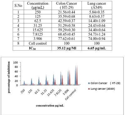
In vitro confirmation of this cytotoxicity of the G. Obtusa extract on Human Lung cancer cell
treating A 549 cancer cells and HT-29 cancer cells, the methanolic extract of G. obtusa shows good cytotoxicity and with an IC50 value of 35.12 μg/mL and 6.65 µg/mL respectively.
[image:4.595.98.498.186.554.2]Table 1: In vitro cytotoxicity effect of Gordonia obtusa extract on Colon and Lung cancer cell lines.
Figure 1: In vitro cytotoxicity of different concentrations of methanolic extract of
Gordonia obtusa.
Green tea is rich in poly phenolic compounds called catechins, which accounts for the one -third of the dry leaves. GT consists of four different types of catechins: epigallocatechin-3-gallate(EGCG), epigallocatechin, epicatechin-3-gallate and epicatechin[6 ].EGCG is the most abundant and powerful antioxidant in green tea for cancer chemoprevention[7]. The previous study showed that EGCG enhanced the effects of ginseng compounds in the inhibition of colon cancer cell growth, indicating that green tea could be an effective synergist with anticancer drugs for cancer chemoprevention[8].Tea catechins appear to inhibit human
0 20 40 60 80 100 perc ent a g e o f inh ibi tio n concentration µg/mL
Colon Cancer ( HT-29)
Lung cancer (A549)
S.No Concentration (μg/mL)
Colon Cancer ( HT-29)
Lung cancer (A549)
1 250 21.56±0.44 5.84±0.35
2 125 35.59±0.68 8.63±0.37
3 62.5 42.59±0.37 14.40±1.09
4 31.25 51.29±0.58 24.43±0.64
5 15.625 59.29±0.30 34.40±0.64
6 7.8125 68.45±0.45 54.73±1.24
7 3.906 77.62±0.61 74.00±0.94
8 Cell control 100 100
melanoma cells; both EGC and EGCG suppressed growth of human melanoma cell line UACC-375[9]. Studies investigating the association of green tea consumption with lung cancer risk have reported inconsistent findings. In support of green tea consumption on reduced lung cancer risk, a meta-analysis was conducted by a literature search in PubMed from1966 to 2008. The overall evaluation of 22 relevant studies suggests that high consumption of green tea but not black tea may be related to the reduction of lung cancer risk[10].
CONCLUSION
The role of plant derived polyphenols in chemoprevention of cancer is an emerged and important area. The study results showed that the methanolic extracts of Gordonia obtusa is promisingly cytotoxic against human colon cancer and human lung cancer cell lines. Therefore, purification of G. obtusa extract in future studies is suggested.
REFERENCES
1. Xu, Z.; Chen, X.; Zhong, Z.; Chen, L.; Wang, Y. Ganoderma lucidum polysaccharides: Immunomodulation and potential anti tumour activities. Am.J. Chin.Med.2011; 39:15-29 2. M.Umadevi, K.P.Sampath kumar, Debjit Bhowmik, S.Duraivel. Traditionally used
Anticancer Herbs in India. Journal of medicinal plant studies 2013;1:59
3. Kuzuhara T, Suganuma M, Fujiki H: Green tea catechin as a chemical chaperone in cancer prevention. Cancer Lett 2008;261:12-20.
4. Parekh J,Nair R and Chanda S. Priliminary screening of some folkloric plants from Western India for potential antimicrobial activity;Indian journal of pharmacology,vol.37,2005,408-409.
5. Mosmann T. Rapid colorimetric assay for cellular growth and survival: application to proliferation and cytotoxicity assays. J Immunol Methods 1983; 65(1-2): 55-63.
6. Balentine DA, Wiseman SA, Bouwens LCM: The chemistry of tea flavonoids. Crit Rev Food Sci Nutr 1997;37:693-704.
7. Lambert, J.D.; Elias, R.J. The antioxidant and pro-oxidant activities of green tea polyphenols: A role in cancer prevention. Arch.Biochem. Biophys.2010, 501, 119-125. 8. Fujiki, H.; Suganuma, M. Green tea: An effective synergist with anticancer drugs for
9. Valcic S. Timmermann BN, Alberts DS, Wachter GA, K rutzsch M, Wymer J, et al. Inhibitory effect of six green tea catechins and caffeine on the growth of four selected human tumour cell lines. Anticancer Drugs 1996;7:461-8.