organic papers
o338
Carlos A. L. Filguieraset al. C4H12N+I5ÿ DOI: 101107/S1600536801004536 Acta Cryst.(2001). E57, o338±o340 Acta Crystallographica Section EStructure Reports
Online
ISSN 1600-5368
Tetramethylammonium pentaiodide
Carlos A. L. Filgueiras,aAdolfo
Horn Jr,aJanet M. S. Skakleb*
and James L. Wardellb²
aInstituto de Quimica, Departamento de
Quimica Inorganica, Universidade Federal do Rio de Janeiro, 21945-970 Rio de Janeiro, RJ, Brazil, andbDepartment of Chemistry,
Univer-sity of Aberdeen, Meston Walk, Old Aberdeen AB24 3UE, Scotland
² Also at Instituto de Quimica, Departamento de Quimica Inorganica, Universidade Federal do Rio de Janeiro, 21945-970 Rio de Janeiro, RJ, Brazil.
Correspondence e-mail: [email protected]
Key indicators
Single-crystal X-ray study
T= 296 K
Mean(I±I) = 0.001 AÊ
Rfactor = 0.052
wRfactor = 0.143
Data-to-parameter ratio = 45.5
For details of how these key indicators were automatically derived from the article, see http://journals.iucr.org/e.
#2001 International Union of Crystallography Printed in Great Britain ± all rights reserved
The title compound, C4H12N+I5ÿ, consists of V-shaped I5ÿ
ions which are linked to adjacent I5ÿions to form a ¯at square
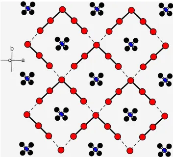
mesh normal to (001), with four I atoms on each edge. The tetramethylammonium ions are located at the centre of each square.
Comment
Various polyiodide ions are known, with the most frequently reported being I3ÿ and I5ÿ (Kloo et al., 2000; Blake et al.,
1998). The structures of a number of tetraorganoammonium salts of I5ÿare in the Cambridge Structural Database (CSD;
Allen & Kennard, 1993) at the Chemical database service of the EPSRC (Fletcheret al., 1996), including those of [EtMe-Ph2N]I5 (213550, refcode FIQLAS; Tebbe & Loukili, 1999),
{2[Me2Ph2N][I5]2I2} (161933, ZIVXOR; Tebbe & Gilles,
1996a), [EtMe3N]I5(Loukili & Tebbe, 1999), [PriMe2PhN]I5I2
(195990, PAZQEM; Tebbe & Loukili, 1998), [Pr4N]I5(Tebbe
& Gilles, 1996b) and [Me4N]I5(130287, DULZOZ; Tsvetkov et al., 1986). However, only the data for cell dimensions and space group are provided for [Me4N]I5 in the CSD. The
structure of [Me4N]I5 had also featured in earlier studies
(Broekemaet al., 1959; Hach & Rundle, 1951), while a listing of cell dimensions and the space group for [Me4N]I5, with a
reference to a dissertation (Loukili, 1998), were provided by Loukili & Tebbe (1999).
Tetraorganoammonium polyiodides are generally prepared from [R4N]I and the requisite number of molar equivalents of
iodine in solvents such as methanol. Tetramethylammonium pentaiodide was isolated in this study from aqueous solutions of [Me4N]I and SnI2 (equimolar) in water, the co-product
being yellow±brown hydrated tin(IV) oxide. Similar reactions between [Me4N]Xand SnX2(X= Cl or Br) in contrast simply
led to [Me4N][SnX3].
The re®nement of the carbon displacement parameters for the tetramethylammonium ion gave very high values, suggesting disorder. This was modelled using two super-imposed orientations of the moleculeviatheSHELX PART
instruction. The C atoms could not be re®ned anisotropically, but the ®nal ratio of the two orientations was 85:15. However,
theRvalue was not improved (5.75% compared to 5.14% for the ordered model) and thus the ordered model has been retained for this report.
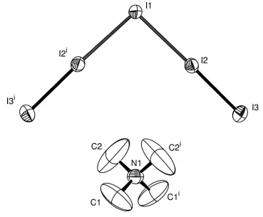
The structure of the title compound, (I) (Fig. 1), consists of isolated tetramethylammonium ions and I5ÿions. The I5units
are linkedvialonger IÐI bonds to form a square mesh normal to (001) (Fig. 2), with I1 coordinated to two I2 atoms at 3.1514 (8) AÊ and two I3 at 3.6453 (9) AÊ [symmetry codes: (i)
1
2ÿx, y+12,12ÿz; (ii)xÿ12,y+12,z], which are within the van
der Waals radius sum of 3.96 AÊ. The tetramethylammonium ions are located at the centre of each square.
This square mesh is similar to that found in (EtMe3N)I5
(Loukili & Tebbe, 1999); however, in this case, the mesh is ¯at and parallel to [010], whereas in (EtMe3N)I5 there is
signi®-cant puckering, likely caused by the relative location of the cation. These are the only such square-mesh formations to be reported to date; other pentaiodide networks form herring-bone, F-shaped and linear conformations, and in a few cases isolated I5ÿanions.
Many pentaiodides are reported in the literature. In addi-tion, pentaiodide ions may form as part of a more complex polyiodide framework. This is clearly seen in the work of Tebbe and co-workers, in which the V angle varies from 83.51 in (EtMe3N)I5 (Loukili & Tebbe, 1999) to 119.60 in
bipyri-dinium heptaiodide, (C10H9N2)I7 (Tebbe & Bittner, 1995),
whereas the `straight limb' angle varies from 172.09in (Me
2
-Ph2N)3I13 (Tebbe & Gilles, 1996a) to 179.57 in (UrEt)2I8
(Grafe-Kavoosian et al., 1998). Bond lengths in the V motif can be divided into the outer (shorter) and inner (longer). The outer bond lengths range from 2.739 to 2.873 AÊ, whereas the inner range from 2.975 to 3.460 AÊ. There is, evidently, a sig-ni®cant (0.1 AÊ) gap between these values which supports the alternative description of the pentaiodide unit as [2(I2)I]ÿ.
Experimental
An equimolar solution of NMe4I and SnI2in water was maintained at
368 K for 2 h with stirring. The initial orange-coloured solution initially produced a near-black coloured precipitate which slowly redissolved to be replaced by a pale-yellow precipitate. The reaction mixture was ®ltered and green±black crystals of the title compound were deposited from the ®ltrate on standing. M.p. 398±400 K: literature value 399±400 K (Gama & Filguieras, 1989). Analysis: C 6.68, H 1.99, N 1.83%; calculated for C4H12I5N: C 6.78, H 1.71, N
1.98%.
Crystal data
C4H12N+I5ÿ
Mr= 708.64
Monoclinic,C2/c a= 13.3110 (10) AÊ
b= 13.5395 (11) AÊ
c= 8.8727 (7) AÊ = 107.801 (2)
V= 1522.5 (2) AÊ3
Z= 4
Dx= 3.092 Mg mÿ3
MoKradiation Cell parameters from 2244
re¯ections = 2.4±29.4
= 10.17 mmÿ1
T= 296 (2) K Prism, dark purple 0.600.500.50 mm
Data collection
Bruker SMART 1000 Area CCD diffractometer
'±!scans
Absorption correction: multi-scan (SADABS; Bruker, 1999)
Tmin= 0.742,Tmax= 0.906 4712 measured re¯ections
2182 independent re¯ections 1622 re¯ections withI> 2(I)
Rint= 0.032
max= 30.9
h=ÿ10!18
k=ÿ18!18
l=ÿ12!11
Re®nement
Re®nement onF2
R[F2> 2(F2)] = 0.052
wR(F2) = 0.143
S= 1.05 2182 re¯ections 48 parameters
H-atom parameters constrained
w= 1/[2(F
o2) + (0.0740P)2
+ 6.1215P]
whereP= (Fo2+ 2Fc2)/3
(/)max< 0.001
max= 1.48 e AÊÿ3
min=ÿ1.47 e AÊÿ3
Extinction correction:SHELXL
Extinction coef®cient: 0.00143 (18)
Acta Cryst.(2001). E57, o338±o340 Carlos A. L. Filguieraset al. C4H12N+I5ÿ
o339
organic papers
Figure 1
The title compound with the atomic numbering scheme. Displacement ellipsoids are drawn at the 30% probability level. H atoms have been omitted for clarity. [Symmetry code: (i)ÿx, y,ÿz+1
2.]
Figure 2
organic papers
o340
Carlos A. L. Filguieraset al. C4H12N+I5ÿ Acta Cryst.(2001). E57, o338±o340The maximum residual electron density was 1.48 e AÊÿ3at 0.89 AÊ
from I3; all residuals >0.8e AÊÿ3were within 1 AÊ of I atoms.
Data collection:SMART(Bruker, 1999); cell re®nement:SAINT
(Bruker, 1999); data reduction: SAINT; program(s) used to solve structure:SHELXS86 (Sheldrick, 1990); program(s) used to re®ne structure: SHELXL97 (Sheldrick, 1997); molecular graphics:
ATOMS (Dowty, 1999) and ORTEP-3 (Farrugia, 1999); software used to prepare material for publication:CIFTAB(Sheldrick, 1997). We wish to acknowledge the use of the EPSRC's Chemical Database Service at Daresbury (Fletcheret al., 1996). JMSS would also like to thank R. A. Howie for helpful suggestions.
References
Allen, F. H. & Kennard, O. (1993).Chem. Des. Autom. News,8, 1, 31±37. Blake, A. J., Devillanova, F. A., Gould, R. O., Li, W. S., Lippolis, V., Parsons, S.,
Radek, C. & SchroÈder, M. (1998).Chem. Soc. Rev.27, 195±205.
Broekema, J., Havinga, E. E. & Wiebenga, E. H. (1959).Acta Cryst.10, 596. Bruker (1999).SADABS,SMARTandSAINT. Bruker AXS Inc., Madison,
Wisconsin, USA.
Dowty, E. (1999).ATOMS for Windows. Version 5.0.7. Shape Software, 521 Hidden Valley Road, Kingsport, TN 37663, USA.
Farrugia, L. J. (1997).J. Appl. Cryst.30, 565±565.
Fletcher, D. A., McMeeking, R. F. & Parkin, D. (1996).J. Chem. Inf. Comput. Sci.36, 746±749.
Gama, G. J & Filguieras, C. A. L. (1989).Quim. Nova,12, 192±196. Grafe-Kavoosian, A., Nafepour, S., Nagel, K. & Tebbe, K. F. (1998). Z.
Naturforsch. Teil B,53, 641±652.
Hach, R. J. & Rundle, R. E. (1951).J. Am. Chem. Soc.73, 4321±4324. Kloo, L., Svensson, P. H. & Taylor, M. J. (2000).J. Chem. Soc. Dalton Trans.
pp. 1061±1065.
Loukili, R. (1998).Untersuchungen an Polyiodiden von Organylammonium-Kationen. Dissertation, University of KoÈln, Germany.
Loukili, R. & Tebbe, K. F. (1999).Z. Anorg. Allg. Chem.625, 650±654. Sheldrick, G. M. (1990).Acta Cryst.A46, 467±473.
Sheldrick, G. M. (1997).SHELXL97 andCIFTAB. University of GoÈttingen, Germany.
Tebbe, K. F. & Bittner, M. (1995).Z. Anorg. Allg. Chem.621, 218±224. Tebbe, K. F. & Gilles, T. (1996a).Z. Anorg. Allg. Chem.622, 138±148. Tebbe, K. F. & Gilles, T. (1996b).Z. Anorg. Allg. Chem.622, 1587±1593. Tebbe, K. F. & Loukili, R. (1998).Z. Anorg. Allg. Chem.624, 1175±1186. Tebbe, K. F. & Loukili, R. (1999).Z. Anorg. Allg. Chem.625, 820±826. Tsvetkov, A. A., Stepin, B. D., Margulis, V. B. & Mamoshin, M. Yu. (1986).Izv.
supporting information
sup-1
Acta Cryst. (2001). E57, o338–o340
supporting information
Acta Cryst. (2001). E57, o338–o340 [doi:10.1107/S1600536801004536]
Tetramethylammonium pentaiodide
Carlos A. L. Filgueiras, Adolfo Horn, Janet M. S. Skakle and James L. Wardell
S1. Comment
Various polyiodide ions are known, with the most frequently reported being I3- and I5- (Kloo et al., 2000; Blake et al.,
1998). The structures of a number of tetraorganoammonium salts of I5- are in the Cambridge Structural Database (CSD;
Allen & Kennard, 1993) at the Chemical database service of the EPSRC (Fletcher et al., 1996), including those of
[EtMePh2N]I5 (213550, refcode FIQLAS; Tebbe & Loukili, 1999), {2[Me2Ph2N][I5]2.I2} (161933, ZIVXOR; Tebbe &
Gilles, 1996a) [EtMe3N]I5 (Loukili & Tebbe, 1999), [PriMe2PhN]I5.I2 (195990, PAZQEM; Tebbe & Loukili, 1998),
[Pr4N]I5 (Tebbe & Gilles, 1996b) and [Me4N]I5 (130287, DULZOZ; Tsvetkov et al., 1986). However, only the data for
cell dimensions and space group are provided for [Me4N]I5 in the CSD. The structure of [Me4N]I5 had also featured in
earlier studies (Broekema et al., 1959; Hach & Rundle, 1951), while a listing of cell dimensions and the space group for
[Me4N]I5, with a reference to a dissertation (Loukili, 1998), were provided by Loukili & Tebbe (1999).
Tetraorganoammonium polyiodides are generally prepared from [R4N]I and the requisite number of molar equivalents
of iodine in solvents such as methanol. Tetramethylammonium pentaiodide was isolated in this study from aqueous
solutions of [Me4N]I and SnI2 (equimolar) in water, the co-product being yellow–brown hydrated tin(IV) oxide. Similar
reactions between [Me4N]X and SnX2 (X = Cl or Br) in contrast simply led to [Me4N][SnX3].
The refinement of the carbon displacement parameters for the tetramethylammonium ion gave very high values,
suggesting disorder. This was modelled using two superimposed orientations of the molecule via the SHELX PART
instruction. The C atoms could not be refined anisotropically, but the final ratio of the two orientations was 85:15.
However, the R value was not improved (5.75% compared to 5.14% for the ordered model) and thus the ordered model
has been retained for this report.
The structure of the title compound (Fig. 1) consists of isolated tetramethylammonium ions and I5- ions. The I5 units are
linked via longer I—I bonds to form a square mesh normal to (001) (Fig. 2), with I1 coordinated to two I2 atoms at
3.1514 (8) Å and two I3 at 3.6453 (9) Å [symmetry codes: (i) 0.5 - x, y + 0.5, 0.5 - z; (ii) x - 0.5, y + 0.5, z], which are
within the van der Waals radius sum of 3.96 Å. The tetramethylammonium ions are located at the centre of each square.
This square mesh is similar to that found in (EtMe3N)I5 (Loukili & Tebbe, 1999); however, in this case, the mesh is flat
and parallel to [010], whereas in (EtMe3N)I5 there is significant puckering, likely caused by the relative location of the
cation. These are the only such square-mesh formations to be reported to date; other pentaiodide networks form
herring-bone, F-shaped and linear conformations, and in a few cases isolated I5- anions.
Many pentaiodides are reported in the literature. In addition, pentaiodide ions may form as part of a more complex
polyiodide framework. This is clearly seen in the work of Tebbe and co-workers, in which the V angle varies from 83.51°
in (EtMe3N)I5 (Loukili & Tebbe, 1999) to 119.60° in bipyridinium heptaiodide, (C10H9N2)I7 (Tebbe & Bittner, 1995),
whereas the `straight limb′ angle varies from 172.09° in (Me2Ph2N)3I13 (Tebbe & Gilles, 1996a) to 179.57° in (UrEt)2I8
(Grafe-Kavoosian et al., 1998). Bond lengths in the V motif can be divided into the outer (shorter) and inner (longer). The
supporting information
sup-2
Acta Cryst. (2001). E57, o338–o340
significant (0.1 Å) gap between these values which supports the alternative description of the pentaiodide unit as [2(I2).I]-.
S2. Experimental
An equimolar solution of NMe4I and SnI2 in water was maintained at 368 K for 2 h with stirring. The initial
orange-coloured solution initially produced a near-black orange-coloured precipitate which slowly redissolved to be replaced by a
pale-yellow precipitate. The reaction mixture was filtered and green–black crystals of the title compound were deposited from
the filtrate on standing. M.p. 398–400 K: literature value 399–400 K (Gama & Filguieras, 1989). Analysis: C 6.68, H
1.99, N 1.83%; calculated for C4H12I5N: C 6.78, H 1.71, N 1.98%.
S3. Refinement
The maximum residual electron density was 1.48 e Å-3 at 0.89 Å from I3; all residuals >0.8 e Å-3 were within 1 Å of I
[image:5.610.119.488.243.548.2]atoms.
Figure 1
The title compound with the atomic numbering scheme. Displacement ellipsoids are drawn at the 30% probability level.
supporting information
sup-3
[image:6.610.128.481.70.394.2]Acta Cryst. (2001). E57, o338–o340 Figure 2
View normal to (001) showing the pentaiodine mesh and the location of the tetramethylammonium ions.
Tetramethylammonium pentaiodide
Crystal data C4H12N+·I5− Mr = 708.64 Monoclinic, C2/c a = 13.311 (1) Å b = 13.5395 (11) Å c = 8.8727 (7) Å β = 107.801 (2)° V = 1522.5 (2) Å3 Z = 4
F(000) = 1232 Dx = 3.092 Mg m−3
Mo Kα radiation, λ = 0.71073 Å Cell parameters from 2244 reflections θ = 2.4–29.4°
µ = 10.17 mm−1 T = 296 K
Prism, dark purple 0.60 × 0.50 × 0.50 mm
Data collection
Bruker SMART 1000 Area CCD diffractometer
Radiation source: fine-focus sealed tube Graphite monochromator
φ–ω scans
Absorption correction: multi-scan (SADABS; Bruker, 1999) Tmin = 0.742, Tmax = 0.906
4712 measured reflections 2182 independent reflections 1622 reflections with I > 2σ(I) Rint = 0.032
θmax = 30.9°, θmin = 2.2° h = −10→18
supporting information
sup-4
Acta Cryst. (2001). E57, o338–o340 Refinement
Refinement on F2 Least-squares matrix: full R[F2 > 2σ(F2)] = 0.052 wR(F2) = 0.143 S = 1.05 2182 reflections 48 parameters 0 restraints
Primary atom site location: heavy-atom method Secondary atom site location: difference Fourier
map
Hydrogen site location: inferred from neighbouring sites
H-atom parameters constrained w = 1/[σ2(F
o2) + (0.074P)2 + 6.1215P] where P = (Fo2 + 2Fc2)/3
(Δ/σ)max < 0.001 Δρmax = 1.48 e Å−3 Δρmin = −1.47 e Å−3
Extinction correction: SHELXL, Fc*=kFc[1+0.001xFc2λ3/sin(2θ)]-1/4 Extinction coefficient: 0.00143 (18)
Special details
Geometry. All e.s.d.'s (except the e.s.d. in the dihedral angle between two l.s. planes) are estimated using the full covariance matrix. The cell e.s.d.'s are taken into account individually in the estimation of e.s.d.'s in distances, angles and torsion angles; correlations between e.s.d.'s in cell parameters are only used when they are defined by crystal symmetry. An approximate (isotropic) treatment of cell e.s.d.'s is used for estimating e.s.d.'s involving l.s. planes.
Refinement. Refinement of F2 against ALL reflections. The weighted R-factor wR and goodness of fit S are based on F2, conventional R-factors R are based on F, with F set to zero for negative F2. The threshold expression of F2 > σ(F2) is used only for calculating R-factors(gt) etc. and is not relevant to the choice of reflections for refinement. R-factors based on F2 are statistically about twice as large as those based on F, and R- factors based on ALL data will be even larger.
Also note that for data up to theta max, the completeness is only 90%. Usually ACTA n would have been used to check the >99% completeness, but in this case with the C-centred cell, ACTA 50 gives meaningless (<50%) completeness values (commented out at the bottom of this file). In addition, several frames of data (approx. 10 in 1500) were lost. However a check with PLATON showed that the data are >99% complete at 27.5 °.
Fractional atomic coordinates and isotropic or equivalent isotropic displacement parameters (Å2)
x y z Uiso*/Ueq
I1 0.0000 0.17657 (6) 0.2500 0.0682 (3) I2 0.16060 (4) 0.01915 (4) 0.19877 (6) 0.0605 (2) I3 0.30588 (5) −0.12546 (5) 0.18005 (8) 0.0750 (3)
N1 0.0000 0.6882 (8) 0.2500 0.075 (3)
C1 0.032 (2) 0.6330 (18) 0.135 (2) 0.210 (14)
H1A −0.0277 0.5980 0.0673 0.316*
H1B 0.0859 0.5868 0.1872 0.316*
H1C 0.0589 0.6774 0.0718 0.316*
C2 0.084 (3) 0.748 (3) 0.333 (4) 0.33 (3)
H2A 0.1427 0.7387 0.2930 0.494*
H2B 0.1050 0.7316 0.4436 0.494*
H2C 0.0626 0.8164 0.3201 0.494*
Atomic displacement parameters (Å2)
U11 U22 U33 U12 U13 U23
supporting information
sup-5
Acta Cryst. (2001). E57, o338–o340
C2 0.38 (5) 0.35 (5) 0.33 (5) −0.26 (5) 0.21 (4) −0.11 (4)
Geometric parameters (Å, º)
I1—I2 3.1485 (8) C1—H1A 0.9600
I1—I2i 3.1486 (8) C1—H1B 0.9600
I2—I3 2.7923 (8) C1—H1C 0.9600
N1—C2i 1.40 (3) C2—H2A 0.9600
N1—C2 1.40 (3) C2—H2B 0.9600
N1—C1 1.433 (17) C2—H2C 0.9600
N1—C1i 1.433 (17)
I2—I1—I2i 94.79 (3) H1A—C1—H1B 109.5
I3—I2—I1 175.11 (2) N1—C1—H1C 109.5
C2i—N1—C2 109 (3) H1A—C1—H1C 109.5
C2i—N1—C1 106.9 (16) H1B—C1—H1C 109.5
C2—N1—C1 108.5 (16) N1—C2—H2A 109.5
C2i—N1—C1i 108.5 (16) N1—C2—H2B 109.5
C2—N1—C1i 106.9 (16) H2A—C2—H2B 109.5
C1—N1—C1i 117 (2) N1—C2—H2C 109.5
N1—C1—H1A 109.5 H2A—C2—H2C 109.5
N1—C1—H1B 109.5 H2B—C2—H2C 109.5