www.wjpr.net Vol 4, Issue 06, 2015.
2043
ISOLATION AND IDENTIFICTION OF MICROORGANISMS
FROM DIFFERENT SOIL SAMPLES OF BILASPUR(C.G).
Sk Erfanul Haque1 and Dr. Shweta Sao*2
1
Research Scholar Depat. of Microbiology, Dr. C.V Raman University, Kota, Bilaspur (C.G). 2
Associate Professor, Head Dept. of Life Sc. Dr. C.V Raman University, Kota, Bilaspur (C.G).
ABSTRACT
Present investigation is the isolation and identification of microorganisms’ i.e bacteria and fungi from different soil samples (soil
samples taken from Ashoknagar, near Maharana Pratap Chowk, Rajkishor Nagar, near C.M.D Chowk.) of Bilaspur in Chhattisgarh. The bacteria identified from soil samples are E.coli, Enterobacter aerogenes, Klebsiella, Proteus vulgaris, Bacillus cereus, Bacillus
subtilis, Clostridium, Staphylococcus aureus, Pseudomonas
aeruginosa, Shigella, and Salmonella and fungi are Aspergillus,
Rhizopus, Mucor, Candida, Penicillium, fusarium etc.Out of them,
some bacteria were commonly found in each sample such as E. coli,
Staphylococcus aureus, Clostridium, some fungi were commonly found
in all samples such as Aspergillus, Rhizopus, Mucor. Pseudomonas aeruginosa was isolated
from garden soilas well as Enterobacter aerogenes, Klebsiella, Proteus vulgaris, were found in garden soil. Staphylococcus aureus, Pseudomonas aeruginosa were found in road side
(traffic area) and Bacillus subtilis, Salmonella, Shigella, Staphylococcus aureus were found in industrial area. Fusarium were found in garden soil. Candida was found in road side soil sample. Aspergillus, Penicillium were observed in industrial area.
KEYWORDS: Culture media, Gram stain, soil samples, lab, microscope, biochemical test.
INTRODUCTION
soil is the outer region of earth-crust consisting of loose material formed by weathering of rock, and gives to plant both mechanical and nutritional support. Soil can be defined as the space time continuum forming the upper part of the earth crust. Thus, soil is a complex product of parental material, or geology, topology, climate, time and biological activity on
Volume 4, Issue 6, 2043-2057. Research Article ISSN 2277– 7105
Article Received on 15 April 2015,
Revised on 05 May 2015, Accepted on 26 May 2015
*Correspondence for
Author
Dr. Shweta Sao
www.wjpr.net Vol 4, Issue 06, 2015.
2044 anthopogenic activity ( Griffin 1972).Microbial life has been present at least 3,500 million years, and the earth itself was only formed 4,600 million year ago. “For much of its history’’
earth was a planet of microbes.
Bacteria are the smallest unicellular prokaryotes (0.5-1×1.0-2.0µm), the most abundant group and usually more numerous than others, the number of which varies between 108 and 1010 cell per gram soil. However, in an agriculture field heir number goes o about 3×10⁹/g soil which accounts for about 3 tones weight per acre. Based on regular presence, bacteria are divided in two groups: (a) Soil indigenous (i.e true resident) or auochthonous, and (b) soil invader or allochthonous.
Moreover, the number and types of bacteria are influenced by soil types and their microenvironment, organic matter, cultivation practices, etc. They are found in high number in cultivation than virgin land, maximum in rhizosphere soil possibly due to aeration and nutrient availability (Rovira,1965; Alexander,1977).
MATERIALS AND METHODS
A) Soil sample: Soils were collected from different area of Bilaspur at the depth of 6-10 cm with polythene bag by sterile method.
B) Laboratory media:- Dehydrated chemically defined medium was used and prepared as per manufacture instrumentation.
Nutrient Agar medium, Potato Dextrose Agar, Mueller Hinton Agar , Mac Conkey Agar, Sabouraud Dextrose Agar, Manitol Salt Agar, Glycerol Yeast Agar, Litmus Milk Broth, Phenol Red Lactose Broth, SIM Agar, Simon Citrate Agar, MR-VP Broth, Starch Agar, Nutrient Gelatin, Urea Broth.
Fungus on sabouraud Bacteria on nutrient agar Mueller Hinton agar. Catalase test Nutrient Agar.
www.wjpr.net Vol 4, Issue 06, 2015.
2045 C) Other Requirements:- Petri plate, test tube, test tube strand, sprit lamp, inoculation loop, spreader, conical flask, culture tube, thread, distil water, cover slip, cotton, digital balance, marker pen, slide, alcohol.
D) Instrumentation:- Autoclave, Hot Air Oven, Laminar Air Flow, Digital Balance, Microscope, Incubator, pH Metter.
METHODS:- To identify the unknown bacteria and fungi from mixed culture by morphological and biochemical methods. It is a systematic and careful process by which we could be identifies bacteria and fungi.
a) Soil Sample Preparation:-Collected some soil samples from our study area and samples were placed in inoculating chamber for air dried. Then the samples were crashed within polythene bag to make fine powder. Weighed 1 gm from each sample. Dissolved in a test tube containing 10 ml of normal saline water. The solution is called stock solution and used to serial dilution.
b) Serial dilution technique
Principle:-The quality of microbes depends on soil nature. The method is based upon the principle that the material containing microorganisms are cultured. Each viable microorganism will developed into a colony. Hence, the microorganisms from soil sample by serial dilution is made easy
Procedure:-Prepared 10 dilution series from 10-1 to 10-10 and 1ml of stock solution transfer to first tube containing 10ml normal saline water labeled as 10-1 and then 1ml of solution transfer 10-1 to 10-2 and up to last tube as the same process and from these selected 10-5, 10-6, 10-7, and10-8 and then 0.5ml of solution taken from selected tube and placed on nutrient media.
c) Identification of microorganisms Gram Staining
www.wjpr.net Vol 4, Issue 06, 2015.
2046 REQUIREMENTS
Primary stain: Crystal Violet (Hucker’s). b) Gram’s Iodine. c) Decolorizing Agent: Ethyl
Alcohol 95% d) Counter sain: Safranin.
Procedure:-Using sterile technique, prepare a smear on the clean slide. Transferring organisms to the slide. Mix and spread both organisms. Allow smears to air-dry and then heat. Gently flood smears with crystal violet and let stand 1minute. Gently wash with tap water. Gently flood smears with the Gram’s iodine mordant and let stand 1minute. Gently wash with tap water. Decolorize with 95% ethyl alcohol. Gently wash with tap water. Counterstain with safranin for 45 seconds. Gently wash with tap water. Blot dry with bibulous paper and examine under oil immersion.
d) BIOCHEMICAL TEST 1) IMViC test.
Principle: IMViC is an acronym that stands for four different tests such as Indole test, Methyl red test, Voges-Proskauer test, Citrate utilization test. IMViC tests are employed in the identification of members of family enterobacteriaceae. Cultures of any members of Enterobacteriace have to grow for 24 to 48 hours at 37°C and the respective tests can be performed.
Indole test: Indole test is performed on sulfide-indole-motility (SIM) medium or in Tryptophan broth. Result is read after adding Kovac’s reagent.
Methyl Red (MR)Test:-Positive methyl red test are indicated by the development of red color after the addition of methyl red reagent. A negative methyl red test is indicated by no color change.
Voges-Proskauer (VP) test:-Negative test is indicated by lack of color change after the addition of Barritt’s A and Barritt’s B reagents. A positive Voges-Proskauer test is indicated
by the development of red-brown color after the addition of Barritt’s A and Barritt’s B reagents.
Citrate utilization test is performed on Simmons citrate agar:
www.wjpr.net Vol 4, Issue 06, 2015.
2047 2) Carbohydrate fermentation test
Principle:-Some organisms are capable of fermenting sugars such as glucose anaerobically, while other use the aerobic pathway.
Procedure:-Aseptically inoculate each labeled carbohydrate broth with bacterial culture. (keep uninoculated tubes as control tubes). Incubate the tubes at 18-24 hours at 37oC. Observe the reaction.
3) H2S production test
Principle:-The SIM medium contains peptone and sodium thiosulfate as the sulfur substrates; ferrous sulfate (FeSO4), which behaves as the H2S indicator, and sufficient agar to make the medium semisolid and thus enhance anaerobic respiration. Regardless of which pathway is used, the hydrogen sulfide gas is colorless and therefore not visible. Ferrous ammonium sulfate in the medium serves as an indicator by combining with the gas, forming an insoluble black ferrous sulfide precipitate that is seen along the line of the stab inoculation and is indicative of H2S production. Absences of the precipitate are evidence of negative reaction.
Procedure:-Take two sterile SIM agar tubes, one named Test and the other Control. Remove the cap of the SIM agar tube named 'Test' and flame the neck of the tube. Inoculate the SIM agar with the inoculation loop containing the inoculums from the culture plate. Again flame the neck of the SIM agar tube and place it in the test tube rack. Inoculate only the broth in the tube named 'Test' using aseptic technique. Leave the broth in the tube named 'Control' uninoculated. Incubate both the tubes (Test and Control) for 24 to 48 hours at 37°C. Remove the SIM agar tubes from the incubator and observe.
4) Litmus milk test
Principle:-The major milk substrates capable of transformation are the milk sugar lactose and the milk proteins casein, lacto-albumin, and lacto-globulin. To distinguish among the metabolic changes produced in milk, a PH indicator, the oxidation-reduction indicator litmus, is incorporated into the medium.
www.wjpr.net Vol 4, Issue 06, 2015.
2048 5) Urease test
Principle:-Urea is a nitrogen containing compound that is produced during decarboxylation of the amino acid arginine in the urea cycle. Some bacteria have the ability to produce an enzyme urease as part of its metabolism to break down urea to ammonia and carbon dioxide.
Procedure:-Take two sterile tube containing urea broths (UB), one named Test and the other Control. Remove the cap of the tube named 'Test' and flame the neck of the tube. Inoculate the tube (UB) with the inoculation loop containing the inoculums from the culture plate. Again flame the neck of the tube (UB) and place it in the test tube rack. Inoculate only the broth in the tube named 'Test' using aseptic technique. Leave the citrate agar in the tube named 'Control' uninoculated. Incubate both the tubes (Test and Control) for 24 to 48 hours at 37°C.
6) Starch hydrolysis test
Principle:-Starch agar is used to demonstrate the hydrolytic activities of these exoenzymes. The medium is composed of nutrient agar supplemented with starch, which serves as the polysaccharide substrate. If the starch has been hydrolyzed, a clear zone of hydrolysis will surround the growth of the bacteria. This is a positive result.
Procedure:-Take two sterile starch agar plates, one named Test and the other Control. Slightly open upper part of starch agar plate named 'Test' and flame the neck of the plate. Inoculate the starch agar late with the inoculation loop containing the inoculums from the bacterial culture plate. Incubate for 48 hours. Flood both plates with iodine. Blue color indicates no hydrolysis, while a clear zone indicates hydrolysis.
7) Catalase test
Principle:-Catalase production can be determined by adding the substrate H2O2 to an appropriately incubated nutrient agar slant culture. If catalase is present, the chemical reaction mentioned is indicated by bubbles of free oxygen gas. This is a positive catalase test; the absence of bubble formation is a negative catalase.
www.wjpr.net Vol 4, Issue 06, 2015.
2049 8) Gelatin hydrolysis test
Principle:-Gelatin is a protein produced by hydrolysis of collagen. Liquefaction is accomplished by some microorganisms capable of producing a gelatinase.
Procedure:-Using sterile technique, inoculate experimental organisms into its appropriately labeled medium by means of loop inoculation. Incubate all the tubes (Test and Control) for 24 to 48 hours at 37°C.
RESULT AND DISCUSSION RESULT
Isolation and identification of bacteria and fungi from different soil samples of Bilaspur (C.G). The present investigation was identified on the basis of their morphological (with the help of Gram staining) and biochemical characteristics’
ISOLATION OF SOME BACTERIA AND FUNGI FROM SOIL SAMPLES
After the four month study from January to May, eleven strains of bacteria and six stains of fungi were observed and isolated from different soil samples of Bilaspur (C.G). The isolated bacteria are E.coli, Enterobacter aerogenes, Klebsiella, Proteus vulgaris, Bacillus cereus, Bacillus subtilis, Clostridium, Staphylococcus aureus, Pseudomonas aeruginosa, Shigella,
and Salmonella and fungi are Aspergillus, Rhizopus, Mucor, Candida, Penicillium, fusarium.
[image:7.595.72.528.702.761.2]IDENTIFICATION OF BACTERIA AND FUNGAL STAIN FROM SOIL SAMPLES The bacteria are isolated on the basis of cultural, morphological, gram staining and biochemical characteristics and fungi are isolated on the basis of cultural, morphological characteristics (Stain and compound microscope).
Table no. 1. Bacteria and fungi were isolated from varies sites of Bilaspur (C.G). Sampling around in each month Sampling site
January(1,2) Ashoknagar (garden soil)
February (1,2) Maharana Pratap Chowk (road side soil) March(1,2) Rajkishor Nagar (agricultural area’s soil)
April(1,2) Near C.M.D Chowk (industrial soil)
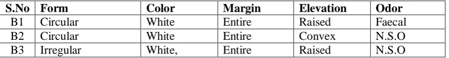
Table no. 2. Cultural characteristics of isolated bacteria from soil samples
S.No Form Color Margin Elevation Odor
B1 Circular White Entire Raised Faecal
B2 Circular White Entire Convex N.S.O
www.wjpr.net Vol 4, Issue 06, 2015.
2050 somewhat
translucent
B4 Circular Grayish Entire Convex N.S.O
B5 Circular Blue-gray Entire Convex N.S.O
B6 Circular White Entire Convex Fruity
B7 Circular Golden Entire Convex N.S.O
B8 Irregular White waxy undulate Flat N.S.O
B9 Irregular White Undulate Flat N.S.O
B10 Irregular Pink Entire Umbonate N.S.O
B11 Circular Grayish Entire Raised N.S.O
Note: N.S.O= No special odor.
Table no. 3. Morphological characteristics of isolated bacteria.
S.No Size(µm) Gram Staining Shape Arrangement
B1 2 - Rod Large clump
B2 1-2 - Rod Single
B3 1-2 - Rod Single
B4 1-3 - Rod Single
B5 1-5 - Rod Single
B6 2-8 - Rod Single
B7 1-5 + Cocci Clump & few
Single
B8 3-10 + Rod Single
B9 3-10 + Rod Single
B10 3-10 + Rod/spindle Single
B11 2-8 - Rod
Note: + indicate gram positive cell & - indicate gram negative cell.
Table no. 4. Biochemical characteristics of isolated bacteria.
S.No Litmus Milk Reaction L ac tos e fe rmenta ti on Indole pr oduc ti on M R R ea cti on VP R ea cti on C it ra te Us e Ur ea se Ac ti vit y C atala se Ac ti vit y Ge latin L iquef ac ti on S tar ch Hydr olys is H2 S p roduc ti on
B1 A AG + + - - - + - - -
B2 A AG - - + + - + - - -
B3 AG AG - - - + + + - - -
B4 Alkine - + + - - - + - - -
B5 Alkine - + + - - + + + - +
B6 Rapid
peptonization - - - - + - + + Rapid - -
B7 A A - + + - - + + - -
B8 Peptonization - - - + - - + + Rapid + -
www.wjpr.net Vol 4, Issue 06, 2015.
2051
B10 Alkine - + - - - -
B11 Alkine - - + - + - + - - -
Note: + indicate positive test & - indicate negative test, A indicate acid production & G indicate gas production.
Indole test. MR-test P-test. Lactose fermentation. H2S test.
Catalase test Urease test
Table no. 5. On the basis of all above characteristics, isolated bacteria were identified as shown in the table.
S.No Identified bacterium
B1 Escherichia coli
B2 Enterobacter aerogenes
B3 Klebsiella sp
B4 Shigella sp
B5 Proteus valgaris
B6 Pseudomonas aeruginosa
B7 Staphylococcus aureus
B8 Bacillus cereus
B9 Bacillus subtilis
B10 Clostridium sp
B11 Salmonella sp
www.wjpr.net Vol 4, Issue 06, 2015.
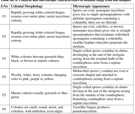
[image:10.595.70.535.79.472.2]2052 Table no. 6. Cultural and microscopic characteristics of isolated fungi from soil samples
S.No Colonial Morphology Microscopic Appearance
F1
Rapidly growing white colored fungus swarms over entire plate; aerial mycelium cottony.
Spores are oval, nonsepate mycelium gives rise to single sporangiophores with globular sporangium containing a
columella; there are no rhizoids.
F2
Rapidly growing white colored fungus swarms over entire plate; aerial mycelium cottony.
Spores are oval, colorless, or brown; nonsepate mycelium gives rise to straight sporaniophores that terminate with black sporangium containing a columella; rootlike hyphae (rhizoids) penetrate the medium.
F3 White colonies become greenish-blue, black, or brown as mature cultures.
Single-celled spores (conidia) in chains developing at the end of the sterigma arising from the terminal bulb of the conidiophores arise from a septate mycelium.
F4 Woolly, white, fuzzy colonies changing color to pink, purple or yellow.
Multicelled spores ( conidia) are oval or crescent-shaped and attached to
conidiophores arising from a septate mycelium.
F5 Mature cultures usually greenish or blue-green.
Single-celled spores (conidia) in chains develop at the end of the sterigma arising from the medula of the conidiophore; branching conidiophore arise from a septate mycelium.
F6 Colonies are small, round, moist, and colorless, with unbroken, even edges.
Yeastlike fungus produces pseudomycelium.
Table no. 7. On the basis of above characteristics, isolated fungi were identified as shown in the table.
F1 Mucor
F2 Rhizopus
F3 Aspergillus
F4 Fusarium
F5 Penicillium
F6 Candia
[image:10.595.71.522.526.722.2]
www.wjpr.net Vol 4, Issue 06, 2015.
2053 Table:8. Monthly average temperature of Bilaspur (C.G).- January13⁰C, February15⁰C, March 20.5, April-27⁰C.
0 20 40
January February March April
Av
e
ra
ge
Te
m
pr
at
ur
e
Temprature
Month
Fig.1. Monthly average temperature of Bilaspur (C.G).
Number of isolated bacteria and fungi from soil samples.- Bacteria- 11, Fungi-6 (fig 2)
Fig.2. Number of isolated bacteria and fungi from soil samples.
Fig.3. Isolated Bacteria, Fungi.
Table:-9. Average Bacterial colonies on Nutrient Agar Medium. Dilution factor 10-5 10-6 10-7 10-8 No. of Bacterial colony 23 7 1 0
N
u
m
b
er
o
f
iso
lat
ed
B
ac
te
ri
a
and
fung
i
fr
om
soi
l
sa
m
pl
es
.
www.wjpr.net Vol 4, Issue 06, 2015.
2054 Dilution factor
Fig.4. Average Bacterial colonies on Nutrient Agar Medium.
Table.10. Average Fungal colonies on Sabouraud Dexrose Agar Medium.
10-5 10-6 10-7 10-8
19 4 1 0
Dilution factor
Fig.5. Average Fungal colonies on Sabouraud Dexrose Agar Medium.
Isolated Gram +ve - 4 and Gram-ve – 7 bacteria.( Fig.6)
0 2 4 6
8 Isolated Bacteria
Fig.6. Total gram +ve and gram –ve Bacteria isolated from soil samples
10-6
10
-6 A verag e Bac teri al c o lo n ies o n N u tri ent A gar M edi u m 10-510
-6 10-710
-6 10-810
-610-5 10-6 10-7 10-8
www.wjpr.net Vol 4, Issue 06, 2015.
2055 DISCUTION
The isolated bacteria are E.coli, Enterobacter aerogenes, Klebsiella, Proteus vulgaris, Bacillus cereus, Bacillus subtilis, Clostridium, Staphylococcus aureus, Pseudomonas
aeruginosa, Shigella, and Salmonella and fungi are Aspergillus, Rhizopus, Mucor, Candida,
fusarium, Penicillium.
Eleven bacteria and six fungi were found from four selected area of Bilaspur(C.G). Out of them, some bacteria were commonly found in each sample such as E. coli, Staphylococcus aureus, Clostridium, some fungi were commonly found in all samples such as Aspergillus, Rhizopus, Mucor.
Pseudomonas aeruginosa was isolated from garden soil as well as Enterobacter aerogenes,
Klebsiella, Proteus vulgaris, were found in garden soil. Staphylococcus aureus,
Pseudomonas aeruginosa were found in road side (traffic area) and Bacillus subtilis,
Salmonella, Shigella, Staphylococcus aureus were found in industrial area.
Fusarium were found in garden soil. Candida was found in road side soil sample. Aspergillus,
Penicillium were observed in industrial area (sugar industry) because of easily and
abundantly available carbon source by product of sugar industries (Ali-2004). Bacterial isolates recovered from all fourth side showed slightly difference in MICs for Cu, Cd, Pb, Zn, Hg, Ni. Similar observation was reported by the two group of researcher in different time. (Kunito et al.-1986; Choudhary and kumar-1996).
In industrial area, road side soil (traffic area) soil samples, the minimum required nutritional condition are not observed due to heavy metal pollution, such as lead, copper, nickel, nilufer, cevic, ayten, kareca. Effect of cadmium, zinc, copper, and fluoranthene on soil bacteria (Turkey). Even through oxigen is not prerequisite for the growth of Pseudomonas
aeruginosa, at least NO3 must be available as respiratory electron acceptor which is minimum
in those soil samples.
Some microbial strains posses’ genetic determinants that confer the resistance. In bacteria, those determinants are often found on plasmid, which have facilitated their study at the molecular level (Cervanter et al-1094).
www.wjpr.net Vol 4, Issue 06, 2015.
2056 microorganisms by harmfully affecting their growth morphology and biochemical activities resulting in decreased biomass diversity (Baath- 1998; Robert-1992; Ahmed-2002).
CONCLUSION
Different type of soil collected from the different area of Bilaspur in C.G and isolated different types of microorganisms (Bacteria and Fungi) such as the bacteria are E.coli, Enterobacter aerogenes, Klebsiella, Proteus vulgaris, Bacillus cereus, Bacillus subtilis,
Clostridium, Staphylococcus aureus, Pseudomonas aeruginosa, Shigella, and Salmonella and
fungi are Aspergillus, Rhizopus, Mucor, Candida, fusarium, Penicillium.
Some microorganisms such as Actinomycetes and economically important fungi to demonstrate the potential of these organisms for bio-control of pathogenic bacteria and other pathogenic organisms which may be helpful in medical microbiology and agricultural field in future.
REFERENCE
1. Campbell, J.I.A., Albrechtsen, M and Sorensen, J. Large pseudomonas phages isolated from barley rhizosphere. FEMS Microbiology E. coli, 1995; 18: 63-74.
2. Chaudhary, P. and Kumar, R. Association of metal tolerance with multiple antibiotic resistance of enteropathogenic organisms isolated from costal region of Deltaic Sundarbans. Ind J. Med Res, 1996; 104: 184-151.
3. Taylor, D.E and Summers, A.O. Association of tellurium resistance and bacteriophage inhibition conferred by plasmids. Journal of Biotechnology, 1979; 13: 1413-1433.
4. Cervantes, C., Ji, G., Ramirez, J.L. and Silver, S. Resistance to arsenic compounds in microorganisms. FEMS Microbiology Reviews, 1994; 15: 355-367.
5. Malik, A. and Ahmed, M. Seasonal variation in bacteria flora of the wastewater and soil in the vicinity of industrial area. Environ Monit and Assess, 2002; 73: 263-273.
6. Robert, H.H. Simultaneous estimates of the diversity and degradative capability 0f heavy
metal affected soil bacterial communities. Bio fert. Soil, 1992; 13: 181-186.
7. Baath, E. Effects of heavy of metal in soil on microbial process and population. Water Air and Siol pollution, 1909; 47: 335-346.
8. Kunito, T., Saeki, K., Nagaoka,K., Oyaizu, H. and Matsumoto, S. Characterization of copper-resistance bacterial community in rhiosphere of highly copper-contaminated soil. Eur. J. Soil. Biol, 2001; 37: 95-102.
www.wjpr.net Vol 4, Issue 06, 2015.
2057 10.Alexander, M. 1977. Soil microbiology. 2nd ed. John Wiley & Sons, Inc. New York. 11.Rovira, A.D. 1965. Plant root exudates and their influence upon soil microorganisms. In