A R T I C L E
Acute transient cognitive dysfunction and acute brain injury
induced by systemic in
fl
ammation occur by dissociable
IL-1-dependent mechanisms
Donal T. Skelly1●Éadaoin W. Griffin1●Carol L. Murray1●Sarah Harney2●Conor O’Boyle1●Edel Hennessy1● Marc-Andre Dansereau1●Arshed Nazmi1●Lucas Tortorelli1●J. Nicholas Rawlins3●David M. Bannerman3● Colm Cunningham 1
Received: 24 April 2017 / Revised: 12 February 2018 / Accepted: 3 April 2018 © The Author(s) 2018. This article is published with open access
Abstract
Systemic inflammation can impair cognition with relevance to dementia, delirium and post-operative cognitive dysfunction. Episodes of delirium also contribute to rates of long-term cognitive decline, implying that these acute events induce injury. Whether systemic inflammation-induced acute dysfunction and acute brain injury occur by overlapping or discrete mechanisms remains unexplored. Here we show that systemic inflammation, induced by bacterial LPS, produces both working-memory deficits and acute brain injury in the degenerating brain and that these occur by dissociable IL-1-dependent processes. In normal C57BL/6 mice, LPS (100 µg/kg) did not affect working memory but impaired long-term memory consolidation. However prior hippocampal synaptic loss left mice selectively vulnerable to LPS-induced working memory deficits. Systemically administered IL-1 receptor antagonist (IL-1RA) was protective against, and systemic IL-1βreplicated, these working memory deficits. Dexamethasone abolished systemic cytokine synthesis and was protective against working memory deficits, without blocking brain IL-1βsynthesis. Direct application of IL-1βto ex vivo hippocampal slices induced non-synaptic depolarisation and irreversible loss of membrane potential in CA1 neurons from diseased animals and systemic LPS increased apoptosis in the degenerating brain, in an IL-1RI-dependent fashion. The data suggest that LPS induces working memory dysfunction via circulating IL-1βbut direct hippocampal action of IL-1βcauses neuronal dysfunction and may drive neuronal death. The data suggest that acute systemic inflammation produces both reversible cognitive deficits, resembling delirium, and acute brain injury contributing to long-term cognitive impairment but that these events are mechanistically dissociable. These data have significant implications for management of cognitive dysfunction during acute illness.
Introduction
Peripheral infections are known to trigger episodes of acute cognitive impairment, including delirium, in older popula-tions and in those with dementia [1, 2]. Sterile infl amma-tion, resulting from tissue trauma or surgery, can also induce post-operative cognitive dysfunction and delirium [3, 4]. Cytokines are key mediators of septic and aseptic inflammation and, given its important role in coordinating CNS responses to systemic inflammation [5, 6], the pro-inflammatory cytokine IL-1βmight be predicted equally to underlie infection- and sterile inflammation-induced cogni-tive dysfunction. Consistent with this idea, IL-1β levels have been associated with delirium in hip fracture patients and in septic encephalopathy [7,8]. Delirium is a profound and acute onset brain dysfunction with impairments in
Corrected: Correction
These authors contributed equally: Donal T. Skelly, Éadaoin W. Griffin, Carol L. Murray, Sarah Harney.
* Colm Cunningham [email protected]
1 School of Biochemistry and Immunology & Trinity College Institute of Neuroscience, Dublin 2, Ireland
2 Department of Physiology, Trinity College Dublin, Dublin 2,
Ireland
3 Department of Experimental Psychology, University of Oxford,
South Parks Road, Oxford, UK
Electronic supplementary materialThe online version of this article (https://doi.org/10.1038/s41380-018-0075-8) contains supplementary material, which is available to authorised users.
123456789
0();,:
123456789
attention and other aspects of cognition. Its high prevalence after surgery and infection emphasises the deleterious con-sequences that systemic inflammation has for cognitive function, particularly in older, cognitively impaired, popu-lations [9, 10]. It is now clear that acute systemic infl am-mation and delirium also significantly increase the risk of long-term cognitive decline and dementia [11–13], and accelerate the course of existing dementia [14,15]. Despite the enormous medical and economic implications [16] of these findings, whether systemic inflammation-induced acute dysfunction and acute brain injury occur by over-lapping or discrete mechanisms has not been invest
It is established that IL-1 impacts on cognitive function [17, 18]. IL-1 disrupts consolidation of context-associated fear memory [19–21] and IL-1 receptor antagonist (IL-1RA) is protective against post-acquisition memory con-solidation deficits induced by both surgery and systemic lipopolysaccharide (LPS) [20,22,23]. However, it is likely that systemic inflammation impacts upon multiple cognitive processes. Furthermore, long-term memory consolidation is manifestly different both from the acute fluctuating short-term memory processes that are affected in delirium and in relevant models [9, 24] and from acute neuronal death events described after systemic inflammation [25]. Given the massive public health burden of dementia and delirium, both of which have been associated with IL-1 [26,27], it is important to characterise differential roles of IL-1βin these processes. In the current study we hypothesised that (1) bacterial endotoxin (LPS; lipopolysaccharide) would have multiple effects on cognition and neuronal integrity and that (2) IL-1 would be causative in these changes.
Materials and methods
Animals
Female C57BL/6 mice at 8–12 weeks of age (Harlan Olac Ltd, UK) were housed in cages of 5 at 21 °C with a 12:12 h light–dark cycle with food and water ad libitum. To test the effects of LPS in cognitive function in young healthy mice (2–4 months) C57BL/6 mice were injected intraperitoneally (i.p.) with 100 (or 200)μg/kg of LPS (Salmonella Equine
abortus, Sigma (L5886)) in sterile saline. To examine
effects of LPS in animals with prior synaptic loss mice were first intrahippocampally inoculated with 1 µl of 10% w/v prion infected- (ME7) or normal brain homogenate (NBH), at mm from bregma: anterior–posterior−2.0, lateral −1.7, depth−1.6) using a Hamilton microsyringe, under anaes-thesia with intra-peritoneal 2,2,2-tribromoethanol. Experi-mental groups (ME7, NBH) were then injected i.p. with
100μg/kg of LPS at 15–16 weeks post inoculation
(demonstrated vulnerability to LPS-induced
working-memory deficits [28]). Control animals received non-pyrogenic saline.
Alternative inflammatory stimuli and anti-inflammatory interventions were also examined: IL-1β (R&D Systems,
Minneapolis, MN, USA, 401-ML/CF) and TNF-α
(Pepro-tech, Rocky Hill, NJ, USA, 305-01A) were injected at 15 µg/kg at 50 µg/kg respectively in sterile saline (i.p.). IL-1RA (10 mg/kg, i.p.; Kineret, Biovitrum, Sweden) was
given immediately before LPS or IL-1β (15 µg/kg);
dexamethasone-21-phosphate (Sigma, D1159) was admi-nistered i.p. at a dose of 2 mg/kg, 60 min before LPS (suf-ficient to robustly suppress the systemic secretion of IL-1β, TNF-α and IL-6 [29]. Samples sizes for all groups in all animal experiments were chosen on the basis of our prior published studies using the ME7+LPS paradigm [25,28, 30] and are specified in the appropriatefigure legends.
IL-1RI−/− mice (B6.129S7-Il1r1<tm1Imx>; kindly pro-vided by Prof. Kingston Mills) have a null mutation inIl1r1 and were backcrossed seven times to C57BL/6 before maintenance as an inbred colony (with C57BL/6 as con-trols). IL-1RI−/− and C57BL/6 mice were not significant different in working memory or contextual fear condition-ing (CFC) tasks [31]. Body temperature was measured using a rectal probe (TH-5 thermoprobe, Physitemp, NJ) 18 h post challenge with LPS (750 µg/kg i.p.) in WT and IL-1RI−/−mice inoculated with ME7 or NBH.
All animal procedures were performed in accordance with Irish Department of Health & Children, Health Pro-ducts Regulatory Authority and UK Home Office regula-tions and all efforts were made to minimise suffering to the animals.
Working memory: Food-rewarded and escape from
water T-maze alternation tasks
animals who had achieved a criterion baseline performance of≥70% (water) or≥80% (food-rewarded) alternation for 2 or more consecutive days. Baseline T-maze performance was ranked and animals were assigned to different treat-ments groups such that all treatment groups had equivalent, or very similar, baseline performance. Experimenters were blind to treatment during scoring of cognitive function. This water-escape task has previously been published [28] and a full description of these methods is available in supple-mentary material.
Contextual fear conditioning
CFC was recorded using a clear perspex box (40 cm × 10 cm × 16 cm) with afloor containing metal rods connected to a shock generator (UGO Basile, Italy). The mice were placed into the box and allowed to explore for 2 min. A tone at 2.9 kHz for 20 s was presented, followed by a shock of 0.4 mA for 2 s. This tone/shock pairing was repeated after 2 min. After a further 30 s of exploration mice were removed to a holding cage. After 30 s in the holding cage saline or LPS (±saline or IL-1RA, at discrete i.p. sites) were admi-nistered before returning the animals to the home cage. IL-1RA was administered at this time (rather than preceding the test of retention) because it is consolidation, rather than retention, of memory that has been shown to be impaired by LPS [32,33]. Fear conditioning was assessed for 5 min in the same location 48 h later. Freezing was defined as the complete absence of movement, except those related to respiration [34]. Auditory fear conditioning was also assessed 48 h post-fear conditioning. Animals were placed in a different context (a novel empty cage) for 6 min and were allowed to explore for 3 min (baseline) before pre-sentation of the tone for 20 s. The time spent freezing during thefinal 3 min was recorded (i.e. during and post tone).
ELISA for cytokines
Under terminal anaesthesia, blood was collected directly from the right atrium into heparinised tubes, was cen-trifuged at 3000 rpm for 15 min at 4 °C and the remaining plasma aliquoted and stored at−20 °C. Samples were then analysed for CCL2 and CXCL1 using R&D systems sandwich-type duo set ELISA kits (DY479, DY453) while IL-1βwas analysed using a Quantikine kit (R&D systems, Minneapolis, MN, USA, MLB00C). A standard protocol was followed as previously described [30] except for IL-1β, which was as per manufacturer’s instructions with minor modifications. To ensure that all cytokines were reliably quantifiable using the appropriate standard curves, samples were serially diluted for CCL2 and CXCL1 (1/9, 1/81 and 1/243). Blood and brain samples were also assayed for the presence of human IL-1RA using an R&D Systems
quantikine assay (DRA00B), performed according to man-ufacturers’ instructions (standards 0–2000 pg/ml). Hippo-campal/thalamic tissue punches were homogenised in 150 mM NaCl, 25 mM Tris-HCl and 1% Triton X100 at pH 7.4 before centrifugation at 14,000 rpm for 10 min. Super-natants were diluted 1 in 2 in assay diluent in wells pre-coated with anti-human IL-1RA polyclonal antibody.
Quantitative PCR
The isolation of total RNA, synthesis of cDNA and analysis of transcription by quantitative PCR were performed as previously described [35]. Briefly, after transcardial perfu-sion, the hippocampus and dorsal thalamus were punched out of 2 mm thick coronal brain sections, snap frozen in
liquid nitrogen and stored at −80 °C. Total RNA was
extracted using Qiagen RNeasy Plus™ mini kits, with
Qiashredders (Qiagen, Crawley, UK, #74134, #79654) according to manufacturer’s instructions. Contaminating gDNA was removed using the Qiagen RNase-free DNase I enzyme (Qiagen #79254). RNA yields were determined by spectrophotometry at 260 and 280 nm and stored at−80 °C. Using a High Capacity cDNA Reverse Transcriptase Kit (Applied Biosystems, Warrington, UK), cDNA was syn-thesised using 200 ng of total RNA in a 10μl reaction volume. 1μl of the reverse transcription reaction was used for quantitative PCR. Reagents were supplied by Applied
Biosystems (SYBR® Green PCR Master Mix; 4364344)
and Roche (FastStart Universal Probe Master [Rox], Lewes, UK; 04914058001). Assays were designed using Primer Express software and published sequences for the genes of interest. All primer sequences were as previously published [25]. Assays were quantified using a relative standard curve, as previously described [35] constructed from cDNA, syn-thesised from 1μg total RNA isolated from mice showing up-regulation of all target transcripts of interest.
Electrophysiology
Transverse hippocampal slices (300 mm) were prepared
from brains of NBH or ME7 mice at 18–19 weeks post
microscope (Olympus BX51 WI, Middlesex, UK) with infra-red differential interference contrast optics (IR-DIC). Patch pipettes were filled with intracellular solution
con-taining (in mM) 130 KMeSO4, 10 KCl, 0.2 EGTA, 10
HEPES, 20 phosphocreatine, 2 Mg2ATP, 0.3 NaGTP, 5
QX-314, 1 TEA (pH 7.3, 290–300 mOsm). Cells were
voltage-clamped at −60 mV and input resistance and
membrane capacitance were measured in response to a 10 mV depolarising voltage pulse. During current clamp recordings, current injection was initially set to maintain membrane potential at−60 mV and was not altered for the duration of experiments. Recordings were made using a Multiclamp 700B (Molecular Devices, Foster City, CA). Signals were filtered at 5 kHz using a 4-pole Bessel filter and were digitised at 10 kHz using a Digidata 1440 analogue-digital interface (Molecular Devices). Data were
acquired and analysed using PClamp 10 and Clampfit
(Molecular Devices). IL-1β was bath-applied to slices at concentrations of 0.1 ng/ml and 1 ng/ml.
TUNEL immunohistochemistry
Immunohistochemistry for apoptotic cells was performed on formalin-perfused, wax embedded tissue from ME7 animals (wild-type and IL-1R1−/−), 18 h post LPS (750 µg/ kg i.p.) using the ‘Dead End’ TUNEL staining method (Promega, Southampton, UK; G3250). Non-specific per-oxidase activity was eliminated by incubating sections in 1% H2O2for 10 min. Sections were washed in 0.85% NaCl for 5 min and PBS washed, before 5 min pre-treatment with proteinase K (10μg/ml). After PBS washing, sections were preincubated with equilibration buffer (10 min) and then with TUNEL buffer (22μl equilibration buffer, 2.5μl nucleotide mix, 0.5μl TdT enzyme per section) for 2 h at 37 °C, labelling apoptotic cells with fluorescein. After reaction termination with 2× sodium citrate (15 min) and PBS washing sections were blocked with 10% normal goat serum (30 min) and incubated with biotinylated anti-fluorescein antibody (5μg/ml). Thereafter the Avidin–Biotin-Complex protocol was performed according to manufacturers instructions, using H2O2 as substrate and diaminobenzidine as chromagen. TUNEL-positive cells that also showed evidence of nuclear condensation were counted in 10 µm coronal sections of ME7 animals treated with LPS in wild-type and IL-1R1−/−mice. At least two sections were counted, by an observer blind to the experimental condi-tions and averaged for each animal.
Statistical analyses
Behavioural data were compared by repeated measures ANOVA with Bonferroni post hoc tests performed after significant main effects. Molecular, electrophysiology and
TUNEL data were analysed by two-way ANOVA, while temperature data were analysed by three-way ANOVA, followed by Bonferroni post hoc tests for a priori selected pairwise comparisons. Sample sizes and variance were similar for all key comparisons.
Results
Differential effects of LPS on different
hippocampal-dependent cognitive tasks
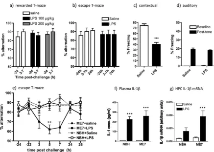
We assessed working memory using T-maze alternation tasks requiring both attention to, and retention in the working memory of (for 25 s), the prior location of an exit in order to determine the new exit location. LPS did not impair working memory in food-rewarded T-maze alterna-tion at 3–7 h post treatment at either 100 or 200 µg/kg i.p. (Fig.1a). To validate thisfinding in an aversively motivated working memory task, not reliant on motivation for appe-titive rewards, we also used an‘escape from shallow water’ T-maze working memory task specifically adapted for use in animals experiencing acute sickness behaviour. LPS (100 µg/kg) had no impact on working memory in C57BL6 mice in this T-maze (Fig.1b).
However, LPS (100 µg/kg i.p.) given immediately post acquisition, was sufficient to significantly decrease freezing in CFC 48 h after exposure to context and foot-shock pairing (Fig.1a,p< 0.001) but had no impact on auditory fear conditioning (Fig.1d). In the CFC task LPS is known to impair consolidation (in the early hours after exposure to shock) rather than the retrieval of that memory 48 h later [32,33]. Therefore, systemic LPS has differential effects on two hippocampal-dependent tasks: disrupting consolidation of contextual memory but not affecting working memory in normal animals.
LPS differentially affects working memory in
vulnerable animals despite equivalent circulating
IL-1
β
animals with neurodegeneration are similarly challenged. Despite the increased susceptibility of ME7 animals to working memory deficits in response to LPS, performance in the CFC task was equally susceptible to LPS disruption in normal and ME7 animals (Fig. S1).
This selective vulnerability of ME7 animals to LPS could potentially be underpinned by differential circulating IL-1β. Peripherally administered LPS (100 µg/kg) induced
robust systemic IL-1β synthesis and IL-1β mRNA
transcription in the hippocampus at 2 h post challenge.
Plasma IL-1β levels were equivalent in ME7 and NBH
animals (Fig. 1f), although brain IL-1β mRNA was exag-gerated in ME7+LPS (Fig. 1g), as previously described [25,28].
Dissociable effects of IL-1RA treatment on
LPS-induced cognitive de
fi
cits
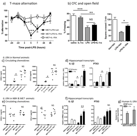
Since acute cognitive impairments following systemic LPS were immediately preceded by robust circulating IL-1β, we hypothesised that systemic IL-1β contributed to these defi -cits and we therefore assessed whether peripheral treatment with the receptor antagonist IL-1RA would protect against the T-maze and CFC deficits described in Fig.1. IL-1RA at 10 mg/kg (i.p.) significantly protected against LPS-induced T-maze deficits in ME7 animals in a time-dependent man-ner (Fig. 2a). There was a significant effect of treatment (F2,234=4.78, p=0.139) and an interaction of treatment and time (F12,234=3.67, p< 0.0001) and Bonferroni post
Fig. 1 LPS-induced effects on working memory, fear conditioning and IL-1β expression. Working memory was assessed by (a) food-rewarded T-maze alternation (all groupsn=7) and (b) escape from shallow water T-maze (both groupsn=9) at 24 h before (−24) and at indicated times post LPS (100 or 200μg/kg i.p.). On theX-axis, 3–7 represents % alternation during 10 trials conducted every 20 min between 3 and 7 h post LPS). Two-way ANOVA analysis found no significant effects of treatment. Contextual (c) and auditory (d) fear conditioning performance (freezing per 5 min) 48 h post challenge with LPS (100μg/kg i.p.). Data are expressed as mean ± SEM and analysed byt-test (***p< 0.0001;n=12 for saline and 16 for LPS; combined from two independent experiments). eWorking memory performance of NBH and ME7 animals 16 weeks post-ME7 inocula-tion, challenged with LPS (100μg/kg i.p.) or saline, assessed by T-maze alternation for 10 trials 24 h pre-challenge, 15 trials between 3–9 h post challenge and 10 trials 24 h post challenge. Full ANOVA
[image:5.595.77.515.52.364.2]Fig. 2 Dissociable effects of systemic IL-1 receptor antagonist (IL-1RA) on T-maze alternation and contextual fear conditioning. a Working memory performance of ME7 animals, 16 weeks post inoculation, challenged with LPS (100μg/kg i.p.) in the presence or absence of IL-1RA (10 mg/kg i.p. immediately following LPS). These data arise from two independent experiments, totallingn=16, except ME7+IL-1RA controls (n=10). Significant Bonferroni post hoc (p < 0.01) after significant two-way ANOVA denoted by ***.b Perfor-mance of normal mice in CFC (time spent freezing 48 h following 0.4 mA foot shock) and in openfield activity following systemic challenge with saline or LPS (100 µg/kg i.p.), in the presence or absence of IL-1RA (10 mg/kg i.p.). All groups weren=11 except WT+saline (n= 7) for CFC experiment andn=5–7 for openfield. Data are expressed as mean ± SEM and were analysed by two-way ANOVA. Significant differences, assessed by Bonferroni post hoc after significant ANOVA are denoted by **p< 0.01 and ***p< 0.001. cEffect of i.p. recom-binant IL-1RA on plasma chemokine (CXCL1, CCL2) induction 2 h post administration of IL-1β(15μg/kg, i.p.) or LPS (100μg/kg, i.p.), analysed by ELISA. Bonferroni post hoc tests (after significant
[image:6.595.69.526.52.504.2]hoc comparison showed a highly significant difference
between ME7+LPS+IL-1RA and ME7+LPS+veh at
5 h (p< 0.001). However, both LPS-treated groups were equally impaired at 7 h, so protection is robust at 5 h, when LPS-induced deficits peak, but does not last.
Conversely, systemic IL-1RA had no protective effect against the LPS-induced CFC deficit despite mitigating the effects of LPS on openfield activity (Fig.2b). LPS robustly induced decreased freezing in C57BL6 mice (main effect of LPS by two-way ANOVA, F3,36=38.68, p< 0.0001). Freezing, 48 h after co-treatment with LPS+IL-1RA (10 mg/kg i.p.), was not significantly different from that induced by LPS (Fig. 2a, no effect of drug: F=0.08). Therefore peripheral IL-1RA is protective against LPS-induced working memory deficits and against suppression of openfield activity but not against LPS-induced impair-ment of consolidation of contextual memory. Although these LPS-induced deficits become manifest across different timescales (i.e. 3–7 h for T-maze and at 48 h for CFC), LPS and IL-1βimpair consolidation of contextual memory in the first hours after exposure to shock and context pairing [33, 36]. Therefore both T-maze and CFC tasks illustrate inflammation-induced disruption of cognitive processes in the hours directly after LPS administration, so it was necessary to apply IL-1RA during this period.
We considered it essential to confirm that i.p. IL-1RA (10 mg/kg) actually blocked IL-1βaction so we assessed its
ability to block well-recognised indices of systemic
and central (hippocampal) inflammatory effects of LPS
(100μg/kg) and IL-1β (15μg/kg). A range of
pro-inflammatory mediators are induced across the timecourse 1–8 h post LPS or IL-1β[28, 37] and here we chose 2 h to capture the peak of most of these. Plasma was prepared 2 h post treatment with recombinant IL-1RA (10 mg/kg, i.p.) and simultaneous injection of IL-1β, LPS or saline. Both LPS and IL-1βincreased plasma CXCL1 and CCL2. IL-1RA had no effect on LPS-induced circulating chemokine but completely blocked IL-1-induced chemokine (p< 0.05, Bonferroni post hoc after significant one way ANOVA, Fig. 2c). Hippo-campal expression of pro-inflammatory genes IL-1β and PTX3 was also increased by both systemic LPS and IL-1β treatments (Fig. 2d). Once again IL-1-induced increases in both IL-1β and PTX3 were inhibited by systemic IL-1RA (p< 0.05), but this treatment failed to block, and in fact even enhanced, LPS-induced hippocampal increases in IL-1β and PTX3 (Fig. 2d). These data indicate that circulating LPS still alters plasma and brain inflammatory profiles despite suc-cessful inhibition of systemic IL-1βaction: LPS signals to the brain despite absence of systemic IL-1βactivity.
We also assessed whether effects of IL-1RA were dif-ferent in ME7 by examining expression of these same indices of LPS and IL-1 actions in NBH and ME7 animals 3 h after treatment with LPS ± IL-1RA. As before, LPS
robustly increased the circulating levels chemokines CXCL1 and CCL2 but IL-1RA had limited impact on these levels, with huge circulating concentrations remaining regardless of treatment (Fig. 2e). Two-way ANOVA ana-lysis followed by Bonferroni post hoc anaana-lysis showed that IL-1RA did not reduce circulating chemokines in ME7 animals: There were no significant main effects or interac-tions for CXCL1, and while there was an interaction between disease and treatment for CCL2 (F1,12=13.39,
p=0.003), Bonferroni post hoc analysis showed that CCL2
was not different between ME7+LPS and ME7+LPS+
IL-1RA.
Hippocampal expression of pro-inflammatory genes
IL-1βand PTX3 was also examined in ME7 and NBH and
disease-associated increases were apparent for both tran-scripts (as previously reported in ref. [38]): main effect of disease (F1,16≥22.85,p≤0.0002). LPS produced significant increases in both transcripts and IL-1RA did not decrease these, and indeed IL-1βmRNA was significantly increased
in ME7+LPS animals when treated with IL-1RA
(Bon-ferroni post hoc,p< 0.01). Therefore, systemic IL-1RA did not reduce hippocampal IL-1β mRNA. Collectively these data demonstrate that even though IL-1RA blocks circu-lating IL-1β actions, LPS still induces brain inflammatory activation, including IL-1β.
Finally we used anti-human IL-1RA ELISA on these in vivo tissues to assess whether peripherally applied human IL-1RA reaches the hippocampus, since it could be hypo-thesised that IL-1RA could show protective effects because of compromised blood brain barrier (BBB) allowing access of IL-1RA selectively to the ME7 hippocampus. Low levels of human IL-1RA were detectable in the hippocampus of all IL-1RA-injected animals but concentrations were
equiva-lent in NBH and ME7 animals (Fig. 2g). These levels
(60–70 pg/ml/mg protein) are almost 7500-fold lower than plasma levels (481,511 ± 95,460 pg/ml). These data indicate that (i) a tiny fraction of IL-1RA penetrates the brain par-enchyma and (ii) that this is not significantly higher in neurodegeneratively diseased (ME7) animals. Moreover, although IL-1RA is known to have a short half-life [22,39], the levels measured in the blood at 3 h post treatment show that IL-1RA remained approximately 2000-fold higher than circulating IL-1βat what is a key stage in inducing working memory dysfunction and contextual memory consolidation.
IL-1
β
, TNF-
α
and redundancy in in
fl
ammation-induced cognitive dysfunction
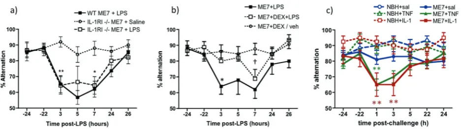
LPS treatment induced working memory impairments in IL-1R1−/− ME7 animals (main effect of treatment:F12,245= 16.61, p< 0.0001) but the deficits were not significantly different to those in wild-type ME7+LPS animals at any time point (All Bonferroni post hoc testsp> 0.05, Fig.3a). Thus, IL-1RI is not indispensable for LPS-induced acute working memory deficits in ME7-inoculated animals. While this may appear contradictory, IL-1RI−/−mice also showed the normal appearance of LPS-induced sickness and weight loss (equivalent at 24 h post LPS in WT and IL-1R1−/− mice: 3.22 g vs 2.96 g) and this is consistent with previous data showing that IL-1R1−/−mice display the full spectrum of innate immune responses to LPS (http://jaxmice.jax.org/ strain/003245.html; ref. [40]). To confirm this we assessed cFOS responses in known IL-1-responsive brain regions after systemic LPS (100 µg/kg i.p.). These data are shown in supplementary table 1. Despite this, systemic IL-1RA was still effective in reducing LPS-induced sickness behaviour (Fig. 2b). Collectively these data support the concept of significant redundancy of specific cytokines in coordination of inflammatory responses to LPS. Therefore, while IL-1 is indisputably important in inducing sickness behaviour responses (ref. [41] and Fig.2b), and here systemic IL-1RA is protective against LPS-induced cognitive deficits, other cytokine systems can mediate roles of IL-1 when animals have developed in the absence of IL-1RI−/−signalling.
To accommodate this redundancy in pro-inflammatory cytokine functions, we used dexamethasone-21-phosphate
(DEX; 2 mg/kg) to block multiple LPS-induced pro-inflammatory cytokines and to assess its impact on
LPS-induced T-maze impairments in IL-1R1−/− ME7 mice
(Fig. 3b). This dose was sufficient to block systemic secretion of IL-1β, TNF-αand IL-6 (Fig. S2 and ref. [29]). DEX alone had no significant impact on T-maze alternation. LPS induced working memory impairments in ME7 ani-mals and this was significantly reduced in animals pre-treated with DEX (Fig. 3b). Repeated measures two-way ANOVA showed a main effect of treatment (F2,30=8.7,
p=0.001) and a treatment × time interaction (F12,180= 2.28, p< 0.01). ME7+LPS was significantly different to both DEX groups at 3 h (p< 0.01; Bonferroni), although
ME7+LPS and ME7+LPS+DEX were no longer
sig-nificantly different at 7 h. Thus DEX protects against LPS-induced deficits but some cognitive impairment eventually occurs despite DEX inhibition of systemic cytokine synthesis.
These data suggested that systemic cytokines contribute to LPS-induced working memory impairments. Therefore, we interrogated the roles of specific systemic cytokines. We treated animals with IL-1β (15 µg/kg i.p.) and assessed for working memory deficits. Moreover, it has been demon-strated that in IL-1RI−/−mice, TNF-αcompensates for the lack of IL-1 signalling and mediates sickness behavioural and weight loss responses to LPS [42]. Therefore, in separate experiments, the impact of systemically adminis-tered TNF-αon T-maze performance was assessed in NBH
Fig. 3 LPS-induced working memory deficits in ME7 animals are intact in IL-1R1−/− mice but are blocked by inhibition of systemic cytokine synthesis and replicated by administration of IL-1βor TNF-α. aT-maze alternation, of WT and IL-1RI−/−animals, 16 weeks post inoculation with ME7, in the presence or absence of systemic LPS challenge (100μg/kg i.p.) was assessed for 10 trials 24 h before acute challenge, 15 trials 3–9 h post challenge and 10 trials 24 h after the challenge. WT ME7+LPS and IL-1R1−/− ME7+LPS were not different at any time. Significant Bonferroni post hoc differences between IL-1R1−/− ME7+LPS and IL-1R1−/− ME7+saline are denoted by *p< 0.05 and **p< 0.01 (n=10 except WT ME7+LPS n=19).bWorking memory performance after systemic LPS (100μg/ kg i.p.) was assessed in the presence or absence of dexamethasone-21-phosphate (2 mg/kg i.p.). Significant post hoc differences between ME7+LPS+veh and ME7+LPS+DEX (by Bonferroni post hoc comparisons after a significant ANOVA) are denoted by *p< 0.01 and
[image:8.595.64.533.54.185.2]and ME7 animals. Impairments were not produced by either cytokine in NBH animals but both IL-1βand TNF-α chal-lenges rapidly induced acute working memory deficits in
ME7 animals that had largely resolved by the 5–7 h
timepoint (Fig. 3c). The IL-1β experiment showed an interaction of disease and treatment (F1,53=5.42,
p=0.0236) and planned Bonferroni pairwise comparisons
showed significant differences between ME7+saline
and ME7+IL-1β at 1 and 3 h and between ME7+saline and ME7+TNF-α at 1 h (p< 0.05). As previously described [43] ME7+TNF-α animals were significantly different from both NBH+TNF-αand from ME7+saline at 1 h after a significant interaction of disease, treatment and time by three-way ANOVA (F6,328=3.09, p< 0.01). Thus, systemic IL-1βand TNF-α cause acute and transient working memory deficits in animals with prior neurode-generative pathology but have no effect on working memory in normal animals. These deficits are somewhat milder than those induced by LPS and recovery is apparent by 5 h.
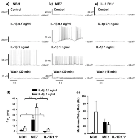
Neuronal sensitivity to IL-1
β
Since equivalent systemic IL-1 levels produce differential cognitive outcomes in ME7 and NBH animals, we predicted that neurons of the degenerating brain might also be more sensitive to equivalent concentrations of IL-1. To address this hypothesis we applied IL-1β directly to ex vivo hip-pocampal slices and performed whole-cell patch clamp recordings from CA1 pyramidal cells from NBH and ME7 animals at 18–19 weeks post inoculation.
CA1 pyramidal neurons from ME7 animals had a sig-nificantly depolarised resting membrane potential (Vm) compared to those from NBH animals (−61 ± 2 mV in NBH and−51 ± mV in ME7,n=13 cells fromfive NBH animals and 24 cells from eight ME7 animals, unpaired t-test, *p< 0.0001, Fig. S3).
We then tested the effects of bath application of IL-1β while recording from cells in current clamp mode, using current injection adjusted to maintain membrane potential at
−60 mV during baseline recordings (Fig. 4a–c). Neurons from ME7 animals were significantly more sensitive to IL-1β, which depolarised resting membrane potential by 21 ± 4 mV at 0.1 ng/ml in ME7 animals (n=13 cells from five animals) but had little effect in NBH animals (1.5 ± 2 mV;
n=5 cells from three animals; p=0.013, Bonferroni post hoc, Fig.4d). At 1 ng/ml, IL-1βdepolarised ME7 animals’ resting membrane potential by 44 ± 7 mV (n=5 cells from three animals) but also had milder effects in NBH animals (6 ± 3 mV;n=5 from three animals;p< 0.001, Bonferroni post hoc, Fig. 4d). ME7 were thus, significantly more sensitive to IL-1β-induced depolarisation at both IL-1β concentrations tested.
As a consequence, IL-1βinduced action potential firing in ME7 CA1 neurons at 0.1 ng/ml, with a maximumfiring rate of 30 ± 9 Hz (0.1 ng/ml,n=13 cells) but did not do so in NBH animals (Fig. 4e). At 1 ng/ml, IL-1β induced spiking at maximum rates of 23 ± 6 Hz (1 ng/ml,n=5 cells, Fig.4e) in ME7 animals and at a highly variable maximum rate of 58 ± 29 Hz in NBH neurons (n=5 cells, Fig. 4e). The lowerfiring rates in ME7 cells can be attributed to the greater depolarisation induced by IL-1β, acting to increase sodium channel inactivation and therefore limiting spike firing frequency. Depolarisation of ME7 neurons persisted even after a more prolonged washout of IL-1β, with cells becoming sufficiently depolarised to inhibit further action potential firing, suggesting that exposure to IL-1β had an irreversible, detrimental effect on these cells.
The effect of IL-1βon ME7 neurons does not appear to be mediated by modulation of excitatory synaptic
trans-mission since 1 ng/ml IL-1β had no effect on evoked
excitatory postsynaptic currents (EPSC amplitude 105 ± 32% of control amplitude, n=4 cells from two ME7 ani-mals, Supplementary Fig. S4). The ability of IL-1β to induce spiking activity in CA1 neurons was dependent on IL-1R1 expression since IL-1β had little effect on resting membrane potential and did not induce action potential firing in slices from IL-1R1−/− animals (n=4 cells from two animals, Fig. 4c–e). Moreover, TNF-α(20 ng/ml) had no effect on membrane potential in slices from IL-1R1−/− animals (−64 ± 2 mV in control and−64 ± 2 mV in TNFα,
n=5 cells from two animals, Supplementary data, S5).
Consequences of exaggerated IL-1 responsiveness:
apoptosis
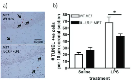
Since neurons in the diseased brain were more sensitive to the effects of IL-1 and failed to recover their resting membrane potential, we hypothesised that the previously reported neu-ronal apoptosis induced by systemic LPS [25] would be mediated by IL-1RI. ME7 animals were challenged with LPS (750 µg/kg i.p.) and euthanised 18 h later, the time at which LPS-induced neuronal apoptosis has previously been demon-strated [25]. TUNEL labelling was performed to assess for apoptotic cell death in 10 µm coronal sections. LPS induced robust apoptosis in wild-type ME7 animals, with respect to ME7+saline animals and the number of apoptotic cells was significantly reduced in IL-1R1−/− ME7+LPS animals
(Fig. 5a, b). Two-way ANOVA showed a main effect of
mediated by IL-1RI. Our prior studies had established that NBH animals were not similarly susceptible to LPS-induced apoptosis [25]. However, we have also verified that LPS did not produce acute apoptosis in NBH animals under the current conditions (Supplementary Figure S6). Similarly, the exag-gerated hypothermic response of ME7 animals to systemic LPS was also mediated by IL-1RI (Fig. S7).
Discussion
We have demonstrated that systemic inflammation impairs cognitive and neuronal function in multiple ways, by dis-sociable IL-1-dependent mechanisms. While LPS had no impact on working memory in normal animals it robustly impaired working memory in animals with existing
Fig. 4 Differential sensitivity of CA1 pyramidal cells to IL-1β in hippocampal slices from NBH and ME7 animals. Traces illustrating current clamp recordings from CA1 pyramidal cells from NBH (a), ME7 (b) and IL-1RI−/−(c) animals during control, baseline recordings and in the presence of IL-1βat concentrations of 0.1 and 1 ng/ml and following washout. The washout period was prolonged in ME7 ani-mals (30 min) in order to maximise the chance of observing a return to resting membrane potential, but this depolarisation appeared irrever-sible. The effects of IL-1βwere quantified by measuring the change in baseline membrane potential (ΔVm), averaged over 10 s, (d) and changes in the maximum action potentialfiring rate (e). Neurons from
[image:10.595.78.517.52.504.2]neurodegenerative pathology. Systemic LPS-induced working memory deficits were mediated by circulating
IL-1β. Systemic LPS also induced IL-1R1-dependent
apoptotic cell death in the neurodegenerating brain and CA1 neurons in hippocampal slices showed heightened vulnerability to IL-1β-induced, non-reversible, loss of membrane potential. The data suggest that systemic inflammation induces both transient cognitive dysfunction and lasting brain injury and that IL-1 contributes to both processes, but by dissociable mechanisms. Since systemic inflammation can trigger episodes of delirium in susceptible individuals [9] and episodes of delirium interact synergis-tically with existing cognitive impairment to accelerate dementia progression [44], understanding the mechanistic basis of both the acute deficits and the lasting injury is of crucial importance. The current data adds to our knowledge on transient and lasting effects of systemic inflammation on the already degenerating brain.
Dissociations in in
fl
ammation-induced,
hippocampal-dependent cognitive impairment
Although impairment of memory consolidation in the CFC paradigm is the best characterised effect of systemic
inflammation on cognitive function in young healthy
rodents (whether induced by LPS, infection or surgical trauma) [17,20,21], we have argued that this consolidation of long-term memory may not be optimal for studying the dynamic and fluctuating attentional and working memory deficits observed during delirium [45]. Hippocampal IL-1β
clearly impairs late-phase LTP [46] and impairs con-solidation of contextual memory both in young and aged
LPS/E. coli-treated rats [47] and in post-operative mice
[20]. However, the consolidation of memory that is central to CFC is associated with impairment in late-phase LTP [48] and deficits in this relatively slow, protein synthesis-dependent, process cannot explain the disruption in dynamic short-term memory and/or attentional processes described here. There is no intuitive role for memory con-solidation in the current T-maze task and the neurological substrates required for CFC and T-maze alternation are manifestly different [49]. Here we show that working
memory and contextual memory consolidation are
differently affected by systemic inflammation: despite the CFC impairment observed with 100 µg/kg LPS in the cur-rent study, neither this nor 200 µg/kg LPS was sufficient to impair working memory in the T-maze in normal animals. Therefore systemic inflammation differentially
affects different hippocampal-dependent tasks, as
has previously been suggested from context discrimination tasks [32, 50]. This dissociation demonstrates that one cannot assume that all IL-1 β effects on cognition are mediated by the same mechanism and research into IL-1β effects on cognitive function must interrogate multiple mechanisms to explain deficits in different cognitive domains.
The same LPS dose that failed to impair working memory in normal animals did induce robust working memory deficits in animals with prior hippocampal synaptic loss. These deficits were ameliorated by peripheral treat-ment with IL-1RA, but this treattreat-ment had no beneficial effect with respect to the CFC deficits already described. Although the LPS-induced deficits become manifest across different timescales (i.e. 3–7 h for T-maze and at 48 h for CFC), it has been clearly demonstrated that LPS and IL-1β impair consolidation of contextual memory and that this occurs in the hours after exposure to shock and context pairing [33]. Treatment after consolidation has been com-pleted, but directly before testing of retention, does not impair memory [32]. Therefore the cognitive process that LPS is impairing in the CFC task occurs in the hours immediately after exposure to the context. Therefore both T-maze and CFC tasks experience inflammation-induced disruption of cognitive processes in the hours directly after LPS administration and it is this rather than the time at which they are tested that is relevant to determine the appropriate time of application of treatments in order to intervene in these processes. These data illustrate that sys-temic inflammation produces impairments in multiple cog-nitive domains and that, at least with respect to these two hippocampal-dependent tasks, there are dissociable IL-1-dependent mechanisms. IL-1β is described to act centrally to impair consolidation in LPS-induced CFC
[image:11.595.56.288.53.193.2]deficits [19–23], but appears to act systemically here to impair working memory, as we discuss below.
Systemic versus central effects
That the working memory impairment induced by LPS is mediated by circulating rather than centrally produced IL-1β is supported by multiple strands of evidence. (1) Peripherally applied IL-1RA was protective against the working memory deficits and CNS concentrations were 1/ 7500 of levels in the plasma at the time of protection against the deficits, consistent with prior data that peripherally administered IL-1RA (17 kDa) shows limited BBB pene-tration [51]. Moreover, although IL-1RA entry to the brain is thought to occur primarily when and where the BBB has been breached [39] we observed similar levels in NBH and ME7 animals arguing against the idea that existing neuro-degenerative disease made the brain more permeable to IL-1RA (2) Cytokine and chemokine analysis shows that IL-1RA blocked IL-1 action in the blood (Fig.2c) but did not prevent LPS-induced changes in hippocampal infl am-matory transcripts, including de novo IL-1 in the brain. This is consistent with data showing direct activation of the brain vasculature by systemic LPS [52] and CNS inflammatory mediator production occurring despite abrogation of sys-temic cytokines via dexamethasone-21-phosphate inhibition [30] or by depletion of peripheral TLR4-positive macro-phages [6,53–55]. (3) DEX was 90% effective in reducing systemic IL-1βsynthesis but not at all effective at blocking
CNS transcription and microglial synthesis of IL-1β
(Fig. S2 and ref. [30]). (4) Systemic administration of IL-1βis sufficient to induce the same deficits as LPS. Thus IL-1RA appears to act peripherally rather than centrally to block IL-1 action with respect to working memory.
Time-dependent protection and redundancy in
in
fl
ammation-induced impairments
The protection afforded by IL-1RA is temporary (Fig.2a). IL-1RA remained at 481.5 ng/ml at 3 h post injection in the current study, compared to 125 pg/ml plasma IL-1β2 h post LPS at 100 µg/kg [37]. Therefore, with a stoichiometric excess of greater than 2000-fold over IL-1β, we are con-fident that the known short half-life of IL-1RA was not sufficient to prevent biologically relevant levels to block effects of circulating IL-1 at a key time in memory dis-ruption. However, it is possible that the rapid renal meta-bolism of IL-1RA [56] could have caused levels to fall below therapeutic efficacy [22] during the later trials (7–9 h post LPS). The protection offered by dexamethasone administration in IL-1R1−/−animals also waned at 7 h post challenge. Given that CNS inflammatory mediator produc-tion persisted even in the presence of systemic IL-1RA or
dexamethasone (Fig. S2), it is plausible that propagation of central mediators such as IL-1βmay have additional effects, or expression of additional inflammatory mediators suffi -cient to disrupt cognition, may also have occurred inde-pendent of systemic IL-1β. One such candidate, which may contribute to systemic IL-1-independent effects, is TNF-α. We show here that systemically administered TNF-α was sufficient, alone, to produce acute impairments and is robustly expressed after systemic challenge with LPS [28] and after inflammatory trauma such as the tibial frac-ture used in POCD models [20,23]. The ability of TNF-αto mimic the effects of IL-1 is important in the light of LPS’ propensity to produce equivalent T-maze impairments in wild-type and in IL-1R1−/−. IL-1RI−/−mice develop in the absence of IL-1 signalling and, despite the acknowledged importance of IL-1 in innate immune and sickness beha-viour responses to LPS (ref. [41] and Fig.2), these mice are known to show normal responses to systemic LPS (http://ja xmice.jax.org/strain/003245.html) [40]. Other cytokines, such as TNF-α, demonstrably compensate for the lack of IL-1 signalling to induce sickness behaviour responses in these mice [42]. Here, reducing systemic cytokines by >90% using DEX [29], produced robust, although tempo-rally restricted, protection against LPS-induced working memory deficits and both TNF-αand IL-1 were sufficient to mimic LPS effects.
It remains unclear exactly how systemic IL-1 (or TNF-α) alters cognitive function in the current paradigm. Based on its rich expression of IL-1RI [57–59], the brain endothelium is an obvious first target and indeed we investigated and refuted the hypothesis that endothelial COX2 mediates this LPS-induced working memory deficit: we found that non-selective COX inhibitors protected against both LPS and IL-1-induced T-maze deficits, but only COX-1 inhibitors protected against LPS-induced T-maze deficits [60]. The rich expression of IL-1 receptors on hippocampal neurons [61] offers compelling support for the direct effect of locally produced IL-1 on memory function and LTP, but this is most relevant for the IL-1-dependence of CFC deficits shown by several previous authors [19, 20, 36]. The demonstration here that the CFC and T-maze tasks are dissociable on a number of levels emphasises that IL-1 exerts effects on different types of memory function through different mechanisms and here, the data support the idea of IL-1 acting first on targets outside the brain. IL-1 can also act directly on vagal afferents or on neurons proximal to the circumventricular organs, which lack a patent blood brain barrier. It is known that peripheral IL-1βcan act directly on these neurons, which are not directly responsive to LPS [53], to induce expression of the immediate early gene cFOS here and in the amygdaloid complex [62,63]. Simi-larly IL-1 has robust effects on peripheral energy
function. Ultimately it will be important to dissect the precise role of IL-1 in this paradigm using cell-specific knockouts of IL-1RI and this is an important target for future experiments.
Direct effects of IL-1
β
on hippocampal neurons
Despite equivalent systemic inflammation in NBH and ME7 animals, differential CNS outcomes were observed in ME7 animals and this may occur in several different ways. Firstly, the degenerating brain is ‘primed’ to show exag-gerated CNS IL-1βresponses to systemic LPS [25,65–67]. This exaggerated CNS IL-1 has been assumed to be
responsible for the exaggerated sickness behaviour
responses to systemic LPS observed in aged and ME7 prion-diseased animals [65,68] and here we demonstrated that exaggerated hypothermia induced by LPS in ME7 animals is very much mitigated in IL-1R1−/− mice, con-firming that IL-1 is a major driver of this heightened sick-ness response (Fig. S7).
Secondly, neurons in the diseased brain may be more susceptible to the effects of inflammatory mediators, and concentrations not deleterious to neuronal function in healthy individuals might disrupt function in diseased
neurons. Here we ‘by-passed’ microglial priming by
applying equal IL-1β concentrations directly to ex vivo hippocampal slices. IL-1β at 0.1 ng/ml (5.9 pM) had no effect on CA1 neurons from NBH animals but was suffi -cient to depolarise and induce maximal action potential firing in ME7 CA1 neurons. These diseased CA1 neurons were significantly more sensitive (low pM) than prior stu-dies of IL-1-induced depolarisation (1 nM: [69,70]) and the depolarisation observed appeared to be non-synaptic, in that IL-1 had no effect on evoked EPSCs or IPSCs (Fig. S4). These effects of IL-1β on CA1 neurons in ME7 animals likely disrupt the precisefiring patterns of CA1 pyramidal cells that underlie the rate and temporal codes mediating hippocampal information processing and could contribute to acutely compromised function in multiple hippocampal-dependent tasks. The ex vivo CA1 spiking activity was dependent on IL-1RI−/−and the observation that IL-1βhad an irreversible, detrimental effect on CA1 cells from ME7 animals also suggests a role for hippocampal IL-1β in systemic inflammation-induced neuronal death. Increased neuronal death after systemic LPS in ME7 animals was independent of circulating IL-1β[30] and here we show that this LPS-induced apoptotic cell death was significantly reduced in IL-1R1−/−mice. Although the specific target of LPS-induced IL-1 in producing cell death was not identified here, we propose that the microglial IL-1βshown here and previously, directly targets proximal neurons and con-tributes to loss of viability. Consistent with this, IL-1β applied directly to ME7 neurons, was sufficient to produce
irreversible depolarisation and this effect was IL-1RI−/− dependent. Thus we present evidence for mechanistic dis-sociation whereby systemic IL-1βhas significant effects on working memory while central IL-1βhas significant effects on neuronal integrity in the vulnerable brain.
The mechanisms by which IL-1 leads to non-reversible membrane depolarisation require further study. IL-1 is widely reported to have pro-convulsant activity and there are data supporting tyrosine kinase phosphorylation of NMDA receptor subunits [71, 72], contributing to IL-1-dependent neuronal death. Recently, altered IL-1R1 acces-sory protein (IL1RAcP) isoform expression in the aged brain was reported to underpin exaggerated effects of IL-1 in the hippocampus [73] it is important to investigate whether this mechanism might contribute to decreased viability of neurons in the degenerating brain upon acute elevations of IL-1.
It is a limitation of this study that higher doses of LPS were used to produce new apoptosis in the brain, than those used to produce working memory dysfunction. Our experience with this model over several studies has been that 100 µg/kg LPS is insufficient to produce mea-surable new pathology (no significant apoptosis or synaptic loss) or to alter the trajectory of disease (all animals return
to baseline performance after acute deficits)
[9, 74]. Conversely higher doses of LPS, which do alter pathology and change trajectory of disease [25, 30, 74], produce acute sickness that is ethically and scientifically incompatible with cognitive testing during the inflammatory episode.
Conclusion
Acknowledgements This work was supported by the Wellcome Trust (SRF 090907). DS was supported by a HRB PhD studentship and Edel Hennessy was supported by a Trinity Foundation Studentship. AN, LT were supported by NIH R01AG050626. We would like to thank Prof. Kingston Mills for the gift of IL-1R1−/−mice, Stuart Allan for the gift of human IL-1ra and Prof. Roger Anwyl for facilitating electro-physiological studies.
Compliance with ethical standards
Conflict of interest The authors declare that they have no conflict of interest.
Open Access This article is licensed under a Creative Commons Attribution 4.0 International License, which permits use, sharing, adaptation, distribution and reproduction in any medium or format, as long as you give appropriate credit to the original author(s) and the source, provide a link to the Creative Commons license, and indicate if changes were made. The images or other third party material in this article are included in the article’s Creative Commons license, unless indicated otherwise in a credit line to the material. If material is not included in the article’s Creative Commons license and your intended use is not permitted by statutory regulation or exceeds the permitted use, you will need to obtain permission directly from the copyright holder. To view a copy of this license, visithttp://creativecommons. org/licenses/by/4.0/.
References
1. Elie M, Cole MG, Primeau FJ, Bellavance F. Delirium risk factors in elderly hospitalized patients. J Gen Intern Med. 1998;13:204–12.
2. George J, Bleasdale S, Singleton SJ. Causes and prognosis of delirium in elderly patients admitted to a district general hospital. Age Ageing. 1997;26:423–7.
3. Kat MG, Vreeswijk R, de Jonghe JF, van der Ploeg T, van Gool WA, Eikelenboom P, et al. Long-term cognitive outcome of delirium in elderly hip surgery patients. A prospective matched controlled study over two and a half years. Dement Geriatr Cogn Disord. 2008;26:1–8.
4. van Munster BC, Korevaar JC, Zwinderman AH, Levi M, Wier-singa WJ, De Rooij SE. Time-course of cytokines during delirium in elderly patients with hip fractures. J Am Geriatr Soc. 2008;56:1704–9.
5. Dantzer R, O’Connor JC, Freund GG, Johnson RW, Kelley KW. From inflammation to sickness and depression: when the immune system subjugates the brain. Nat Rev Neurosci. 2008;9:46–56. 6. Gosselin D, Rivest S. MyD88 signaling in brain endothelial
cells is essential for the neuronal activity and glucocorticoid release during systemic inflammation. Mol Psychiatry. 2008; 13:480–97.
7. Cape E, Hall RJ, van Munster BC, de Vries A, Howie SE, Pearson A, et al. Cerebrospinal fluid markers of neuroinflammation in delirium: a role for interleukin-1βin delirium after hip fracture. J Psychosom Res. 2014;77:219–25.
8. Serantes R, Arnalich F, Figueroa M, Salinas M, Andres-Mateos E, Codoceo R, et al. Interleukin-1 beta enhances GABAA receptor cell-surface expression by a phosphatidylinositol 3-kinase/Akt pathway: relevance to sepsis-associated encephalopathy. J Biol Chem. 2006;281:14632–43.
9. Davis DH, Skelly DT, Murray C, Hennessy E, Bowen J, Norton S, et al. Worsening cognitive impairment and neurodegenerative pathology progressively increase risk for delirium. Am J Geriatr Psychiatry. 2015;23:403–15.
10. Fong TG, Davis D, Growdon ME, Albuquerque A, Inouye SK. The interface between delirium and dementia in elderly adults. Lancet Neurol. 2015;14:823–32.
11. Davis DH,Muniz Terrera G,Keage H,Rahkonen T,Oinas M,Mat-thews FE, et al. Delirium is a strong risk factor for dementia in the oldest-old: a population-based cohort study. Brain. 2012;135:2809–16.
12. MacLullich AM, Beaglehole A, Hall RJ, Meagher DJ. Delirium and long-term cognitive impairment. Int Rev Psychiatry. 2009;21:30–42.
13. Pandharipande PP, Girard TD, Jackson JC, Morandi A, Thompson JL, Pun BT, et al. Long-term cognitive impairment after critical illness. N Engl J Med. 2013;369:1306–16.
14. Fong TG, Jones RN, Shi P, Marcantonio ER, Yap L, Rudolph JL, et al. Delirium accelerates cognitive decline in Alzheimer disease. Neurology. 2009;72:1570–5.
15. Holmes C, Cunningham C, Zotova E, Woolford J, Dean C, Kerr S, et al. Systemic inflammation and disease progression in Alz-heimer’s disease. Neurology. 2009;73:768–74.
16. Leslie DL, Marcantonio ER, Zhang Y, Leo-Summers L, Inouye SK. One-year health care costs associated with delirium in the elderly population. Arch Intern Med. 2008;168:27–32.
17. Cunningham C, Sanderson DJ. Malaise in the water maze: untangling the effects of LPS and IL-1 beta on learning and memory. Brain Behav Immun. 2008;22:1117–27.
18. Yirmiya R, Goshen I. Immune modulation of learning, memory, neural plasticity and neurogenesis. Brain Behav Immun. 2011;25:181–213.
19. Barrientos RM, Higgins EA, Sprunger DB, Watkins LR, Rudy JW, Maier SF. Memory for context is impaired by a post context exposure injection of interleukin-1 beta into dorsal hippocampus. Behav Brain Res. 2002;134:291–8.
20. Cibelli M, Fidalgo AR, Terrando N, Ma D, Monaco C, Feldmann M, et al. Role of interleukin-1 beta in postoperative cognitive dysfunction. Ann Neurol. 2010;68:360–8.
21. Goshen I, Kreisel T, Ounallah-Saad H, Renbaum P, Zalzstein Y, Ben-Hur T, et al. A dual role for interleukin-1 in hippocampal-dependent memory processes. Psychoneuroendocrinology. 2007;32:1106–15.
22. Barrientos RM, Hein AM, Frank MG, Watkins LR, Maier SF. Intracisternal interleukin-1 receptor antagonist prevents post-operative cognitive decline and neuroinflammatory response in aged rats. J Neurosci. 2012;32:14641–8.
23. Terrando N, Rei Fidalgo A, Vizcaychipi M, Cibelli M, Ma D, Monaco C, et al. The impact of IL-1 modulation on the devel-opment of lipopolysaccharide-induced cognitive dysfunction. Crit Care. 2010;14:R88.
24. Brown LJ, Ferner HS, Robertson J, Mills NL, Pessotto R, Deary IJ, et al. Differential effects of delirium onfluid and crystallized cognitive abilities. Arch Gerontol Geriatr. 2011;52:153–8. 25. Cunningham C, Wilcockson DC, Campion S, Lunnon K, Perry
VH. Central and systemic endotoxin challenges exacerbate the local inflammatory response and increase neuronal death during chronic neurodegeneration. J Neurosci. 2005;25:9275–84. 26. Heneka MT, Kummer MP, Stutz A, Delekate A, Schwartz S,
Vieira-Saecker A, et al. NLRP3 is activated in Alzheimer’s dis-ease and contributes to pathology in APP/PS1 mice. Nature. 2013;493:674–8.
27. Holmes C, El-Okl M, Williams AL, Cunningham C, Wilcockson D, Perry VH. Systemic infection, interleukin 1 beta, and cognitive decline in Alzheimer’s disease. J Neurol Neurosurg Psychiatry. 2003;74:788–9.
29. Teeling JL, Cunningham C, Newman TA, Perry VH. The effect of non-steroidal anti-inflammatory agents on behavioural changes and cytokine production following systemic inflammation: implications for a role of COX-1. Brain Behav Immun. 2010;24:409–19.
30. Murray CL, Skelly DT, Cunningham C. Exacerbation of CNS inflammation and neurodegeneration by systemic LPS treatment is independent of circulating IL-1 beta and IL-6. J Neuroinflamm. 2011;8:50.
31. Murray CL, Obiang P, Bannerman D, Cunningham C. Endogen-ous IL-1 in cognitive function and anxiety: a study in IL-1RI-/-mice. PLoS ONE. 2013;8:e78385.
32. Czerniawski J, Guzowski JF. Acute neuroinflammation impairs context discrimination memory and disrupts pattern separation processes in hippocampus. J Neurosci. 2014;34: 12470–80.
33. Pugh CR, Kumagawa K, Fleshner M, Watkins LR, Maier SF, Rudy JW. Selective effects of peripheral lipopolysaccharide administration on contextual and auditory-cue fear conditioning. Brain Behav Immun. 1998;12:212–29.
34. Fanselow MS. Contextual fear, gestalt memories, and the hippo-campus. Behav Brain Res. 2000;110:73–81.
35. Cunningham C, Campion S, Teeling J, Felton L, Perry VH. The sickness behaviour and CNS inflammatory mediator profile induced by systemic challenge of mice with synthetic double-stranded RNA (poly I:C). Brain Behav Immun. 2007;21:490–502.
36. Pugh CR, Nguyen KT, Gonyea JL, Fleshner M, Wakins LR, Maier SF, et al. Role of interleukin-1 beta in impairment of contextual fear conditioning caused by social isolation. Behav Brain Res. 1999;106:109–18.
37. Skelly DT, Hennessy E, Dansereau MA, Cunningham C. A sys-tematic analysis of the peripheral and CNS effects of systemic LPS, IL-1 beta, [corrected] TNF-alpha and IL-6 challenges in C57BL/6 mice. PLoS ONE. 2013;8:e69123.
38. Cunningham C, Wilcockson DC, Boche D, Perry VH. Compar-ison of inflammatory and acute-phase responses in the brain and peripheral organs of the ME7 model of prion disease. J Virol. 2005;79:5174–84.
39. Greenhalgh AD, Galea J, Denes A, Tyrrell PJ, Rothwell NJ. Rapid brain penetration of interleukin-1 receptor antagonist in rat cere-bral ischaemia: pharmacokinetics, distribution, protection. Br J Pharmacol. 2010;160:153–9.
40. Glaccum MB, Stocking KL, Charrier K, Smith JL, Willis CR, Maliszewski C, et al. Phenotypic and functional characterization of mice that lack the type I receptor for IL-1. J Immunol. 1997;159:3364–71.
41. Konsman JP, Veeneman J, Combe C, Poole S, Luheshi GN, Dantzer R. Central nervous action of interleukin-1 mediates acti-vation of limbic structures and behavioural depression in response to peripheral administration of bacterial lipopolysaccharide. Eur J Neurosci. 2008;28:2499–510.
42. Bluthe RM, Laye S, Michaud B, Combe C, Dantzer R, Parnet P. Role of interleukin-1beta and tumour necrosis factor-alpha in lipopolysaccharide-induced sickness behaviour: a study with interleukin-1 type I receptor-deficient mice. Eur J Neurosci. 2000;12:4447–56.
43. Hennessy E, Gormley S, Lopez-Rodriguez AB, Murray C, Cun-ningham C. Systemic TNF-alpha produces acute cognitive dys-function and exaggerated sickness behavior when superimposed upon progressive neurodegeneration. Brain Behav Immun. 2017;59:233–44.
44. Davis DH, Muniz Terrera G, Keage H, Stephan BC, Fleming J, Ince PG, et al. Association of delirium with cognitive decline in late life: a neuropathologic study of 3 population-based cohort studies. JAMA Psychiatry. 2017;74:244–51.
45. Cunningham C, Maclullich AM. At the extreme end of the psychoneuroimmunological spectrum: delirium as a mala-daptive sickness behaviour response. Brain Behav Immun. 2013;28:1–13.
46. Tong L, Prieto GA, Kramar EA, Smith ED, Cribbs DH, Lynch G, et al. Brain-derived neurotrophic factor-dependent synaptic plas-ticity is suppressed by interleukin-1 via p38 mitogen-activated protein kinase. J Neurosci. 2012;32:17714–24.
47. Frank MG, Barrientos RM, Hein AM, Biedenkapp JC, Watkins LR, Maier SF. IL-1RA blocksE. coli-induced suppression of Arc and long-term memory in aged F344xBN F1 rats. Brain Behav Immun. 2010;24:254–62.
48. Chapman TR, Barrientos RM, Ahrendsen JT, Maier SF, Patterson SL. Synaptic correlates of increased cognitive vulnerability with aging: peripheral immune challenge and aging interact to disrupt theta-burst late-phase long-term potentiation in hippocampal area CA1. J Neurosci. 2010;30:7598–603.
49. Bannerman DM, Sprengel R, Sanderson DJ, McHugh SB, Raw-lins JN, Monyer H, et al. Hippocampal synaptic plasticity, spatial memory and anxiety. Nat Rev Neurosci. 2014;15:181–92. 50. Czerniawski J, Miyashita T, Lewandowski G, Guzowski JF.
Systemic lipopolysaccharide administration impairs retrieval of context-object discrimination, but not spatial, memory: evidence for selective disruption of specific hippocampus-dependent memory functions during acute neuroinflammation. Brain Behav Immun. 2015;44:159–66.
51. Gutierrez EG, Banks WA, Kastin AJ. Blood-borne interleukin-1 receptor antagonist crosses the blood-brain barrier. J Neu-roimmunol. 1994;55:153–60.
52. Singh AK, Jiang Y. How does peripheral lipopolysaccharide induce gene expression in the brain of rats? Toxicology. 2004;201:197–207.
53. Chakravarty S, Herkenham M. Toll-like receptor 4 on non-hematopoietic cells sustains CNS inflammation during endotox-emia, independent of systemic cytokines. J Neurosci. 2005;25:1788–96.
54. Chen Z, Jalabi W, Shpargel KB, Farabaugh KT, Dutta R, Yin X, et al. Lipopolysaccharide-induced microglial activation and neu-roprotection against experimental brain injury is independent of hematogenous TLR4. J Neurosci. 2012;32:11706–15.
55. Serrats J, Schiltz JC, Garcia-Bueno B, van Rooijen N, Reyes TM, Sawchenko PE. Dual roles for perivascular macrophages in immune-to-brain signaling. Neuron. 2010;65:94–106.
56. Cawthorne C, Prenant C, Smigova A, Julyan P, Maroy R, Herholz K, et al. Biodistribution, pharmacokinetics and metabolism of interleukin-1 receptor antagonist (IL-1RA) using [18F]-IL1RA and PET imaging in rats. Br J Pharmacol. 2011;162:659–72. 57. Ericsson A, Liu C, Hart RP, Sawchenko PE. Type 1 interleukin-1
receptor in the rat brain: distribution, regulation, and relationship to sites of IL-1-induced cellular activation. J Comp Neurol. 1995;361:681–98.
58. Liu X, Yamashita T, Chen Q, Belevych N, McKim DB, Tarr AJ, et al. Interleukin 1 type 1 receptor restore: a genetic mouse model for studying interleukin 1 receptor-mediated effects in specific cell types. J Neurosci: Off J Soc Neurosci. 2015;35:2860–70.
59. Wong ML, Licinio J. Localization of interleukin 1 type I receptor mRNA in rat brain. Neuroimmunomodulation. 1994;1:110–5. 60. Griffin EW, Skelly DT, Murray CL, Cunningham C.
Cyclooxygenase-1-dependent prostaglandins mediate suscept-ibility to systemic inflammation-induced acute cognitive dys-function. J Neurosci. 2013;33:15248–58.
62. Engler H, Doenlen R, Engler A, Riether C, Prager G, Niemi MB, et al. Acute amygdaloid response to systemic inflammation. Brain Behav Immun. 2011;25:1384–92.
63. Nadjar A, Combe C, Laye S, Tridon V, Dantzer R, Amedee T, et al. Nuclear factor kappaB nuclear translocation as a crucial marker of brain response to interleukin-1. A study in rat and interleukin-1 type I deficient mouse. J Neurochem. 2003; 87:1024–36.
64. Ota K, Wildmann J, Ota T, Besedovsky HO, Del Rey A. Interleukin-1beta and insulin elicit different neuroendocrine responses to hypoglycemia. Ann N Y Acad Sci. 2009;1153:82–8. 65. Godbout JP, Chen J, Abraham J, Richwine AF, Berg BM, Kelley KW, et al. Exaggerated neuroinflammation and sickness behavior in aged mice following activation of the peripheral innate immune system. FASEB J. 2005;19:1329–31.
66. Palin K, Cunningham C, Forse P, Perry VH, Platt N. Systemic inflammation switches the inflammatory cytokine profile in CNS Wallerian degeneration. Neurobiol Dis. 2008;30:19–29.
67. Pott-Godoy MC, Tarelli R, Ferrari CC, Sarchi MI, Pitossi FJ. Central and systemic IL-1 exacerbates neurodegeneration and motor symptoms in a model of Parkinson’s disease. Brain. 2008;131:1880–94.
68. Combrinck MI, Perry VH, Cunningham C. Peripheral infection evokes exaggerated sickness behaviour in pre-clinical murine prion disease. Neuroscience. 2002;112:7–11.
69. Coogan A, O’Connor JJ. Inhibition of NMDA receptor-mediated synaptic transmission in the rat dentate gyrus in vitro by IL-1 beta. Neuroreport. 1997;8:2107–10.
70. Ferri CC, Ferguson AV. Interleukin-1 beta depolarizes para-ventricular nucleus parvocellular neurones. J Neuroendocrinol. 2003;15:126–33.
71. Balosso S,Maroso M,Sanchez-Alavez M,Ravizza T,Frasca A,Bartfai T, et al. A novel non-transcriptional pathway mediates the proconvulsive effects of interleukin-1 beta. Brain. 2008; 131:3256–65.
72. Viviani B, Bartesaghi S, Gardoni F, Vezzani A, Behrens MM, Bartfai T, et al. Interleukin-1beta enhances NMDA receptor-mediated intracellular calcium increase through activation of the Src family of kinases. J Neurosci. 2003;23:8692–700.
73. Prieto GA, Snigdha S, Baglietto-Vargas D, Smith ED, Berchtold NC, Tong L, et al. Synapse-specific IL-1 receptor subunit
recon-figuration augments vulnerability to IL-1 beta in the aged hippo-campus. Proc Natl Acad Sci USA. 2015;112:E5078–87. 74. Cunningham C, Campion S, Lunnon K, Murray CL, Woods JF,
Deacon RM, et al. Systemic inflammation induces acute beha-vioral and cognitive changes and accelerates neurodegenerative disease. Biol Psychiatry. 2009;65:304–12.