warwick.ac.uk/lib-publications
Manuscript version: Author’s Accepted ManuscriptThe version presented in WRAP is the author’s accepted manuscript and may differ from the published version or Version of Record.
Persistent WRAP URL:
http://wrap.warwick.ac.uk/111473 How to cite:
Please refer to published version for the most recent bibliographic citation information. If a published version is known of, the repository item page linked to above, will contain details on accessing it.
Copyright and reuse:
The Warwick Research Archive Portal (WRAP) makes this work by researchers of the University of Warwick available open access under the following conditions.
© 2018 Elsevier. Licensed under the Creative Commons Attribution-NonCommercial-NoDerivatives 4.0 International http://creativecommons.org/licenses/by-nc-nd/4.0/.
Publisher’s statement:
Please refer to the repository item page, publisher’s statement section, for further information.
Signaling transduction regulated by 5-hydroxytryptamine 1A receptor and
orexin receptor 2 heterodimers
Qin-Qin Wanga, Chun-Mei Wanga, Bao-Hua Chenga, Chun-Qing Yanga, Bo Baia*, Jing Chena,b* aNeurobiology Key Laboratory of Jining Medical University in Colleges of Shandong, Jining,
272067, P.R. of China; wq19870917@163.com (Q.-Q.W.); wangchunmei410@163.com (C.-M.W.); chengbh1979@163.com (B.-H.C.); ycqangel@126.com (C.-Q.Y.)
bDivision of Biomedical Sciences, Warwick Medical School, University of Warwick, Coventry,
CV4 7AL, UK.
*Corresponding authors: Division of Biomedical Sciences, Warwick Medical School, University of Warwick, Coventry, Tel: +44 2476968693
E-mail addresses: bobai@mail.jnmc.edu.cn; Jing.Chen@warwick.ac.uk.
Abstract
As G-protein-coupled receptors (GPCRs), 5-hydroxytryptamine 1A receptor
(5-HT1AR) and orexin receptor 2 (OX2R) regulate the levels of the cellular
downstream molecules. The heterodimers of different GPCRs play important roles in
various of neurological diseases. Moreover, 5-HT1AR and OX2R are involved in the
pathogenesis of neurological diseases such as depression with deficiency of
hippocampus plasticity. However, the direct interaction of the two receptors remains
elusive. In the present study, we firstly demonstrated the heterodimer formation of
5-HT1AR and OX2R. Exchange protein directly activated by cAMP (Epac) cAMP
bioluminescence resonance energy transfer (BRET) biosensor analysis revealed that
the expression levels of cellular cAMP significantly increased in HEK293T cells
transfected with the two receptors compared with the 5-HT1AR group. Additionally,
the cellular level of calcium was upregulated robustly in HEK293T cells
co-transfected with 5-HT1AR and OX2R group after agonist treatment. Furthermore,
western blotting data showed that 5-HT1AR and OX2R heterodimer decreased the
levels of phosphorylation of extracellular signal-regulated kinase (ERK) and
cAMP-response element-binding protein (CREB). These results not only unraveled
the formation of 5-HT1AR and OX2R heterodimer but also suggested that the
heterodimer affected the downstream signaling pathway, which will provide new
Key words:GPCRs, 5-HT1AR, OX2R, heterodimer, ERK, CREB
Introduction
G-protein-coupled receptors (GPCRs) are critical in regulating a variety of
physiological processes including cell growth, cell survival, cell migration and
metabolism [1-3]. Previous research showed that deficiency in the function of GPCRs
resulted in the progresses of the pathology of brain disorders [4, 5], suggesting the
important role of GPCRs in the brain and the possibility of GPCRs as therapeutic
targets for neurological diseases. More importantly, evidence has revealed that
different GPCRs formed heterodimer and were involved in different physiological and
pathological processes. The heteroreceptor of Dopamine D2 receptor (Drd2) and
Disrupted in Schizophrenia 1 (DISC1) increased the glycogen synthase kinase
(GSK)-3 signaling regulated by Drd2, which leading to the antipsychotic-like effects
[6]. Moreover, the heteroreceptor of galanin receptor 1 (GalR1) and 5-HT1AR has
been proposed to be involved in the antidepressant effects caused by zinc [7, 8]. These
observations provide new insights into the cellular mechanism during the
pathogenesis of neurological diseases [9, 10].
5-hydroxytryptamine receptors play important roles in cellular signaling regulation.
There are seven subtypes of 5-hydroxytryptamine receptors including
5-hydroxytryptamine 1A receptor (5-HT1AR), 5-hydroxytryptamine 2 receptor
(5-HT2R), 5-hydroxytryptamine 3 receptor (5-HT3R), 5-hydroxytryptamine 4
receptor (5-HT4R), 5-hydroxytryptamine 5 receptor (5-HT5R), 5-hydroxytryptamine
6 receptor (5-HT6R) and 5-hydroxytryptamine 7 receptor (5-HT7R) [11, 12]. As one
member of these receptors, 5-HT1AR has been reported to express widely in the brain
areas including hippocampus, hypothalamus and midbrain. Additionally, increasing
amounts of evidence indicated that 5-HT1AR is critical for lots of physiological
function such as cognition and emotion control [13, 14]. 5-HT1AR mainly binds to
the Gi protein resulting in the reduction of adenylate cyclase (AC) activation, and
cAMP calcium levels after ligand stimulation [15]. Moreover, 5-HT1AR has been
proposed to form heterodimers with other GPCRs and is involved in the regulation of
cellular signaling pathway. Previous studies have suggested that 5-HT1AR
homodimer activation reduced the level of cellular cAMP, while the heterodimers of
5-HT1AR and dopaminergic receptor 2 (Drd2) led to potentiation of cAMP levels
following the agonist 8-OHDPAT treatment [10]. Additionally, co-activation of
5-HT1AR and fibroblast growth factor receptor 1 (FGFR1) heterodimer by
8-OHDPAT and FGF2 induced the upregulation of phosphorylated extracellular
signal-regulated kinase (p-ERK) and increased the neurite densities of primary
hippocampal neurons [16]. These results demonstrated the important role of
5-HT1AR in GPCRs heterodimer functions.
Analogously, orexin receptor 2 (OX2R) expresses extensively in adult brain
including cerebral cortex, hippocampus and cerebellum [17]. As a member of GPCRs,
OX2R binds to the Gs protein and Gq protein resulting in the activation of adenylate
cyclase (AC), and further leads to the upregulation of the expression levels of
p-CREB, p-ERK, cellular cAMP and calcium levels after ligand orexin A or orexin B
stimulation [18-20]. More interestingly, the expression of OX2R gradually decreased
with age [17], indicating the potential role of OX2R in aging. Current research
showed that orexin/OX2R signaling mainly regulated the processes of sleep, reward,
feeding, pain and endocrine [21-24]. Behavior experiments demonstrated that mice
with OX2R deficiency displayed an increase of despair, suggesting the important role
of OX2R in brain disorders [25].
Although 5-HT1AR and OX2R are belonged to the GPCRs family, activate the
downstream signaling pathway to regulate various of physiological processes [13, 14,
17], whether these two receptors could form heterodimers has been less studied.
In the present study, we firstly found 5-HT1AR and OX2R form heterodimer in
HEK293T cells and in hippocampus. Furthermore, co-activation of 5-HT1AR-OX2R
heterodimer led to a significant increase of cellular cAMP and calcium influx
compared with the 5-HT1AR transfected group in HEK293T cells. In contrast,
p-ERK and p-CREB were decreased robustly following the agonists stimulation in
5-HT1AR and OX2R co-transfected group. Our findings provide new insights into
role of 5-HT1AR and OX2R in the brain function.
RESULTS
The heterodimer of 5-HT1AR and OX2R in hippocampus
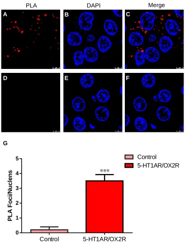
To explore the existence of 5-HT1AR-OX2R heterodimers, we initially carried out
Proximity Ligation Assay (PLA) on slices of rat hippocampus. Goat anti-5-HT1AR
and rabbit anti-OX2R antibodies were used to perform PLA assay. After incubation of
5-HT1AR and OX2R antibodies, we detected stable fluorescence signal in the
hippocampus slices (Figure 1A-C). In contrast, the control group was performed with
only 5-HT1AR primary antibody followed by the incubation of both secondary
antibodies and no signal was found in the control group (Figure 1D-F). DAPI was
used to stain the nuclei (Figure 1B and Figure 1E). And the merged images of PLA
signal and DAPI showed that 5-HT1AR and OX2R formed the heterodimers (Figure
1C and Figure 1F). The statistic results also demonstrated that 5-HT1AR and OX2R
formed heterodimers in hippocampus (Figure 1G). This is the first evidence
Figure 1. Direct interaction between 5-HT1AR and OX2R in the hippocampus of the adult rat.
(A-C) Representative photomicrographs staining with antibodies against 5-HT1AR and OX2R by PLA.
Red dots represent the interaction of 5-HT1AR and OX2R (A). The nuclei were stained with DAPI (blue) (B). The merged photomicrograph in (C). Scale bars=5 μm.
ttsfadsdxf9aa">61</key></foreignraphs of control group incubating with 5-HT1AR primary antibody
followed by the incubation with both the secondary antibodies by PLA (D). The nuclei were stained with DAPI (blue) (E). The merged photomicrograph in (F). Scale bars=5 μm.
(G) Statistics for PLA dots in (A-F).
5-HT1AR was co-localized with OX2R in the membrane of HEK293T cells
PLA DAPI Merge
D A
E F
B C
Control 5-HT1AR/OX2R 0
1 2 3 4 5
P
L
A
F
o
c
i/
N
u
c
le
n
s
Control
5-HT1AR/OX2R
***
We next verified the co-localization of 5-HT1AR and OX2R. We used cell model to
examine the expression pattern of 5-HT1AR and OX2R. 5-HT1AR-ECFP and
OX2R-Venus plasmids were co-transfected into the HEK293T cell. The cells were
transferred into the slides in the 12-well plate 24 h after the plasmids transfection.
Sequentially, the fluorescence signals were analyzed using confocal microscopy 24 h
following the cell passage. The results showed that the fluorescence signals of ECFP
and Venus were significantly merged in the HEK293T cell membrane (Figure 2A-C).
To exclude the possibility of ECFP affecting the localization of 5-HT1AR, we then
transfected HEK293T cells using 5-HT1AR plasmid without any tags. Instead,
immunostaining of 5-HT1AR by its antibody was performed to indicate the
expression of 5-HT1AR after 48 h following the transfection. As shown in Figure 2,
the fluorescence signals of 5-HT1AR antibody and Venus were also largely merged in
HEK293T cells (Figure 2D-F). These results provide the evidence that 5-HT1AR is
[image:7.595.92.519.402.693.2]co-localized with OX2R.
Figure 2. Co-localization of 5-HT1AR and OX2R in the cell membrane of HEK293T cells.
(A-C) Representative photomicrographs showing the co-localization of 5-HT1AR and OX2R in
HEK293T cells transfected with OX2R-Venus (green, A) and 5-HT1AR-ECFP (cyan, B). The
co-localization was indicated by C.
OX2R-Venus
A 5-HT1AR-ECFP Merge
OX2R-Venus 5-HT1AR Merge
B C
(D-F) Confocal microscopy image of the co-localization of 5-HT1AR and OX2R in HEK293T cells
transfected with 5-HT1AR and OX2R-Venus. OX2R-Venus indicated the expression pattern of
OX2R (D). Anti-5-HT1AR antibody was used to indicate the expression pattern of 5-HT1AR (E). The co-localization was indicated by F. Scale bars=10 μm.
The direct interaction between 5-HT1AR and OX2R in HEK293T cells
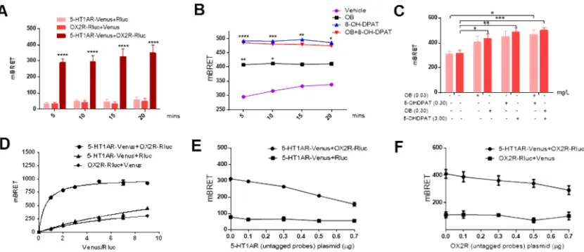
BRET assay is very efficient to evaluate the direct interaction of different proteins
[26-28]. To further evaluate the existence of 5-HT1AR-OX2R heterodimer, we used
BRET analysis to determine whether 5-HT1AR interacted with OX2R directly.
5-HT1AR-Venus and/or OX2R-Rluc plasmids were introduced into HEK293T cells.
For BRET experiment, the substrate coelenterazine H (COH) was added into the
medium at the final concentration of 5 M. Then the 5-HT1AR agonist 8-OHDPAT
and/or the OX2R agonist orexin B (OB) were incubated at the final concentration of 3
mg/L (8-OHDPAT) or 0.3 mg/L (OB) respectively. The analysis was performed
immediately using the Tristar LB941 plate reader. The data were collected at every 5
mins in duration of 20 mins. And the results showed that the mBRET values were
higher in the HEK293T cells with co-transfection of 5-HT1AR-Venus and
OX2R-Rluc plasmids compared to the control group in all the indicated periods of
detected-time (Figure 3A). Meanwhile, the 5-HT1AR agonist 8-OHDPAT and/or the
OX2R agonist OB were added respectively to investigate the role of 5-HT1AR and
OX2R activation in regulation of the interaction of these two receptors. As shown in
Figure 3B, the mBRET values were potentiated after 8-OHDPAT and/or OB treatment,
indicating that the direct interaction of 5-HT1AR and OX2R could be enhanced
following activation of the heterodimer. Besides, the direct interaction of the two
receptors could also be enhanced in HEK293T cells of transfection of the two factors
with the increase of the concentration of the agonists (Figure 3C).
To verify the specificity of the interaction of 5-HT1AR and OX2R, saturation and
competitive assays were performed. For saturation experiment, we transfected the
HEK293T cells with a constant amount of OX2R-Rluc and the increasing amount of
5-HT1AR-Venus. For competition experiment, a constant amount of 5-HT1AR-Venus
were transfected into the HEK293T cells. The empty plasmid pCDNA3.1 was used to
keep the constant amount of the total plasmids of every well. The cells were
transferred into 96-well microplate 24 h after transfection of the plasmids. The
substrate coelenterazine H at the final concentration of 5 M was added into the
medium. The 5-HT1AR agonist 8-OHDPAT and/or the OX2R agonist OB were then
added. The analysis was carried out immediately using the Tristar LB941 plate reader.
mBRET values were collected to make the saturation and competition curves (Figure
3D-F). The results showed that the mBRET value was elevated in a dose-dependent
with the increasing amounts of 5-HT1AR-Venus plasmid and reached a saturation
stage when the ratio of the amount of 5-HT1AR-Venus to OX2R-Rluc was 7,
indicating that the interaction between 5-HT1AR and OX2R was not due to the
overexpression of these two receptors and the interaction between the two receptors
was specific (Figure 3D). Consistently, the results from competitive assay showed that
the value of mBRET decreased gradually with the increasement of the amount of
5-HT1AR or OX2R with no tags (Figure 3E-F). Together, these results suggested that
[image:9.595.99.514.451.630.2]5-HT1AR and OX2R directly interacted in HEK293T cells.
Figure 3. Formation of 5-HT1AR and OX2R heterodimer determined by BRET assay in HEK293T cells.
(A-C) Show the interaction of 5-HT1AR and OX2R in HEK293T cells with co-transfected
5-HT1AR-Venus and OX2R-Rluc without (A) or with the agonist 8-OHDPAT and OB treatment
(B-C). P values were obtained by copmaring the “5-HT1AR-Venus and OX2R-Rluc” group to
the “5-HT1AR-Venus and Rluc” group in (A). P values were obtained by copmaring the “OB”
group or the “8-OHDPAT” group or the “OB+8-OHDPAT” group to the “Vehicle” group in (B).
transfected with the constant amount of OX2R-Rluc and the increasing amount of
5-HT1AR-Venus. BRET values were collected at different ratios of the amount of
5-HT1AR-Venus to OX2R-Rluc. All the data were used to make the saturation curve by
GraphPad.
(E-F) The competition curve of the heterodimerization of 5-HT1AR and OX2R. HEK293T cells were
transfected with the constant amount of OX2R-Rluc and 5-HT1AR-Venus plasmids and the
increasing amount of 5-HT1AR (E) or OX2R (F) without any tags.
Data were presented as mean ± S.E.M.. *P < 0.05, ** P < 0.01, *** P < 0.001, **** P < 0.0001.
The co-immunoprecipitation of 5-HT1AR and OX2R
To further confirm the interaction of 5-HT1AR and OX2R, we performed
immunoprecipitation experiment. The plasmids of Myc-5-HT1AR and HA-OX2R
were co-transfected into HEK293T cells. As shown in Figure 4A and B, the levels of
5-HT1AR and OX2R protein were greatly upregulated in HEK293T cells after the
transfection of myc-5-HT1AR and HA-OX2R plasmids (Figure 4A-B). Moreover, the
immunoprecipitation results revealed a 5-HT1AR band in the products after
pulling-down OX2R by HA antibody (Figure 4C). This finding was highly consistent
with the data from PLA and BRET analysis and provided further evidence for the
[image:10.595.110.461.472.676.2]existence of 5-HT1AR-OX2R heterodimers.
Figure 4. Direct protein-protein interaction of 5-HT1AR and OX2R determined by co-immunoprecipitation assay.
(A) Representative immunoblots of 5-HT1AR and β-actin in HEK293T cells transfected with Myc-5-HT1AR. The 5-HT1AR and β-actin antibodies to evaluate the over expression of 5-HT1AR. Moc k Myc-5-HT T1A R HA-OX2 R Cotr ansf ectio n IB:5-HT1AR IP:HA IB:5-HT1AR Cell Lysates IB:HA C
Myc-5-HT1AR - +
β-actin
5-HT1AR
A
HA-HOX2R - +
(B) Representative immunoblots of OX2R and β-actin in HEK293T cells transfected with HA-OX2R. The OX2R and β-actin antibodies to evaluate the over expression of OX2R.
(C) Representative immunoblots of immunoprecipites from the total lysates of HEK293T cells
transfected with Myc-5-HT1AR or/and HA-OX2R and probed with anti-5-HT1AR or HA.
5-HT1AR-OX2R heterodimer changed the cellular cAMP and calcium levels
Cellular cAMP and calcium were critical in regulating cell viability, inflammatory
response, oxidative stress reaction and reward learning in the central nervous system
[29-36]. Dysfunction of cellular cAMP and calcium contributed to neurodegenerative
and psychiatric disorders [37]. Recent studies indicated the interaction of cAMP and
calcium as the potential therapeutic targets for the treatment of neurological diseases
[37]. As reported, OX2R activation regulated cellular cAMP and calcium levels [18,
19]. However, 5-HT1AR activation reduced the cellular cAMP levels, while having
no effect on calcium influx [10]. Moreover, the heterodimer of different GPCRs led to
the changes of levels of cAMP and calcium [10, 38]. To explore whether
5-HT1AR-OX2R heterodimers affected the levels of cAMP and calcium, cAMP
BRET biosensor and Fluo-4NW assay kit were performed respectively.
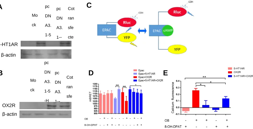
We also investigated the expression levels of 5-HT1AR and OX2R in HEK293T
cells after transfection. And the results showed that the expression levels of the two
receptors in 5-HT1AR or OX2R group were comparable to that of the co-transfected
group (Figure 5A-B). Epac cAMP BRET biosensor consisting of Rluc with Epac tag
in the N-terminal and YFP at the C-terminal was effective to detect the cellular cAMP
levels. When the levels of cAMP increase, cAMP binds to the Epac resulting in the
conformational changes of the sensor, leading to the increase of the distance between
YFP and Rluc, and resulting in the decrease of the signals (Figure 5C). The cAMP
biosensor YFP-Epac-Rluc and 5-HT1AR or/and OX2R were introduced into
HEK293T cells. And the cAMP biosensor YFP-Epac-Rluc alone was introduced into
HEK293T cells was as the control group. The cells were transferred into 96-well
microplate for BRET experiment. 24 h later, the 5-HT1AR agonist 8-OHDPAT and/or
the OX2R agonist OB were added into the medium at the final concentration of 3
Tristar LB941 plate reader. The results showed that 8-OHDPAT and/or OB treatment
alone did not change the basal cAMP levels HEK293T cells. And consistent with the
previous findings, the levels of cAMP were reduced by 8-OHDPAT in 5-HT1AR
transfected group, while OB treatment increased the levels of cAMP in OX2R
transfected group [10]. However, there is no significant difference of cAMP levels in
HEK293T-5-HT1AR/OX2R group after 8-OHDPAT or OX2R treatment alone. While
the cAMP levels increased significantly in HEK293T-5-HT1AR/OX2R group after
both 8-OHDPAT and OB challenge compared with no agonist treatment (Figure 5D).
Additionally, the levels of cAMP decreased significantly in HEK293T-5-HT1AR cells
after treatment with 8-OHDPAT (Supplementary Figure 1A). While cAMP and
calcium levels increased robustly in HEK293T-OX2R cells after treatment with OB
(Supplementary Figure 1B). While there were no significant changes of cAMP and
calcium after treatment with different concentrations of 8-OHDPAT or OB in
HEK293T-5-HT1AR group and HEK293T-5-OX2R group (Supplementary Figure
1A-B). Besides, the cAMP levels increased significantly in
HEK293T-5-HT1AR/OX2R group after treatment with 8-OHDPAT and OB compared
with the untreated group (Supplementary Figure 1C). Additionally, the upregulation of
cAMP depended on the concentrations of 8-OHDPAT and OB (Supplementary Figure
1C). These results suggested that the 5-HT1AR-OX2R heterodimer affected the levels
of cellular cAMP in a new way which was different from the monomers of 5-HT1AR
or OX2R (Figure 5D).
Fluo-4NW is a calcium indicator and Fluo-4NW kit has been taken as the effective
and convenient way to evaluate the levels of cellular calcium. The results of
Fluo-4NW assay demonstrated that 8-OHDPAT treatment had no effect on the
calcium levels in 5-HT1AR group, while OB challenge increased the levels of
calcium in OX2R group (Figure 5E). 8-OHDPAT treatment had no effect on the
calcium levels in HEK293T-5-HT1AR/OX2R group compared to the 5-HT1AR group
with 8-OHDPAT treatment (Figure 5E). However, the levels of calcium increased
robustly of HEK293T-5-HT1AR/OX2R group after 8-OHDPAT and OB challenge
the OX2R transfected group with OB treatment (Figure 5E). Taken together, these
results above demonstrated that 5-HT1AR-OX2R heterodimers function as a new
receptor which was different from the original 5-HT1AR or OX2R receptor in
regulation of cellular calcium levels, which providing new insights into the
[image:13.595.95.511.201.413.2]understanding of the role of 5-HT1AR and OX2R (Figure 5E).
Figure 5 Evaluation of cellular cAMP and calcium levels by 5-HT1AR-OX2R heterodimer activation.
(A-B) Representative immunoblots of the total lysates of HEK293T cells transfected with 5-HT1AR
or/and OX2R and probed with anti-5-HT1AR (A) or OX2R (B).
(C) Show the schematic diagram of the EPAC BRET biosensor.
(D) Show the cellular cAMP levels in HEK293T cells transfected with 5-HT1AR and/or OX2R
using EPAC BRET biosensor.
(E) Show the cellular calcium levels HEK293T cells transfected with 5-HT1AR and/or OX2R using
Fluo-4NW assay kit.
Data were expressed as mean ± S.E.M. *P < 0.05, ** P < 0.01, *** P < 0.01.
5-HT1AR-OX2R heterodimer downregulated p-ERK levels
As one member of mitogen-activated protein kinases (MAPK) family, extracellular
signal-related kinase (ERK) plays vital roles in cell migration, cell proliferation, cell
differentiation and apoptosis [39]. Early studies showed that enhancement of p-ERK
protected dopaminergic neurons in substantia nigra from death caused by neurotoxin
MPTP [40], indicating the important role of ERK in brain disorders. Moreover,
previous research demonstrated that both 5-HT1AR and OX2R regulated the levels of
p-ERK [15, 20].
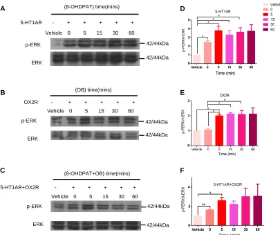
We then investigated whether the heterodimer of 5-HT1AR and OX2R would affect
the levels of ERK and p-ERK. Western blotting was performed to determine the levels
of total ERK and p-ERK of 5-HT1AR-OX2R heterodimers after agonist stimulation.
The results showed that there was no difference of the levels of total ERK after
agonist challenge among HEK293T-5-HT1AR group, HEK293T-OX2R group and
HEK293T-5-HT1AR/OX2R group (Figure 6A-F). However, the levels of p-ERK
increased approximately four folds 5 mins after 8-OHDPAT challenge and reached the
peak at 30 mins in HEK293T-5-HT1AR group (Figure 6A). The levels of p-ERK
increased approximately two folds 5 mins after OB challenge and reached the peak at
15 mins in HEK293T-OX2R group (Figure 6B). Moreover, the levels of p-ERK
increased significantly 5 mins in HEK293T-5-HT1AR/OX2R group after both
8-OHDPAT and OB treatment. However, there was no change of p-ERK levels from
15 to 60 mins in HEK293T-5-HT1AR/OX2R group after the 8-OHDPAT and OB
treatment (Figure 6 C). Besides, the levels of p-ERK levels in HEK293T-5-HT1AR
group were increased gradually 10 mins after treatment with the increasement of the
concentration of 8-OHDPAT (Figure 7A). Also, the levels of p-ERK levels of
HEK293T-OX2R cells were increased gradually 10 mins after treatment with the
increase of the concentration of OB (Figure 7B). While there was an increase trend of
the levels of p-ERK in HEK293T-OX2R group after treatment with different
concentrations of 8-OHDPAT and OB (Figure 7C). The statistical data also confirmed
the results (Figure 7D-F). Additionally, the results of further experiments showed that
there were no significant changes of p-ERK levels in HEK293T-HT1AR/OX2R group
10 mins following treatment with 8-OHPAT or OB compared with the untreated group
(Supplementary Figure 2A-B), indicating that the 5-HT1AR-OX2R heterodimers
functioned in a way which was different from the original receptors. Altogether, these
findings demonstrated that the expression levels of p-ERK decreased in
HEK293T-5-HT1AR/OX2R group compared with HEK293T-5-HT1AR or
Figure 6. The heterodimer of 5-HT1AR and OX2R downregulated the phosphorylation of ERK.
(A) Representative immunoblots of p-ERK in HEK293T cells transfected with 5-HT1AR following
8-OHDPAT treatment.
(B) Representative immunoblots of p-ERK in HEK293T cells transfected with OX2R after OB
stimulation.
(C) Representative immunoblots of p-ERK in HEK293T cells transfected with 5-HT1AR and OX2R
after 8-OHDPAT and OB challenge by western blotting.
(D) Statistics for p-ERK expression in (A).
(E) Statistics for p-ERK expression in (B).
(F) Statistics for p-ERK expression in (C).
Data were expressed as mean ± S.E.M..*P< 0.05,** P< 0.01,*** P< 0.01.
p-ERK ERK (OB) time(mins) (8-OHDPAT) time(mins) (8-OHDPAT+OB) time(mins) A B C p-ERK ERK p-ERK ERK D E F
Vehicle 0 5 15 30 60
5-HT1AR - + + + + +
Vehicle 0 5 15 30 60
OX2R - + + + + +
Vehicle 0 5 15 30 60
5-HT1AR+OX2R - + + + + +
Figure 7. The effect of concentrations of 8-OHFPAT and OB on levels of the phosphorylation of ERK.
(A) Representative immunoblots of p-ERK in HEK293T cells transfected with 5-HT1AR
following 8-OHDPAT treatment with different concentrations.
(B) Representative immunoblots of p-ERK in HEK293T cells transfected with OX2R after OB
stimulation with different concentrations.
(C) Representative immunoblots of p-ERK in HEK293T cells transfected with 5-HT1AR and
OX2R after 8-OHDPAT and OB challenge with different concentrations.
(D) Statistics for p-ERK expression in (A).
(E) Statistics for p-ERK expression in (B).
(F) Statistics for p-ERK expression in (C).
Data were expressed as mean ± S.E.M. *P < 0.05, ** P < 0.01, *** P < 0.01.
5-HT1AR-OX2R heterodimer downregulated p-CREB levels
CREB plays important roles in the maintenance of immune homeostasis and
regulating of learning and memory in the central nervous system [41, 42]. It has been
reported that the behavior and cognition ability of mice is regulated by CREB in
Defect of CREB contributed to the progresses of brain disorders such as depression
[44]. Previous studies revealed that GPCR induced the upregulation of
phosphorylated CREB (p-CREB) through enhancing the activation of adenylyl
cyclase (AC). While the levels of p-CREB decreased when GPCR coupling to the Gi
protein causing the reduction of the activation of adenylyl cyclase (AC) [45].
Additionally, as the Gi-coupled GPCR, 5-HT1AR has been reported to reduce
p-CREB levels after 8-OHDPAT stimulation [10, 45]. To uncover whether
5-HT1AR-OX2R heterodimers affect the levels of p-CREB, western blotting was
performed to determine the expression levels of total CREB and p-CREB of
5-HT1AR-OX2R heterodimers after agonist challenge. The results showed that there
was no difference in the levels of total CREB among HEK293T-5-HT1AR group,
HEK293T-OX2R group and HEK293T-5-HT1AR/OX2R group (Figure 8A-F).
However, the levels of p-CREB decreased significantly in
HEK293T-5-HT1AR/OX2R group compared with HEK293T-5-HT1AR or
HEK293T-OX2R group without agonist treatment (Figure 8A-C). Furthermore, the
expression levels of p-CREB in HEK293T-5-HT1AR/OX2R group tended to decrease
after the treatment of 8-PHDPAT and OB (Figure 8C). Additionally, the levels of
p-CREB in HEK293T-5-HT1AR group were decreased gradually 10 mins after
treatment with the increase of the concentration of 8-OHDPAT (Figure 9A). While the
levels of p-CREB HEK293T-OX2R group were increased gradually 10 mins after
treatment with the increase of the concentration of OB (Figure 9B). And the levels of
p-CREB in HEK293T-5-HT1AR/OX2R group decreased significantly after treatment
with high concentration of 8-OHDPAT and OB compared with the untreated group
(Figure 9C). The statistical data also confirmed the results (Figure 9D-F). Therefore,
all these results demonstrated that the expression levels of p-CREB decreased in
HEK293T-5-HT1AR/OX2R group compared with HEK293T-5-HT1AR or
HEK293T-OX2R group after agonist treatment (Figure 8-9).
Taken together, our findings demonstrated that 5-HT1AR and OX2R formed the
heterodimers and affected downstream signaling molecular which was different from
Figure 8. The heterodimer of 5-HT1AR and OX2R downregulated the phosphorylation of p-CREB.
(A)Representative immunoblots of p-CREB in HEK293T cells transfected with 5-HT1AR following
8-OHDPAT treatment.
(B)Representative immunoblots of p-CREB in HEK293T cells transfected with OX2R after OB
stimulation.
(C)Representative immunoblots of p-CREB in HEK293T cells transfected with 5-HT1AR and OX2R
after 8-OHDPAT and OB challenge.
(D)Statistics for p-CREB expression in (A).
(E) Statistics for p-CREB expression in (B).
(F) Statistics for p-CREB expression in (C).
Data were expressed as mean ± S.E.M..*P< 0.05,** P< 0.01,*** P< 0.01.
p-CREB
CREB
Vehicle 0 5 15 30 60
OX2R - + + + + +
(OB) time(mins) p-CREB
CREB
Vehicle 0 5 15 30 60
5-HT1AR - + + + + +
(8-OHDPAT) time(mins)
Vehi
cle 0 5 15 30 60 0.0 0.5 1.0 1.5 p -C R E B /C R E B Vehicle 0 5 15 30 60 5-HT1AR ** * *
Vehicle 0 5 15 30 60 5-HT1AR +OX2R - + + + + +
Figure 9. The effect of concentrations of 8-OHFPAT and OB on levels of the phosphorylation
of CREB.
(A) Representative immunoblots of p-CREB in HEK293T cells transfected with 5-HT1AR
following 8-OHDPAT treatment with different concentrations.
(B) Representative immunoblots of p-CREB in HEK293T cells transfected with OX2R after OB
stimulation with different concentrations.
(C) Representative immunoblots of p-CREB in HEK293T cells transfected with 5-HT1AR and OX2R
after 8-OHDPAT and OB challenge with different concentrations.
(D) Statistics for p-CREB expression in (A).
(E) Statistics for p-CREB expression in (B).
(F) Statistics for p-CREB expression in (C).
Data were expressed as mean ± S.E.M. *P < 0.05, ** P < 0.01, *** P < 0.01.
Discussion
In the present study, we found the heterodimer formation of 5-HT1AR and OX2R
and the heterodimer affected the downstream signaling. Firstly, the formation of
5-HT1AR-OX2R heterodimer was observed in hippocampus of adult rat by PLA.
co-localized in HEK293T cells. BRET assay results revealed the heterodimer
formation of 5-HT1AR-OX2R and the agonist treatment enhanced the direct-direct
interaction of the two receptors. Thirdly, co-immunoprecipitation experiment further
demonstrated the protein direct-direct interaction between 5-HT1AR and OX2R,
which was consistence with the results of BRET assay. Lastly, compared to the
5-HT1AR transfected group after 8-OHDPAT stimulation, western blotting displayed
that the levels of cellular calcium and cAMP were upregulated significantly and the
expression levels of p-CREB and p-ERK were downregulated in the
HEK293T-5-HT1AR/OX2R group following 8-OHDPAT and OB treatment.
Altogether, these findings provided direct evidence of the formation of
5-HT1AR-OX2R heterodimer and highlight the potential role of the heterodimer in
[image:21.595.105.507.362.537.2]physiological and pathological conditions in the central nervous system(Figure 10).
Figure 10. The schematic representation of 5-HT1AR-OX2R heterodimer-induced downstream signaling.
The heterodimer of 5-HT1AR and OX2R significantly upregulated the levels of cellular cAMP and
calcium, and downregulated the levels of p-ERK and p-CREB after activation by the agonists.
The homodimer or the heterodimer of GPCRs have been studied for many years
[38, 46]. As members of GPCRs, 5-HT1AR and OX2R are both widely expressed in
hippocampus [13, 17]. Given the similar localization and characteristics of 5-HT1AR
and OX2R in brain, 5-HT1AR and OX2R probably form heterodimer in the central
5-HT1AR and OX2R by PLA. PLA assay has been reported to be a very efficient
method to detect the direct interaction between different proteins[47]. And our PLA
data demonstrated that 5-HT1AR and OX2R formed heterodimers in hippocampus
(Figure 1A-G). Additionally, we also found that there were very few PLA dots in
cortex. The different pattern of the PLA data in different brain areas probably resulted
from the distinct expression pattern and different functions of the two receptors in
various brain regions.
Considerable studies showed that the heterodimers formed by different GPCRs
could affect the downstream signaling pathway [47-49]. In our current study, we also
explored the signaling transduction affected by 5-HT1AR-OX2R heterodimer using
cAMP BRET biosensor, Fluo-4NW kit and western blotting analysis. The levels of
cellular cAMP and calcium increased robustly of HEK293T-5-HT1AR/OX2R group
following 8-OHDPAT and OB challenge, indicating the heterodimer of
5-HT1AR-OX2R affect the expression levels of the second messengers. ERK and
CREB were the key molecules downstream of 5-HT1AR and OX2R. In addition to
the involvement in physiological processes including cell proliferation, cell migration
and memory [50-54], ERK and CREB also play important roles in pathological
processes including hyperalgesia, ischemic stroke, Alzheimer’s disease and pain
[55-58]. As described above, 5-HT1AR has been reported to decrease the levels of
cAMP, leading to the downregulation of p-CREB after ligand stimulation. In contrast,
OX2R induced the upregulation of cAMP levels, leading to the increasement of
p-CREB after OB stimulation. Our results revealed that 5-HT1AR-OX2R heterodimer
induced the downregulation of p-ERK and p-CREB after 8-OHDPAT and OB
challenge compared with the 5-HT1AR transfected group treated with 8-OHDPAT.
Interestingly, the expression level of p-CREB decreased significantly in HEK293T
cells with overexpression of 5-HT1AR and OX2R compared with the control group
(Figure 8-9). And the levels of p-CREB in HEK293T-5-HT1AR/OX2R group tend to
further decrease after the challenge of 8-OHDPAT and OB (Figure 8-9). These results
suggested that the 5-HT1AR and OX2R heterodimer regulated the expression levels
The results above indicated that 5-HT1AR-OX2R heterodimer affected the
downstream signaling transduction and the expression levels of cellular molecules
including calcium, cAMP, p-ERK and p-CREB (Figure 5-9). The mechanism
underlying this regulation remains unclear. Several suggestions from the above results
are listed as below. On one hand, the heterodimer of 5-HT1AR and OX2R as a novel
receptor, which was different from original 5-HT1AR or OX2R, induced the
downstream signaling after co-activation by 8-OHDPAT and OB. On the other hand,
as described above, the heterodimer of 5-HT1AR and OX2R in HEK293T cells led to
the upregulation of cAMP and calcium after 8-OHDPAT and OB stimulation
compared to the 5-HT1AR group following 8-OHDPAT stimulation (Figure 5C-E). It
was noteworthy that the increasement of calcium levels in the co-transfected group
treated with 8-OHDPAT and OB was still lower than OX2R group after OB treatment,
while 5-HT1AR alone induced the downregulation of cAMP and no changes of
calcium levels after ligand treatment (Figure 5C-E). 5-HT1AR and OX2R
heterodimer led to the downregulation of cellular p-ERK and p-CREB compared to
the OX2R group after OB stimulation. Here, we found 5-HT1AR downregulated the
levels of p-CREB and OX2R upregulated the levels of p-CREB (Figure 8A-B).
Activation of 5-HT1AR-OX2R heterodimers induced the upregulation of cAMP,
while leading to the downregulation of p-CREB (Figure 5D and Figure 8). As
mentioned above, 5-HT1AR mainly binds to the Gi protein, leading to the decrease
of adenylate cyclase (AC) activation, and further leads to the downregulation of levels
of p-CREB and cellular cAMP after ligand stimulation [15]. And OX2R mainly binds
to the Gs protein and Gq protein resulting in the activation of adenylate cyclase (AC),
and further leads to the upregulation of the expression levels of p-CREB, p-ERK,
cellular cAMP and calcium levels after ligand stimulation [18-20]. In the study, we
found activation of 5-HT1AR-OX2R heterodimers increased the levels of cAMP and
calcium, also led to the downregulation of p-ERK and p-CREB (Figure 6 and Figure
8). Thus, the signaling changes of the heterodimers after ligand treatment might result
from the activation of Gs and Gq signaling, which would cause the upregulation of
of p-CREB by activating protein kinase A (PKA). And calcium had positive effects on
the expression levels of p-CREB through regulating calmodulin-dependent kinases
(CaMK) [59]. Besides, the signaling pathway of cAMP could be regulated by CaMK
and mitogen-activated protein kinase (MAPK) [59]. Thus, the downregulation of
p-CREB in 5-HT1AR/OX2R group after ligands stimulation might result from
increasement of cAMP and calcium levels.
cAMP/PKA pathway was a very important regulator of neurodegeneration, learning
and memory [60]. Previous studies showed that enhancement of calcium-CREB
signaling pathway improved the ability of learning and memory of rats [61].
Additionally, the results from a genome-wide association study demonstrated that
CREB interacting with some of risk genes of mental disorders, suggesting that CREB
probably be identified as one of the risk factors for psychiatric disorders including
schizophrenia and major depression disorder [62]. Besides, enhancement of
p-ERK/p-CREB signaling ameliorated the death of dopaminergic cell line SH-SY5Y
cells after neurotoxin MPP+challenge and dopaminergic neurons in substantia nigra
(SN) by neurotoxin MPTP treatment [40, 63]. Together, these findings revealed that
cAMP-calcium-ERK-CREB signaling was a very important regulator of neurological
disease. Importantly, the expression levels of 5-HT1AR decreased significantly in
depression model [64]. And OX2R knockout mice presented as an increase of despair,
indicating the important role of OX2R in mental disorders such as depression [25].
Our data revealed that the heterodimer of 5-HT1AR and OX2R modulated
cAMP-calcium-ERK-CREB pathway, which providing a new therapeutic target for
neurological diseases.
As a GPCR, 5-HT1AR forms heterodimer with other GPCRs involved in the
regulation of hippocampus neurogenesis [16, 65]. Deficiency of hippocampus
neurogenesis has been observed in neurological diseases such as depression and
Alzheimer’s disease [5, 66], indicating the important role of neurogenesis of
hippocampus in the brain. 5-HT1AR has been reported to regulate the neurogenesis of
hippocampus [67, 68]. Moreover, lines of evidence showed that 5-HT1AR was
depression [64, 69-73]. Recent research demonstrated that 5-HT1AR and FGFR1
formed heterodimer and enhanced the plasticity of hippocampal neurons [16].
Interestingly, intracerebroventricular co-injection of 8-OHDPAT and FGF2 resulted in
reduction in immobility and increased swimming time of adult rats compared with the
vehicle group or 8-OHDPAT group or FGF2 group, indicating the potential role of
heterodimer of 5-HT1AR with other GPCRs as the therapeutic targets for depression
[16]. Thus, further animal experiments and clinical analyses are needed to uncover the
function of the 5-HT1AR-OX2R heterodimer in the regulation of plasticity of
hippocampus. OX2R has been reported to be involved in the processes of depression.
OX2R deficiency enhanced the symptom of despair, indicating that OX2R might have
good effects on depression [25]. As described above, 5-HT1AR was also participated
in the pathology of depression. Behavior test revealed that the upregulation of
5-HT1AR expression improved the performance of depressed rats [74]. This data
suggested that 5-HT1AR had beneficial effects on depression. Herein, we found the
formation of 5-HT1AR and OX2R heterodimer, which functioned in a different way
from 5-HT1AR or OX2R alone, indicating the potential roles of the 5-HT1AR-OX2R
Supplementary Figure 1. Levels of cAMP and calcium in HEK293T-5-HT1AR-OX2R cells after treatment with different concentration of 8-OHDPAT and OB.
(A-C) Show the cellular cAMP levels in HEK293T cells transfected with 5-HT1AR (A) or OX2R (B)
or 5-HT1AR/OX2R (C) using EPAC BRET biosensor following 8-OHDPAT and/or OB
treatment with different concentrations.
(D) Show the cellular calcium levels HEK293T cells transfected with 5-HT1AR and/or OX2R using
Fluo-4NW assay kit following 8-OHDPAT and/or OB treatment with different concentrations..
Supplementary Figure 2. Evaluation of cellular p-ERK levels by 5-HT1AR-OX2R heterodimer activation in HEK293T cells treated with 8-OHDPAT or OB.
(A) Show the cellular p-ERK levels in HEK293T cells transfected with 5-HT1AR and OX2R
following treatment with 8-OHDPAT of different concentrations.
(B) Show the cellular p-ERK levels in HEK293T cells transfected with 5-HT1AR and OX2R
following treatment with OB of different concentrations.
Materials and methods
Peptides, regents and antibodies
Human orexin B was purchased from Phoenix Pharmaceuticals (003-31). The
5HT1AR agonist 8-OHDPAT was obtained from sigma (H8520) and the Duolink PLA
kit was obtained from Sigma (Duolink In Situ Detection Reagents Red). The
transfection regent LipofectamineTM2000 was purchased from Invitrogen (11668-019).
Anti-CREB and anti-phospho-CREB antibodies were purchased from Cell Signaling
Technology (anti-CREB antibody:9197, anti-p-CREB antibody:9198). Anti-HA
antibody was purchased from Cell Signaling Technology (#3724). Anti-Myc antibody
obtained from Merck Millipore (AB3094). Anti-5-HT1AR antibody for western
blotting and immunofluorescence was purchased from Cell Signaling Technology
(#9897). Anti-5-HT1AR antibody for PLA was obtained from Sigma (SAB2501759).
Anti-β-actin antibody was purchased from ZSGB-BIO (TA-09). Anti-p-ERK antibody
was obtained from Cell Signaling Technology (#9101). Anti-ERK antibody was
purchased from Cell Signaling Technology (#4696).
Plasmid constructs
For immunoprecipitation, a full-length human 5-HT1AR (Gene ID: 3350) was
subcloned into the mammalian expression vector pcDNA3.1-Myc-His A using Xha℃
and EcoR℃. A full-length human OX2R (Gene ID: 3062) was subcloned into the
expression vector pcDNA3.1-3HA using Xho℃ and Kpn℃. pcDNA3.1-Myc-His A,
pcDNA3.1-OX2R, pcDNA3.1-5-HT1AR and pcDNA3.1-3HA were gifts from Dr.
Chen Jing (Division of Biomedical Sciences, Warwick Medical School, University of
Warwick, Coventry, CV4 7AL, UK). For immunofluorescence and BRET, a
full-length human 5-HT1AR was subcloned into the N-terminus of pECFP-N1
(Clontech, #6900-1) using Hind℃ and BamH℃. The OX2R-Rluc plasmid was
constructed by subcloning the coding sequence of human OX2R without the stop
codon into pRluc-N1 (BioSignal Packard, Inc., Montreal, Canada). Human OX2R
was inserted into the N-terminus of pVenus-N1 (Addgene, #61854) using Hind℃ and
BamH℃.
Immunofluorescence
Immunofluorescence analysis was carried out as described previously [75]. In brief,
cell cultures were fixed by paraformaldehyde (PFA) for 10 mins and blocked for 2 h
in blocking solution at room temperature following by primary antibody incubation
overnight at 4℃. The samples were washed three times with phosphate-buffered saline
(PBS) and then incubated with the secondary antibody for 1 h at room temperature.
The secondary antibody used as follows: CY3-conjugated goat anti-rabbit IgG
Leica DMRE laser scanning confocal microscope at the 550nm (Lecia, Milton Keynes,
UK). Besides, the Venus signal was collected at the 480 nm. The pixel size of the
imaging was 1024× 1024.
Western blotting and Quantification
The procedure of western blotting was according to the instructions described
before [47]. Briefly, HEK293T cells expressing 5-HT1AR and/or OX2R were lysed in
RIPA (50Mm Tris (PH 7.4), 150Mm NaCl, 1% Triton X-100, 1% sodium
deoxycholate, 0.1% SDS) and the proteins extracted with different molecular weights
were separated by SDS-PAGE gels. The proteins were then transferred to the PVDF
membranes. The membranes were incubated with the primary antibodies overnight at
4℃ after blocking with 5% nonfat milk for 2 h at room temperature. The primary
antibodies used as follows: Myc (1:1000), HA (1:1000), 5-HT1AR (1:2000), CREB
(1:1000), p-CREB (1:1000), ERK (1:1000), p-ERK (1:1000), β-actin (1:5000). The
next day, membranes were incubated with the corresponding secondary antibodies for
1 h at room temperature after washing with TBST for 3 times. The secondly
antibodies were used as follows: HRP-conjugated goat anti-mouse IgG (1:10000,
ZSGB-BIO, ZB-2305), HRP-conjugated goat anti-rabbit IgG (1:10000, ZSGB-BIO,
ZB-2301). Peroxidase activity was determined with the chemiluminescent substrate
(Multi Sciences, P1425). The band of β-actin was considered as the loading control.
BRET assay for analysis of the interaction of 5-HT1AR and OX2R
BRET assay was performed according to the procedures described previously [47].
The 5-HT1AR-Venus and/or OX2R-Rluc were introduced into HEK293T cells. After
24 h transfection, cells were digested in 0.02% trypsin and plated into a 96-well
microplate (Corning 3600) for another 24 h. The substrate coelenterazine H was
added into the medium at the final concentration of 5 M. The 5-HT1AR agonist
8-OHDPAT or the OX2R agonist OB was then added at the final concentration of 3
Tristar LB941 plate reader (Berthold Technologies, Bad Wildbad, Germany) and the
time of duration was under 30 mins because of the activity of the substrate. And the
filtered light emission was sequentially recorded at 460 nm and 535 nm respectively
by MikroWin 2000 software. The data were presented as “BRET ratio” or “mBRET”
(mBRET=1000×BRET ratio). The value of mBRET was calculated through
subtracting the ratio of the control group.
For saturation experiment, we transfected the HEK293T cells in 12-well plate with
a constant amount of OX2R-Rluc and the increasing amount of 5-HT1AR-Venus. The
empty plasmid pCDNA3.1 was used to keep the constant amount of the total plasmid
of each well. mBRET values were collected at different ration of 5-HT1AR-Venus to
OX2R-Rluc to plot the saturation curves.
For competition experiment, we transfected the HEK293T cells in 12-well plate
with a constant amount of 5-HT1AR-Venus and OX2R-Rluc. Besides, the increasing
amount of 5-HT1AR without any tags was added to the reaction system to compete
with 5-HT1AR-Venus. And the empty plasmid pCDNA3.1 was also added to keep the
constant amount of the total plasmid of each well. mBRET values were collected to
plot the competition curves.
Co-immunoprecipitation
To analyze the interaction between 5-HT1AR and OX2R, co-immunoprecipitation
assay was performed according to the protocol of Pierce Anti-HA-Agarose (Thermo
Scientific, 26181). In brief, approximately 5×10⁶HEK293T cells were harvested after
myc-5-HT1AR and/or HA-OX2R transfection for 24 h. The cells were lysed in 150μl
of RIPA buffer. And the supernatant was collected after centrifugation at 12000 g for
15 mins. The pellet resin was then obtained using the 50μl of Pierce Anti-HA Agarose
by centrifugation at 12000 g for 5 seconds. Add 50 μl of the supernatant into the
collected pellet resin at a final volume of 200 μl by adding TBS followed by
incubation on a rocking platform overnight at 4℃. Next day, after centrifugation at
three times. Finally, the immunoprecipitation products were analyzed by western
blotting.
PLA
The standard procedure for PLA was described before [47]. Briefly, 5 μm
paraffin-embedded slices were incubated with anti-5-HT1AR and anti-OX2R
antibodies overnight at 4℃ after blocked with the blocking solution for 1 h at room
temperature. The slices were incubated with plus and minus probes for 1 h at 37℃
after washed with Buffer A for three times. The slices were then incubated with the
amplification-polymerase solution at 37℃ for 3 h. The signals were analyzed by the
Leika TCS SP5.
BRET measurement for cAMP
The measurement for cAMP using Epac cAMP BRET biosensor analysis was
according to the procedures described previously [47, 76]. In brief, HEK293T cells
were transfected with the cAMP biosensor YFP-Epac-Rluc. And the cAMP biosensor
YFP-Epac-Rluc alone was transfected into HEK293T cells was as the control group.
Cells were digested and plated into a 96-well microplate (Corning 3600) for 24 h
following adding the substrate coelenterazine H into the medium at the final
concentration of 5 uM. The agonist 8-OHDPAT or OB or both 8-OHDPAT and OB
were added to the medium. The data were collected using the Tristar LB941 plate
reader (Berthold Technologies, Bad Wildbad, Germany) and presented as “mBRET”.
The data were collected at every 5 mins in duration of 20 mins.
Cell transfection
HEK293T cells were cultured in the Dulbecco’s modified Eagle’s medium (DMEM,
Invitrogen) medium containing 10% fetal bovine serum (FBS) and 0.1% NaHCO3
(Sigma-Aldrich) in humidified atmosphere containing 5% CO2at 37℃. For BRET
assay, 1 g plasmid was using for each well of 24-well plate. For western blotting
introduced into the HEK293T cells according to the protocol of Lipofectamine 2000
reagent.
Statistical analysis
The data were analyzed using GraphPad Prism v5.0. All the data were presented as
means ± SEM. The treatment group was compared with the control group by student’s
t-test or one-way ANOVA followed by a post hoc test including Sidak's multiple,
Student-Newman-Keul's test or Dunnet test comparisons test. Differences were
considered to be significant when the P value less than 0.05. All the experiments were
repeated three times at least.
Conclusions
Taken together, we found that 5-HT1AR and OX2R could form heterodimers in
vitro and in vivo and further affect the expression of the downstream signaling
molecular including cAMP, calcium, p-ERK and p-CREB, which will not only
promote our understanding of the role of 5-HT1AR and OX2R in the physiological
processes but also provide new molecular mechanisms underlying the neurological
diseases.
Author’s contribution
Q.-Q.W, B.B and J.C conceived and designed the experiments; Q.-Q.W conducted
the experiments and wrote the manuscript; C.-M.W and B.-H.C performed the data
analysis; C.-Q.Y contributed to the cell culture; B.B and J.C supervised the project
and wrote the manuscript.
Acknowledgments
The project was supported by Supporting Fund for Teachers’ research of Jining
Medical University (JY2017KJ036), Natural Science Foundation of Shandong
Province (ZR2018MC005), Research Fund for the Doctoral Program of Jining
81501018)and Science and technology plan project of Shandong higher education
institutions (J15LE19).
Competing interests
The authors declare no conflicts of interest.
References
1. Sexton, P.M. and A. Christopoulos, To Bind or Not to Bind: Unravelling GPCR
Polypharmacology.Cell, 2018. 172(4): p. 636-638.
2. Erlandson, S.C., C. McMahon, and A.C. Kruse,Structural Basis for G Protein-Coupled
Receptor Signaling.Annu Rev Biophys, 2018.
3. Wei, L., et al.,Rosiglitazone Inhibits Angiotensin II-Induced Proliferation of Glomerular
Mesangial Cells via the Galphaq/Plcbeta4/TRPC Signaling Pathway. Cell Physiol
Biochem, 2017. 44(6): p. 2228-2242.
票 픚䈀Shao, W., et al., Suppression of neuroinflammation by astrocytic dopamine D2
receptors via alphaB-crystallin.Nature, 2013. 494(7435): p. 90-4.
5. Drevets, W.C., et al., Serotonin-1A receptor imaging in recurrent depression:
replication and literature review.Nucl Med Biol, 2007. 34(7): p. 865-77.
6. Su, P., et al., A dopamine D2 receptor-DISC1 protein complex may contribute to
antipsychotic-like effects.Neuron, 2014. 84(6): p. 1302-16.
7. Tena-Campos, M., et al., Zinc Is Involved in Depression by Modulating G
Protein-Coupled Receptor Heterodimerization. Mol Neurobiol, 2016. 53(3): p.
8. Fuxe, K., et al.,On the existence and function of galanin receptor heteromers in the
central nervous system.Front Endocrinol (Lausanne), 2012. 3: p. 127.
9. Lee, J.Y., et al., Crystal structure of the human sterol transporter ABCG5/ABCG8.
Nature, 2016. 533(7604): p. 561-4.
10. Lukasiewicz, S., et al.,Dopamine D2 and serotonin 5-HT1A receptor interaction in the
context of the effects of antipsychotics - in vitro studies.J Neurochem, 2016. 137(4): p.
549-60.
11. Peng, Y., et al., 5-HT2C Receptor Structures Reveal the Structural Basis of GPCR
Polypharmacology.Cell, 2018. 172(4): p. 719-730 e14.
12. Basak, S., et al.,Cryo-EM structure of 5-HT3A receptor in its resting conformation.Nat
Commun, 2018. 9(1): p. 514.
13. Lian, P., et al., Higher-Affinity Agonists of 5-HT1AR Discovered through Tuning the
Binding-Site Flexibility.J Chem Inf Model, 2015. 55(8): p. 1616-27.
14. Garcia-Garcia, A.L., et al., Serotonin Signaling through Prefrontal Cortex 5-HT1A
Receptors during Adolescence Can Determine Baseline Mood-Related Behaviors.
Cell Rep, 2017. 18(5): p. 1144-1156.
15. Rojas, P.S. and J.L. Fiedler, What Do We Really Know About 5-HT1A Receptor
Signaling in Neuronal Cells?Front Cell Neurosci, 2016. 10: p. 272.
16. Borroto-Escuela, D.O., et al.,Fibroblast growth factor receptor 1- 5-hydroxytryptamine
1A heteroreceptor complexes and their enhancement of hippocampal plasticity. Biol
Psychiatry, 2012. 71(1): p. 84-91.
levels in the mouse brain.Neurosci Lett, 2002. 332(3): p. 190-4.
18. Wang, C., et al., C-terminus of OX2R significantly affects downstream signaling
pathways.Mol Med Rep, 2017. 16(1): p. 159-166.
19. Tang, J., et al., The signalling profile of recombinant human orexin-2 receptor. Cell
Signal, 2008. 20(9): p. 1651-61.
20. Guo, Y. and P. Feng, OX2R activation induces PKC-mediated ERK and CREB
phosphorylation.Exp Cell Res, 2012. 318(16): p. 2004-13.
21. Yelin-Bekerman, L., et al.,Hypocretin neuron-specific transcriptome profiling identifies
the sleep modulator Kcnh4a.Elife, 2015. 4: p. e08638.
22. Korim, W.S., I.J. Llewellyn-Smith, and A.J. Verberne,Activation of Medulla-Projecting
Perifornical Neurons Modulates the Adrenal Sympathetic Response to Hypoglycemia:
Involvement of Orexin Type 2 (OX2-R) Receptors. Endocrinology, 2016. 157(2): p.
810-9.
23. Weller, C.M., et al.,Cluster headache and the hypocretin receptor 2 reconsidered: a
genetic association study and meta-analysis.Cephalalgia, 2015. 35(9): p. 741-7.
24. Yin, J., et al., Crystal structure of the human OX2 orexin receptor bound to the
insomnia drug suvorexant.Nature, 2015. 519(7542): p. 247-50.
25. Scott, M.M., et al., Hcrtr1 and 2 signaling differentially regulates depression-like
behaviors.Behav Brain Res, 2011. 222(2): p. 289-94.
26. Harikumar, K.G., et al.,Bioluminescence Resonance Energy Transfer (BRET) Assay
for Determination of Molecular Interactions in Living Cells.Bio Protoc, 2017. 7(22).
without G protein activation.J Biol Chem, 2018.
28. Bon, N., et al., Phosphate (Pi)-regulated heterodimerization of the high-affinity
sodium-dependent Pi transporters PiT1/Slc20a1 and PiT2/Slc20a2 underlies
extracellular Pi sensing independently of Pi uptake. J Biol Chem, 2018. 293(6): p.
2102-2114.
29. Jambor, T., et al., Parallel effect of 4-octylphenol and cyclic adenosine
monophosphate (cAMP) alters steroidogenesis, cell viability and ROS production in
mice Leydig cells.Chemosphere, 2018.
30. Gu, J., et al., Maresin 1 attenuates mitochondrial dysfunction through the
ALX/cAMP/ROS pathway in the cecal ligation and puncture mouse model and sepsis
patients.Lab Invest, 2018.
31. Veremeyko, T., et al.,Cyclic AMP Pathway Suppress Autoimmune Neuroinflammation
by Inhibiting Functions of Encephalitogenic CD4 T Cells and Enhancing M2
Macrophage Polarization at the Site of Inflammation.Front Immunol, 2018. 9: p. 50.
32. Louis, T., et al., Cyclic AMP-dependent plasticity underlies rapid changes in odor
coding associated with reward learning. Proc Natl Acad Sci U S A, 2018. 115(3): p.
E448-E457.
33. Araujo, L.B., et al.,Effects of mineral trioxide aggregate, BiodentineTM and calcium
hydroxide on viability, proliferation, migration and differentiation of stem cells from
human exfoliated deciduous teeth.J Appl Oral Sci, 2018. 26: p. e20160629.
34. de Seabra Rodrigues Dias, I.R., et al., The Calcium-Induced Regulation in the
Autoimmunity.Front Pharmacol, 2017. 8: p. 962.
35. Liao, C., Y. Zheng, and Y. Guo, MYB30 transcription factor regulates oxidative and
heat stress responses through ANNEXIN-mediated cytosolic calcium signaling in
Arabidopsis.New Phytol, 2017. 216(1): p. 163-177.
36. Conceicao, E.P., et al.,Effect of Early Overfeeding on Palatable Food Preference and
Brain Dopaminergic Reward System at Adulthood: Role of Calcium Supplementation.
J Neuroendocrinol, 2016. 28(5).
37. Bergantin, L.B. and A. Caricati-Neto,Challenges for the pharmacological treatment of
neurological and psychiatric disorders: Implications of the Ca(2+)/cAMP intracellular
signalling interaction.Eur J Pharmacol, 2016. 788: p. 255-260.
38. Bai, B., et al.,Heterodimerization of human apelin and bradykinin 1 receptors: novel
signal transduction characteristics.Cell Signal, 2014. 26(7): p. 1549-59.
39. Sun, Y., et al., Signaling pathway of MAPK/ERK in cell proliferation, differentiation,
migration, senescence and apoptosis.J Recept Signal Transduct Res, 2015. 35(6): p.
600-4.
40. Luo, D., et al.,N-Propargyl Caffeamide (PACA) Ameliorates Dopaminergic Neuronal
Loss and Motor Dysfunctions in MPTP Mouse Model of Parkinson's Disease and in
MPP(+)-Induced Neurons via Promoting the Conversion of proNGF to NGF. Mol
Neurobiol, 2018. 55(3): p. 2258-2267.
41. Ko, Y.H., et al.,6,7,4'-Trihydroxyisoflavone, a major metabolite of daidzein, improves
learning and memory via the cholinergic system and the p-CREB/BDNF signaling
42. Wang, B., et al., Methamphetamine modulates the production of interleukin-6 and
tumor necrosis factor-alpha via the cAMP/PKA/CREB signaling pathway in
lipopolysaccharide-activated microglia.Int Immunopharmacol, 2018. 56: p. 168-178.
43. Sharma, P., S. Sharma, and D. Singh, Apigenin reverses behavioural impairments
and cognitive decline in kindled mice via CREB-BDNF upregulation in the
hippocampus.Nutr Neurosci, 2018: p. 1-10.
44. Tsuchimine, S., N. Sugawara, and N. Yasui-Furukori, Increased levels of CREB in
major depressive patients with antidepressant treatment.Psychiatry Res, 2018.
45. Cheng, Z., et al.,Luciferase Reporter Assay System for Deciphering GPCR Pathways.
Curr Chem Genomics, 2010. 4: p. 84-91.
46. Abe, M., et al., Homodimer formation by the ATP/UTP receptor P2Y2 via disulfide
bridges.J Biochem, 2018.
47. Ji, B., et al., Novel signaling of dynorphin at kappa-opioid receptor/bradykinin B2
receptor heterodimers.Cell Signal, 2017. 31: p. 66-78.
48. Scarlett, K.A., et al., Agonist-induced CXCR4 and CB2 Heterodimerization Inhibits
Galpha13/RhoA-mediated Migration.Mol Cancer Res, 2018.
49. Liu, H., et al., Heterodimerization of the kappa opioid receptor and neurotensin
receptor 1 contributes to a novel beta-arrestin-2-biased pathway. Biochim Biophys
Acta, 2016. 1863(11): p. 2719-2738.
50. Zhang, Y., et al., Sorafenib inhibited cell growth through the MEK/ERK signaling
pathway in acute promyelocytic leukemia cells.Oncol Lett, 2018. 15(4): p. 5620-5626.
of C6 glioma by modulating ERK signaling and gene expression.Neurosci Lett, 2018.
674: p. 42-48.
52. Zhang, X., et al., Diethylstilbestrol regulates mouse gubernaculum testis cell
proliferation via PLC-Ca(2+) -CREB pathway. Cell Biochem Funct, 2018. 36(1): p.
13-17.
53. Gao, J., et al.,Harmine suppresses the proliferation and migration of human ovarian
cancer cells through inhibiting ERK/CREB pathway. Oncol Rep, 2017. 38(5): p.
2927-2934.
54. Sen, T., et al., Activation of PERK Elicits Memory Impairment through Inactivation of
CREB and Downregulation of PSD95 After Traumatic Brain Injury. J Neurosci, 2017.
37(24): p. 5900-5911.
55. Yu, L.N., et al., EphrinB-EphB Signaling Induces Hyperalgesia through ERK5/CREB
Pathway in Rats.Pain Physician, 2017. 20(4): p. E563-E574.
56. Li, Z., et al., Resveratrol protects CA1 neurons against focal cerebral ischemic
reperfusion-induced damage via the ERK-CREB signaling pathway in rats.Pharmacol
Biochem Behav, 2016. 146-147: p. 21-7.
57. Zhang, L., et al., Tyrosine phosphatase STEP61 negatively regulates amyloid
beta-mediated ERK/CREB signaling pathways via alpha7 nicotinic acetylcholine
receptors.J Neurosci Res, 2013. 91(12): p. 1581-90.
58. Wang, H., et al., Antinociceptive effects of oxymatrine from Sophora flavescens,
through regulation of NR2B-containing NMDA receptor-ERK/CREB signaling in a mice
59. Carlezon, W.A., Jr., R.S. Duman, and E.J. Nestler,The many faces of CREB.Trends
Neurosci, 2005. 28(8): p. 436-45.
60. Li, L., et al., The effects of Chinese medicines on cAMP/PKA signaling in central
nervous system dysfunction.Brain Res Bull, 2017. 132: p. 109-117.
61. Ge, C.L., et al., Tongqiao Huoxue Decoction ameliorates learning and memory
defects in rats with vascular dementia by up-regulating the Ca(2+)-CaMKII-CREB
pathway.Chin J Nat Med, 2015. 13(11): p. 823-830.
62. Xiao, X., et al.,The cAMP responsive element-binding (CREB)-1 gene increases risk
of major psychiatric disorders.Mol Psychiatry, 2017.
63. Gu, X., et al., Photoactivation of ERK/CREB/VMAT2 pathway attenuates
MPP(+)-induced neuronal injury in a cellular model of Parkinson's disease.Cell Signal,
2017. 37: p. 103-114.
64. Wang, X.L., et al., Treatment with Shuyu capsule increases 5-HT1AR level and
activation of cAMP-PKA-CREB pathway in hippocampal neurons treated with serum
from a rat model of depression.Mol Med Rep, 2018. 17(3): p. 3575-3582.
65. Borroto-Escuela, D.O., et al.,Dynamic modulation of FGFR1-5-HT1A heteroreceptor
complexes. Agonist treatment enhances participation of FGFR1 and 5-HT1A
homodimers and recruitment of beta-arrestin2.Biochem Biophys Res Commun, 2013.
441(2): p. 387-92.
66. Zhen, J., et al., Deep Brain Magnetic Stimulation Promotes Neurogenesis and
Restores Cholinergic Activity in a Transgenic Mouse Model of Alzheimer's Disease.
67. Rojas, P.S., et al., Dual effect of serotonin on the dendritic growth of cultured
hippocampal neurons: Involvement of 5-HT1A and 5-HT7 receptors.Mol Cell Neurosci,
2017. 85: p. 148-161.
68. Noto, B., et al., Increased adult neurogenesis in mice with a permanent
overexpression of the postsynaptic 5-HT1A receptor. Neurosci Lett, 2016. 633: p.
246-251.
69. Dupre, K.B., et al., Effects of 5-HT1A receptor stimulation on D1 receptor
agonist-induced striatonigral activity and dyskinesia in hemiparkinsonian rats. ACS
Chem Neurosci, 2013. 4(5): p. 747-60.
70. Meadows, S.M., et al.,Characterizing the differential roles of striatal 5-HT1A auto- and
hetero-receptors in the reduction of l-DOPA-induced dyskinesia. Exp Neurol, 2017.
292: p. 168-178.
71. Dupre, K.B., et al.,Striatal 5-HT1A receptor stimulation reduces D1 receptor-induced
dyskinesia and improves movement in the hemiparkinsonian rat.Neuropharmacology,
2008. 55(8): p. 1321-8.
72. Zheng, G., et al., Revealing vilazodone's binding mechanism underlying its partial
agonism to the 5-HT1A receptor in the treatment of major depressive disorder. Phys
Chem Chem Phys, 2017. 19(42): p. 28885-28896.
73. Riad, M., et al.,Chronic fluoxetine rescues changes in plasma membrane density of
5-HT1A autoreceptors and serotonin transporters in the olfactory bulbectomy rodent
model of depression.Neuroscience, 2017. 356: p. 78-88.
enhance hippocampal serotonin system in a rat model of depression. Neurosci Lett,
2016. 615: p. 66-71.
75. Wang, Q., et al.,Elevated Hapln2 Expression Contributes to Protein Aggregation and
Neurodegeneration in an Animal Model of Parkinson's Disease.Front Aging Neurosci,
2016. 8: p. 197.
76. Barak, L.S., et al.,Pharmacological characterization of membrane-expressed human
trace amine-associated receptor 1 (TAAR1) by a bioluminescence resonance energy