CYTOLOGY OF SALIVARY GLAND LESIONS WITH HISTOPATHOLOGICAL CORRELATION: A
THREE YEAR STUDY IN A TERTIARY CARE HOSPITAL
1,*
Samoon Nuzhat,
2Syed Besina Yasin,
6Umara Jan,
1,3,5
Senior Resident, Department of Pathology, SKIMS Soura, J&K, India
2Professor and Head, Department of Pathology, SKIMS Soura, J&K, India
4,Senior Resident, Department of Neurosurgery, SKIMS Soura, J&K, India
6,7,8,9Postgraduate Resident,
ARTICLE INFO ABSTRACT
Back ground:
They are easily accessible for FNAC with lower risk factors compared to surgical biopsy.
FNAC was done using 20 cc syringes and reporting was done according to Milan’s system for reporting
recategorised according to Milan’s system for reporting salivary gland cytology. Histopathological examination was done on routine H & E (Haematoxylin and eosin) stained paraff
stains and IHC was done where ever needed.
salivary gland lesions and histopathogical correlation was done in 43 cases as two were non diagnostic , two cases of metastatic melanoma and
carcinoma were not included. Male to female ratio was 1:1.7. Pleomorphic adenoma was the most frequent benign neoplasm while mucoepidermoid carcinoma was the most frequent malignant lesion. A statistical analysis w
Sensitivity of 90% was observed, whereas the specificity was 95.65%. The positive and negative predictive values were 94.7% and 92%, respectively.
patient compliance and cost effective, continues to have high diagnostic accuracy and is thus helpful for guiding management. Neoplasms with classic cytomorphology are easily diagnosed; however, in difficult cases showing overlapping features, the u
Copyright © 2019Samoon Nuzhat et al. This is an open use, distribution, and reproduction in any medium, provided
INTRODUCTION
Salivary gland neoplasms represent less than 1% of all tumors and 2-6.5% of all head and neck tumors
Ethunandan, 2009). They are rarely subjected to incisional or needle biopsy techniques because of the risks of fistula formation or tumour implantation . The initial diagnostic workup of a salivary gland nodule uses a multi approach. Initial imaging using USG and/or MRI enables localization of the lesion within the salivary gland and provides information regarding the imaging characte including the contours of the lesion. Imaging assists with surgical planning for larger tumors within the salivary gland. However, to clarify the malignant potential of a lesion, FNAC remains the preferred diagnostic test.
*Corresponding author: Samoon Nuzhat,
ISSN: 0975-833X
Article History:
Received 16th December, 2018
Received in revised form
13th January, 2019
Accepted 17th February, 2019
Published online 31st March, 2019
Citation: Samoon Nuzhat, Syed Besina Yasin, Bhat Nazia, Bhat Irfan Hussain, Huzaifa Nazier, Umara Jan, Hilal, Sabiya Hafiz and Saba Gul
“Cytology of Salivary gland lesions with Histopathological corre
Research, 11, (03), 2368-2373.
Key Words:
Salivary Gland, Milan’s System for Reporting Salivary Gland Cytology,
Histopathology.
RESEARCH ARTICLE
CYTOLOGY OF SALIVARY GLAND LESIONS WITH HISTOPATHOLOGICAL CORRELATION: A
THREE YEAR STUDY IN A TERTIARY CARE HOSPITAL
Syed Besina Yasin,
3Bhat Nazia,
4Bhat Irfan Hussain,
Umara Jan,
7Hilal,
8Sabiya Hafiz and
9Saba Gul
Senior Resident, Department of Pathology, SKIMS Soura, J&K, India
Professor and Head, Department of Pathology, SKIMS Soura, J&K, India
Senior Resident, Department of Neurosurgery, SKIMS Soura, J&K, India
Postgraduate Resident, Department of Pathology SKIMS Soura, J&K, India
ABSTRACT
Back ground: Salivary gland lesions form about 2-6.5% of all head and neck neoplasms in adults. They are easily accessible for FNAC with lower risk factors compared to surgical biopsy.
FNAC was done using 20 cc syringes and reporting was done according to Milan’s system for reporting salivary gland cytology. Cases from the year 2016 and 2017 were retrieved and recategorised according to Milan’s system for reporting salivary gland cytology. Histopathological examination was done on routine H & E (Haematoxylin and eosin) stained paraff
stains and IHC was done where ever needed. Results: In the present study we had 47 cases of salivary gland lesions and histopathogical correlation was done in 43 cases as two were non diagnostic , two cases of metastatic melanoma and a single case of metastaic squamous cell carcinoma were not included. Male to female ratio was 1:1.7. Pleomorphic adenoma was the most frequent benign neoplasm while mucoepidermoid carcinoma was the most frequent malignant lesion. A statistical analysis was performed for the 43 cases histopathology of which was available. Sensitivity of 90% was observed, whereas the specificity was 95.65%. The positive and negative predictive values were 94.7% and 92%, respectively. Conclusions:
patient compliance and cost effective, continues to have high diagnostic accuracy and is thus helpful for guiding management. Neoplasms with classic cytomorphology are easily diagnosed; however, in difficult cases showing overlapping features, the use of the Milan system could be beneficial.
open access article distributed under the Creative Commons Attribution provided the original work is properly cited.
Salivary gland neoplasms represent less than 1% of all tumors 6.5% of all head and neck tumors (Auclair, 1997; . They are rarely subjected to incisional or needle biopsy techniques because of the risks of fistula r implantation . The initial diagnostic workup of a salivary gland nodule uses a multi-modal approach. Initial imaging using USG and/or MRI enables localization of the lesion within the salivary gland and provides information regarding the imaging characteristics including the contours of the lesion. Imaging assists with surgical planning for larger tumors within the salivary gland. to clarify the malignant potential of a lesion, FNAC
Senior Resident, Department of Pathology, SKIMS Soura ,J&K
Approximately 80% of the salivary gland tumors are found in the Parotid gland and 10 to 15% in the submandibular gland . Around 80% Parotid tumors and 50% of submandibular tumors are benign (Paparella's
tumors are infrequent, accounting for 10 to 15% of all salivary neoplasms and are fundamentally located in the palate (50%), lips (15%), buccal mucosa (12%), tongue (5%) and floor of mouth (5%), among other regions
Salivary gland tumors highest incid
and 4th decade for benign tumors and 4th and 5th decades for the malignant tumors (Ahrnad
The aim of this study was to analyse various salivary gland tumors on Fnac with histopathological correlation.
high accuracy in distinguishing between benign and malignant salivary gland lesions; however, its precision varies when it
International Journal of Current Research
Vol. 11, Issue, 03, pp.2368-2373, March, 2019
DOI: https://doi.org/10.24941/ijcr.34937.03.2019
Samoon Nuzhat, Syed Besina Yasin, Bhat Nazia, Bhat Irfan Hussain, Huzaifa Nazier, Umara Jan, Hilal, Sabiya Hafiz and Saba Gul
Cytology of Salivary gland lesions with Histopathological correlation: A three year study in a tertiary care hospital
Available online at http://www.journalcra.com
CYTOLOGY OF SALIVARY GLAND LESIONS WITH HISTOPATHOLOGICAL CORRELATION: A
THREE YEAR STUDY IN A TERTIARY CARE HOSPITAL
Bhat Irfan Hussain,
5Huzaifa Nazier,
Senior Resident, Department of Pathology, SKIMS Soura, J&K, India
Professor and Head, Department of Pathology, SKIMS Soura, J&K, India
Senior Resident, Department of Neurosurgery, SKIMS Soura, J&K, India
of Pathology SKIMS Soura, J&K, India
6.5% of all head and neck neoplasms in adults. They are easily accessible for FNAC with lower risk factors compared to surgical biopsy. Methods: FNAC was done using 20 cc syringes and reporting was done according to Milan’s system for
salivary gland cytology. Cases from the year 2016 and 2017 were retrieved and recategorised according to Milan’s system for reporting salivary gland cytology. Histopathological examination was done on routine H & E (Haematoxylin and eosin) stained paraffin sections. Special In the present study we had 47 cases of salivary gland lesions and histopathogical correlation was done in 43 cases as two were non a single case of metastaic squamous cell carcinoma were not included. Male to female ratio was 1:1.7. Pleomorphic adenoma was the most frequent benign neoplasm while mucoepidermoid carcinoma was the most frequent malignant lesion. as performed for the 43 cases histopathology of which was available. Sensitivity of 90% was observed, whereas the specificity was 95.65%. The positive and negative
Conclusions: FNAC being a simple, rapid, high patient compliance and cost effective, continues to have high diagnostic accuracy and is thus helpful for guiding management. Neoplasms with classic cytomorphology are easily diagnosed; however, in
se of the Milan system could be beneficial.
ribution License, which permits unrestricted
Senior Resident, Department of Pathology, SKIMS Soura ,J&K.
Approximately 80% of the salivary gland tumors are found in the Parotid gland and 10 to 15% in the submandibular gland . Around 80% Parotid tumors and 50% of submandibular Paparella's, 1991). Minor salivary gland accounting for 10 to 15% of all salivary neoplasms and are fundamentally located in the palate (50%), lips (15%), buccal mucosa (12%), tongue (5%) and floor of mouth (5%), among other regions (Speight, 2002; Yih, 2005). Salivary gland tumors highest incidence observed in the 3rd and 4th decade for benign tumors and 4th and 5th decades for Ahrnad, 2002; Bashirs et al., 2013). The aim of this study was to analyse various salivary gland tumors on Fnac with histopathological correlation. FNA has high accuracy in distinguishing between benign and malignant salivary gland lesions; however, its precision varies when it
INTERNATIONAL JOURNAL OF CURRENT RESEARCH
Samoon Nuzhat, Syed Besina Yasin, Bhat Nazia, Bhat Irfan Hussain, Huzaifa Nazier, Umara Jan, Hilal, Sabiya Hafiz and Saba Gul. 2019.
comes to subtyping neoplasms (Colella et al., 2010; Jain et al., 2013; Jayaram, 1994; Kocjan, 1990; Schindler et al., 2001; Chakrabarti, 2010). To develop a standardized terminology for reporting salivary gland cytopathology, the American Society of Cytopathology and the International Academy of Cytology initiated a project to propose an international classification scheme (the Milan system) for reporting salivary gland FNA(American Society of Cytopathology, 2016).
MATERIALS AND METHODS
The data for the present study was collected from the record section of the department of Pathology of our hospital. Three year period from January 2016 to January 2019 was taken for the present study, retrospective of two years and a prospective study of one year. A total of 48 cases were evaluated. Information about age, gender, tumor location and tumor size were determined for each salivary tumor type. The frequencies of different benign and malignant salivary tumors in both major and minor glands were identified. The histopathological diagnosis of surgical specimens was compared with the preoperative FNA diagnosis of salivary gland lesions, and the sensitivity, specificity, positive predictive value, negative predictive value, and overall accuracy of FNA for differentiating between benign and malignant disease were evaluated. The cytological diagnoses were also categorized according to the Milan System for Reporting Salivary Gland Cytopathology as follows: nondiagnostic, nonneoplastic, atypical, benign neoplasm, neoplasm of uncertain malignant potential (NUMP), suspicious for malignancy, or positive for malignancy.
RESULTS
In the present study a total of 48 cases were evaluated. The age of the patients ranged from 17 to 68, and the mean age was 39.5 years. The male to female ratio was 1:1.7.
The parotid gland was the most common site involved (29 cases / 60.41%), and it was followed by the submandibular gland (16 cases /33.3%) and the minor salivary gland (4 cases / 8.3%). There were no post-FNA complications in any of these cases. Nondiagnostic aspirations were found for (2 cases / 4.2%). The cytological diagnoses offered in different cases are listed in (Table 1 and table 2). Non neoplastic lesions accounted for (5 cases /10.4%), whereas (38 lesions / 79.2%) were neoplastic. (3 cases /6.25 %) were classified as cystic lesions. Of the 38 neoplastic lesions, (17 cases /35.4%) were benign, and (18 cases /37.5%) were malignant. 5 cases/10.4% were classified as suspicious of a malignant lesion. Follow-up histopathology was available for 43 cases (table 3) of the 48 patients (89.6%), and there was discordance in 4 of these cases (Table 4). A case reported as benign cystic lesion turned out to be a warthins tumor on histopathology. Two cases diagnosed as pleomorphic adenoma on cytology, one turned out as adenoid cystic carcinoma and other one as chordoma on histopathology a case diagnosed as adenoid cystic carcinoma on cytology came out to be a pleomorphic adenoma on histopathology. The cytological diagnosis was true-positive in 18 of 43 cases (41.85%) and true-negative in 23 of 43 cases (53.48%). There were 2 false-negative results (4.16%) and we had a single false-positive case (2.08%). A statistical analysis was performed for the 43 cases of which histopathology was available; a sensitivity of 90% was observed, whereas the specificity was 95.65%. The positive and negative predictive values were 94.7% and 92%, respectively (Table 5)
DISCUSSION
[image:2.595.40.552.494.825.2]Benign salivary gland tumors were more common in age group of 51 to 60 years with a mean age of 43.63 years and the peak age incidence observed for malignant salivary gland tumors was 41 to 60 years with a mean of 51.54 years.
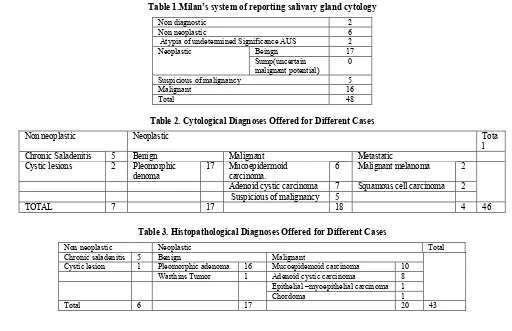
Table 1.Milan’s system of reporting salivary gland cytology
Non diagnostic 2
Non neoplastic 6
Atypia of undetermined Significance AUS 2
Neoplastic Beingn 17
Sump(uncertain
malignant potential) 0
Suspicious of malignancy 5
Malignant 16
Total 48
Table 2. Cytological Diagnoses Offered for Different Cases
Non neoplastic Neoplastic Tota
l
Chronic Saladenitis 5 Benign Malignant Metastatic
Cystic lesions 2 Pleomorphic
denoma 17 Mucoepidermoid carcinoma. 6 Malignant melanoma 2
Adenoid cystic carcinoma 7 Squamous cell carcinoma 2
Suspicious of malignancy 5
TOTAL 7 17 18 4 46
Table 3. Histopathological Diagnoses Offered for Different Cases
Non neoplastic Neoplastic Total
Chronic saladenitis 5 Benign Malignant
Cystic lesion 1 Pleomorphic adenoma 16 Mucoepidemoid carcinoma 10
Warthins Tumor 1 Adenoid cystic carcinoma 8
Epithelial –myoepithelial carcinoma 1
Chordoma 1
Chatterjee et al. (Maj, 2000) observed large number of benign cases in third decade followed by fourth decade. Malignancy reported in his study was maximum in fifth decade. Potdar and Paymaster (Potdar, 1969) reported an age range of 9 to 81 years with average age for benign tumors as 40.1 years and for malignant tumors as 46.3 years.
In the present study a male preponderance was noted with a male: female ratio of 2.3:1. This is in agreement with the series reported by Potdar and Paymaster (Potdar, 1969), and Spiro et al. (1990). who reported a male preponderance in their series. However this was in contrast to series reported by Dandapat et al. (1991) and Rewsuwan et al. (2006).
Table 4.Comparisons of Cytological Diagnoses With Histopathology (n=43)
Fnac diagnosis Histopathological diagnosis
Chronic
sailadeitis Cysts Pleomophic adenoma Warthins tumor Mucoepidermoid carcinoma Adenoid cystic
carcinoma
Epithelilal myoepithelial
carcinoma Chordoma
Chronic sailadenitis (5) 5
Cysts (2) 1 1
Suspicious of malignancy (5) 3 1 1
Pleomorphic adenoma (17) 15 1 1
Mucoepidermoid carcinoma (6) 6
Adenoid cystic carcinoma (7) 1 6
Squamous cell carcinoma .(1) 1
[image:3.595.42.563.104.485.2]Total (43) 5 1 16 1 10 8 1 1
Table 5. Statistical analysis
True positive (18) False positive (01) Positive predictive value TP/TP+FP=94.7% False negative (02) True negative(23) Negative predictive value TN/TN+FN=92% Sensitivity TP/TP+FN=90% Specificity TN/TN+FP=95.65%
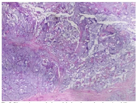
Fig 1. Photomicrograph shows mixed tumor of benign epithelial cells
[image:3.595.278.549.213.480.2]with blue myxoid matrix. Pleomorphic adenoma. (H&E ,10X) areas and intermediate cells .Mucoepidermoid carcinoma.(H&E , 40X) Fig 2.Photomicrograph shows squamoid areas, keratinization , cystic
Fig 3. Photomicrograph shows small uniform epithelial cells with hyperchromatic nuclei and coarse chromatin, dispersed and adhering to a large, hyaline stromal globules. Adenoid
[image:3.595.302.540.512.685.2]cystic carcinoma. (MGG, 40X)
Fig 4.Photomicrograph shows gland like spaces filled with pink hyaline material representing Adenoid cystic carcinoma. (H&E,
40X)
[image:3.595.46.283.514.686.2]Parotid was the commonest site of neoplasia (65%) in this series followed by submandibular gland (25%) and minor salivary glands (10%).This is in conformity with other workers, viz., Dandapat et al. (1991) Spiro et al. (1991) Gore
et al. (20) and Richardson et al. (1975). All the cases
diagnosed as pleomorphic adenoma on fnac were correctly diagnosed on histopathology as well. Pleomorphic adenoma is a mixed tumor of benign epithelial cells with blue myxoid matrix. (Fig1). Pleomorphic adenoma was the most frequent benign neoplasm (34%). Literature also reveals that primary tumors of sublingual salivary glands are extremely uncommon. Richardson et al. (1975), Vargas et al. (Pablo Agustin Vergas, Rene Gerhard, 2002), and Nagarkar et al. (2004), Pleomorphic adenoma was the most common benign salivary gland tumor encountered in parotid, submandibular and minor salivary glands. Similar findings were observed in the present study where pleomorphic adenoma was the most common benign salivary gland tumor at all locations. Mucoepidermoid carcinoma was the most common malignant neoplasm . 6 cases were diagnosed on Fnac with histopathological correlation. 3 cases were diagnosed as malignant lesion not specified on Fnac showed mucoepidermoid carcinoma on histopathological examination characterized by the presence of the three different cell types:mucous,sqamoid,and intermediate,present in variable proportions (Fig 2). A cytologic diagnosis of mucoepidermoid carcinoma requires a background of mucus
[image:4.595.32.290.66.235.2]and debris and a variable population of cells. In our study a case of high grade mucoepidemoid carcinoma was diagnosed as squamous cell carcinoma on Fnac. Adenoid cystic carcinoma is relatively uncommon tumour of salivary glands and are characterised by a prolonged clinical course and a fatal outcome. It was first described as ‘cylindroma’ by Billroth in 1859. Half of these tumors: occur in glandular areas other than the major salivary glands, principally in the hard palate, but they also arise in the tongue, minor salivary glands and lacrimal glands (Mohd Atha, 2012). 6 cases of Adenoid cystic carcinoma diagnosed on histology (Fig.4) were correctly diagnosed as Adenoid cystic carcinoma on Fnac however 1 case was diagnosed as pleomorphic adenoma and other as non specific malignant lesion. One case of pleomorphic adenoma was incorrectly diagnosed as adenoid cystic carcinoma on fnac. The distinction between pleomorphic adenoma and adenoid cystic carcinoma on Fnac may be difficult on account of several features-myxoid acellular material may be found in both and hyaline globules characteristic of adenoid cystic carcinoma (Fig 3) may also be seen in pleomorphic adenoma. Two cases of metastatic deposits of malignant melanoma (Fig 6) one in parotid gland and one submandibular gland were diagnosed on Fnac only as they were known cases of malignant melanoma no futher histopathological investigation was done. A single case of Chordoma (Fig 7) was diagnosed on Histopathology hich was diagnosed was pleomorphic
Fig 5. Photomicrograph shows presence of papillary projections into cystic spaces which have an epithelial lining composed of two layers of
[image:4.595.305.541.67.237.2]cells with oncocytic features and underling chronic inflammatory cell infiltrate. Warthins tumor (H&E, 40 X)
Fig 6. Photomicrograph shows metastatic tumor cells with intracellular as well as extacellular melanin pigment deposition. Malignant melanoma
.(MGG, 40X)
Fig 7. Photomicrograph shows lobules of vacuolated, eosinophilic to clear tumor cells embedded in a myxoid matrix.
Chordoma.(H&E, 40X)
Fig. 8. Photomicrograph shows epithelial cells with abundant eosinophillic cytoplasm with a few clear myoepithelial cells .
[image:4.595.46.282.282.462.2] [image:4.595.306.540.283.459.2]adenoma on Fnac. One case of epithelial myoepithelial carcinoma was diagnosed on histopathology which was diagnosed as malignant lesion not specified on cytology. There were 2 cases reported as cystic lesions; one case turned out to be a warthin tumor (fig 5)on histopathology, however, no histopathological or significant clinical follow-up was available of the other case. Salivary gland cystic lesions can be either nontumorous or tumorous (benign or malignant). Retention cysts, mucoceles, and lymphoepithelial cysts are the common nontumorous cysts, whereas MEC, Warthin’s tumor, acinic cell carcinoma, cystic PA, and cystadenoma are examples of tumorous cystic lesions. In cystic lesions, fluid is aspirated, and the cellularity of the smear is generally low; therefore, malignant cells can be missed, and this can lead to a false-negative report. Postevacuation FNA with multiple passes from different planes is helpful for substantially reducing sampling errors. (Rajwanshi, 2006). The sensitivity of FNA, as mentioned in different previous studies, varies from 54% to 98% with high specificity values of 88% to 99% for separating benign lesions from malignant lesions (Kim et al., 2013; Stewart, 2000; Orell, 1995; Ashraf, 2010; Daneshbod et al., 2009; Jafari et al., 2009; Schmidt et al., 2011). Similarly, in the current study, the sensitivity was 90%, and the specificity was 95.5%.
Conclusion
Salivary gland FNA is a safe, minimally invasive, cost efficient, and effective diagnostic technique for salivary gland lesions. It edges over frozen sections because it proves the nature of the lesion before surgery and thus acts as a useful triage tool and prevents patients with non neoplastic lesions from undergoing surgery. Most of the salivary gland lesions can be accurately diagnosed by FNA with adequate sampling and cytopathologist experience. It can be used to differentiate benign from malignant lesions preoperatively thus helping further surgical management of the patient.
Conflict of interest: None\
Funding: None
REFERENCES
Ahrnad S., Lateef M., Ahmad R. 2002. Clinicopathological study of primary salivary gland tumors in Kashmir. JK-Practitioner., 9:231-3.
American Society of Cytopathology. The Milan System for Reporting Salivary Gland Cytopathology. http://www. cytopathology.org/the-milansystem-for-reporting-salivary-gland-cytopathology/. Accessed March 31, 2016
Ashraf A., Shaikh AS., Kamal F., Sarfraz R., Bukhari MH. 2010. Diagnostic reliability of FNAC for salivary gland swellings: a comparative study. Diagn Cytopathol., 38:499-504.
Auclair P.L. Ellis G.L. 1997. Major Salivary Glands In: Silverberg SG, Delellis R.A., Frable W.J., editors. Principles and Practice of Surgical Pathology and Cytopathology, 31d ed. Edinburgh, Churchill Livingstone.,
1461-1515.
Bashirs S., Mustafa F., Malla H., Khan A., Rasool M., Sharma S. 2013. Histopathological Spectrum of Salivary Gland Tumors: A 10 Year Experience. Sch. J. App. Med. Sci., 1(6):1070-1074.
Brookstone MS., Huvos AG., Spiro RH. 1990. Central adenoid cystic carcinoma of the mandible‖ – J Oral Maxillofac
Surg., 48: 1329 – 1333.
Chakrabarti S., Bera M., Bhattacharya PK. et al., 2010. Study of salivary gland lesions with fine needle aspiration cytology and histopathology along with immunohistochemistry. J Indian Med Assoc., 108:833-836. Colella G., Cannavale R., Flamminio F., Foschini MP. 2010.
Fine-needle aspiration cytology of salivary gland lesions: a systematic review. J Oral Maxillofac Surg., 68:2146-2153. Dandapat MC., Rath BK., Patnaik BK., Dash SN., 1991.
Tumors of salivary glands. Indian J Surg., 53:200.
Daneshbod Y., Daneshbod K., Khademi B. 2009. Diagnostic difficulties in the interpretation of fine needle aspirate samples in salivary lesions: diagnostic pitfalls revisited.
Acta Cytol., 53:53-70.
Ethunandan M., Davies B., Pratt CA., Puxeddu R., Brennan PA. 2009. Primary epithelial submandibular salivary gland tumors- Review of management in a district general hospital setting. Oral Oncol., 45:173-6.
Gore DO., Annamunthodo H., Harland A. 1964. Tumors of salivary gland origion. Surg Gynecol Obstet., 119:1290. Jafari A., Royer B., Lefevre M., Corlieu P., Perie S., St Guily
JL. 2009. Value of the cytological diagnosis in the treatment of parotid tumors. Otolaryngol Head Neck Surg.,
140:381-385.
Jain R., Gupta R., Kudesia M., Singh S. 2013. Fine needle aspiration cytology in diagnosis of salivary gland lesions: a study with histologic comparison. Cytojournal., 10:5. Jayaram G., Verma AK., Sood N., Khurana N. 1994. Fine
needle aspiration cytology of salivary gland lesions. J Oral
Pathol Med., 23: 256-261.
Kim BY., Hyeon J., Ryu G. et al., 2013. Diagnostic accuracy of fine needle aspiration cytology for high-grade salivary gland tumors. Ann Surg Oncol., 20:2380-2387.
Kocjan G., Nayagam M., Harris M. 1990. Fine needle aspiration cytology of salivary gland lesions: advantages and pitfalls. Cytopathology., 1:269-275.
Maj T Chatterjee and PK Panda, 2000. A Pathological study of benign and malignant tumors of salivary glands; MJAFI; 56(4).
Mohd Atha, Sodhi, K.S., Kala, S., Maurya, M.R.K., Chauhan, S., M.S..Tripathi, R.K. Parvez Khan, Sanjeev Pandey, 2012. Adenoid cystic carcinoma lacrimal gland. Journal of
Medical Sciences., 15(1):76-77.
Nitin M. Nagarkar, Sandeep Bansal, Arjun Dass, Surinder K. Singhal, Harsh Mohan, 2004. Salivary gland tumors- Our Experience; Indian Journal of Otolaryngology and Head
and Neck Surgery, 56(1).
Orell SR. 1995. Diagnostic difficulties in the interpretation of fine needle aspirates of salivary gland lesions: the problem revisited. Cytopathology., 6:285-300.
Pablo Agustin Vergas, Rene Gerhard, Vergilius J. F. Araujo Filiho Ines Vieira de Castro, 2002. Salivary gland tumors in Brazillian population: A retrospective study of 124 cases;REV. HOSP. CLIN. FAC. MED. S. PAULO 57(6):271-276.
Paparella's Otolaryngology, 1991. W. B. Saunders, Vol. III, 3rd Edition, 20 :2099-2127.
Pastore A., Borin M., Malagutti N. et al., 2013. Preoperative assessment of salivary gland neoplasms with fine needle aspiration cytology and echography: a retrospective analysis of 357 cases. Int J Immunopathol Pharmacol. 26:965-971.
Postema RJ., van Velthuysen ML., van den Brekel MW., Balm AJ., Peterse JL. 2004. Accuracy of fine-needle aspiration cytology of salivarygland lesions in the Netherlands Cancer Institute. Head Neck. 26:418-424.
Potdar GG., Paymaster JC. 1969. Tumors of salivary glands.
Am J Surg., 118:440.
Rajwanshi A., Gupta K., Gupta N. et al., 2006. Fine-needle aspiration cytology of salivary glands: diagnostic pitfalls— revisited. Diagn Cytopathol., 34:580-584.
Richardson GS., Dickason WL., Gaisford JC. 1975. Tumors of salivary glands; An analysis of 752 cases. Plastic Reconstr
Surg., 55:131.
Schindler S., Nayar R., Dutra J., Bedrossian CW. 2001. Diagnostic challenges in aspiration cytology of the salivary glands. Semin Diagn Pathol., 18:124-146.
Schmidt RL., Hall BJ., Wilson AR., Layfield LJ. 2011. A systematic review and meta-analysis of the diagnostic accuracy of fine-needle aspiration cytology for parotid gland lesions. Am J Clin Pathol., 136:45-59.
Speight PM., Barrett AW. 2002. Salivary gland tumours. Oral
Dis., 8:229-40.
Stewart CJ., MacKenzie K., McGarry GW., Mowat A. 2000. Fine-needle aspiration cytology of salivary gland: a review of 341 cases. Diagn Cytopathol., 22:139-146.
Sunida Rewsuwan, Jongkolnee Settakorn, Pongsak Mahanupab, 2006. Salivary gland tumors in Maharaj Nakorn Chiang Mai Hospital: A retrospective study of 198 cases; Chiang Mai Med Bull., 45:4553.
Yih WY., Kratochvil FJ., Stewart JC. 2005. Intraoral minor salivary gland neoplasms: review of 213 cases. J Oral
Maxillofac Surg., 63:805-10.