ISSN Print: 2156-8553
DOI: 10.4236/as.2019.107075 Jul. 31, 2019 985 Agricultural Sciences
Synthesis of a Wheat/Maize Hybrid CENH3
Gene, the Genetic Transformation of Wheat, Its
Chromosomal Localization and Effects on
Chromosome Behaviors in Wheat/Maize
Somatic Hybrids
Xiaoyu Yang
1,2*, Jianhui Li
3, Weichang Yu
1*1Guangdong Provincial Key Laboratory for Plant Epigenetics, College of Life Sciences and Oceanography, Shenzhen University, Shenzhen, China
2Key Laboratory of Optoelectronic Devices and Systems of Ministry of Education and Guangdong Province, College of Optoelectronic Engineering, Shenzhen University, Shenzhen, China
3School of Life Sciences, State Key Laboratory of Agrobiotechnology, The Chinese University of Hong Kong, Hong Kong, China
Abstract
Centromere-specific histone H3 (CENH3) replaces the canonical histone H3 in nucleosomes of functional centromeres, and plays important roles in faithful chromosome segregation during cell division. CENH3 is also impor-tant in the recognition of alien centromeres and determines the accommoda-tion or eliminaaccommoda-tion of alien chromosomes in interspecific or intergenic hybri-dization. In this study, a maize full length CENH3 with a yellow fluorescent protein (YFP) tag at C-terminus (ZmCENH3-YFP) and a synthetic hybrid wmCENH3 with the N-terminus from wheat CENH3 and the histone fold domain (HFD) from maize tagged with a red fluorescent protein (RFP) at the C-terminus (wmCENH3-RFP) were transformed to wheat by biolistics transfor-mation. Transgenic wheat plants with both ZmCNEH3-YFP and wmCENH3- RFP genes were identified by PCR. The expression of ZmCENH3-YFP was not observed, while the expression of wmCENH3-RFP could be detected by RT-PCR, direct fluorescence microscopy, and immunostaining with anti-RFP antibody. The expressed wmCENH3-RFP was localized to nuclei as dotted patterns, indicating its targeting to wheat centromeres. Somatic hybridization was performed between wmCENH3-RFP transgenic wheat and transgenic maize that expressed a ZmCENH3-YFP gene to investigate chromosome be-haviors in somatic hybrids. Cytological and FISH analyses of somatic hybrid How to cite this paper: Yang, X.Y., Li, J.H.
and Yu, W.C. (2019) Synthesis of a Wheat/ Maize Hybrid CENH3 Gene, the Genetic Transformation of Wheat, Its Chromosomal Localization and Effects on Chromosome Behaviors in Wheat/Maize Somatic Hybr-ids. Agricultural Sciences, 10, 985-1014.
https://doi.org/10.4236/as.2019.107075
Received: June 22, 2019 Accepted: July 28, 2019 Published: July 31, 2019
Copyright © 2019 by author(s) and Scientific Research Publishing Inc. This work is licensed under the Creative Commons Attribution International License (CC BY 4.0).
DOI: 10.4236/as.2019.107075 986 Agricultural Sciences cells showed the formation of micronuclei and lagging chromatin in both somatic hybridizations with or without the wmCENH3-RFP transgene, indi-cating that ectopically expressed wmCENH3 could not overcome chromosome elimination in wheat/maize somatic hybrids. Immunostaining of wmCENH3- RFP and ZmCENH3-YFP in early stage somatic hybrid cells indicated that both wmCENH3-RFP and ZmCENH3-YFP proteins were expressed, but their binding patterns changed from the commonly observed dotted patterns to dif-fused ones, suggesting that the inactivation of CENH3 might be a factor for chromosome elimination in wheat/maize somatic hybridization.
Keywords
Centromeric Histone H3, Genetic Transformation, Chromosome Elimination, Somatic Hybridization, Wheat, Maize
1. Introduction
Centromeric histone H3 (CENH3) is a variant to replace canonical histone H3 nucleosome in functional centromere for kinetochore assembly. CENH3 is only 50% identical to canonical histone H3 in the histone fold domain (HFD) [1]. It plays an important role in chromosome behaviors during cell divisions, espe-cially in chromosome elimination and adaptation. For example, haploid Arabi-dopsis plant could be produced by chromosome elimination when a wide type plant was crossed with a cenh3 mutant expressing an engineered CENH3 or na-turally occurring CENH3 from distant related plant species [2] [3]. The chro-mosome elimination is always on chrochro-mosomes from the hybridization parent that expresses the divergent CENH3. Chromosome elimination has also been reported in the unstable interspecific barley hybrids caused by the loss of CENH3 protein and centromere inactivation [4]. In contrast, in the oat/maize addition lines, oat CENH3 can nucleate functional kinetochores on maize chromosomes while the maize CENH3 gene is silenced [5]. Thus, maize chro-mosomes can be retained in the oat genetic background.
Two regions of CENH3 protein are highly variable: N-terminal tail for kine-tochore assembly and the loop 1 region which interacts with nucleosomal DNA and is crucial for centromere targeting and the recognition between CENH3 and centromeric DNA sequence [6] [7]. Domain swap experiments showed that chimeric CENH3 with the N-terminal tail from Lepidium oleraceum and the HFD from A. thaliana caused chromosome missegregation and genome elimina-tion, while the reversely swapped chimeric CENH3 with the A. thaliana N-terminal tail and the L.oleraceum HFD acted normally as the Arabidopsis CENH3[3]. Thus, the evolution of CENH3 could contribute to hybridization barrier that prevents genetic cross between distantly related plant species.
DOI: 10.4236/as.2019.107075 987 Agricultural Sciences morphologically and ethologically floral isolation [10], and failure in pollen-pistil interactions [11]. Technologies have been developed to overcome hybridization barriers to broaden genetic resources for plant breeding. For example, artificial hybridization could overcome some habitat, temporal and ethological barriers
[8]; gametic barriers could be solved via in vitro fertilization technology, in which male and female gametes were isolate from reproductive organs of paren-tal plants, and fused to produce zygotes through calcium-mediated, electrofusion or microinjection, and ion measurement methods [12]. Post-zygotic barriers in-clude hybrid non-viability, weakness and sterility [13] [14], hybrid breakdown
[15], and hybrid necrosis [16]. Post-zygotic barriers are mainly caused by aborted embryo or endosperm, abnormal chromosome behaviors, and selective chromo-some elimination [17] [18]. Some techniques have been developed to tackle post-zygotic barriers. For example, chromosome doubling technique was devel-oped to solve hybrid sterility [19], and embryo rescue technique was developed to recover unviable and weak hybrid embryos from abortion [20]. Somatic hybridiza-tion technique was also developed to generate somatic hybrids between sexually incompatible plants that could not be realized by conventional genetic cross.
Recent studies have revealed molecular mechanisms of chromosome elimina-tion in hybridizaelimina-tion barrier. The importance of CENH3 in chromosome elimi-nations hints that CENH3 might be manipulated to overcome chromosome eli-minations. In wheat/maize hybrids from genetic cross and somatic hybridiza-tion, maize chromosomes are quickly eliminated during the first several cell cycles [21]-[26]. Chen et al. [27] reported an attempt to ectopically express a maize CENH3 gene (ZmCENH3) in transgenic wheat. However, although the ZmCENH3 could be transcribed at a low level, ZmCENH3 protein was not de-tected by both western blot and immunostaining. Chromosome elimination was not suppressed in genetic crosses between transgenic wheat and maize. To over-come the suppression of ZmCENH3 transgene expression in transgenic wheat, we synthesized a hybrid wmCENH3gene, which has the N-terminus before loop 1 domain from wheat TaCENH3, the C-terminal HFD after loop 1 from maize ZmCENH3, and a red fluorescent protein (RFP) tag at the C-terminus. The synthesized wmCENH3 gene was cloned into a gene expression cassette under a strong maize ubiquitin promoter [28], and transformed into wheat by biolistic transformation. Transgenic wheat was generated and the influence of ectopically expressed wmCENH3 on chromosome behaviors in wheat/maize somatic hybr-ids was analyzed.
2. Materials and Methods
2.1. Plant Materials
contain-DOI: 10.4236/as.2019.107075 988 Agricultural Sciences ing 4.4 g/L MS mixture with vitamins, 30 g/L sucrose, 0.2 g/L casein, 0.146 g/L glutamine, 2 mg/L 2, 4-D, 3 g/L Gelrite (pH 5.7). The induced wheat calli were kept in an incubator with air temperature of 28˚C in darkness and subcultured every 4 weeks.
Transgenic ZmCENH3-YFP maize seeds were kindly provided by Prof. James Birchler (University of Missouri, Columbia). Seeds were sowed in soil and kept in growth chamber with photoperiod of 16 h, light intensity of 400 - 500 µmol/m2/s, relative humidity of 70%, air temperature of 28˚C during the day and
of 25˚C during the night.
2.2. Sequence Analysis
To analyze the similarity of three different CENH3 proteins, amino acid se-quences of TaCENH3, ZmCENH3 and a synthetic wmCENH3 were aligned with the canonical histone H3 of rice using the CLC Sequence Viewer 7.6 software program (https://www.qiagenbioinformatics.com/products/clc-sequence-viewer/).
2.3. Constructs for Wheat Transformation
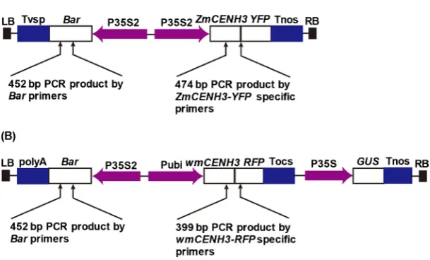
The maize CENH3 gene with a YFP tag was constructed in pTF101, and was kindly provided by Prof. James Birchler (University of Missouri, Columbia). The pTF101-ZmCENH3-YFP (Figure 1(A)) had streptomycin resistance in E. coli, and two expression cassettes: a maize full length CENH3 gene with a YFP tag at C-terminus driven by 2 × 35S promoter from cauliflower mosaic virus (CaMV), and a Bar gene driven by 2 × 35S promoter as the selectable marker.
A wmCENH3-RFP gene was synthesized and cloned into the pCAMBIA3301 vector to construct pCAMBIA3301-wmCENH3-RFP (Figure 1(B)). This con-struct had kanamycin resistance in E. coli, and three expression cassettes on T-DNA region: a wmCENH3-RFP gene with wheat TaCENH3 N-terminus be-fore Loop1 domain, the maize ZmCENH3 C-terminus after Loop1, and a RFP gene tag at C-terminus, driven by a maize ubiquitin promoter [28]; a Bar gene driven by 2 × 35S promoter from cauliflower mosaic virus as the selectable marker; and a β-glucuronidase (GUS) gene [30] driven by 35S promoter as the reporter gene.
2.4. Biolistic Transformation of Wheat and Plant Regeneration
Plasmid DNAs were extracted by alkaline extraction method [31], and purified with QIAprep Spin miniprep Kit according to the manufacturer’s protocol. 1 μg plasmid DNA of pCAMBIA3301-wmCENH3-RFP or pTF101-ZmCENH3-YFP was coated to 1.5 mg (1 μm diameter) gold particles (Bio-Rad) according to the protocol of Gold Particle Preparation for Bombardment developed by the Iowa State University Plant Transformation FacilityDOI: 10.4236/as.2019.107075 989 Agricultural Sciences
Figure 1. CENH3 constructs for wheat transformation. (A) pTF101-ZmCNEH3-YFP construct contains a Bialaphos resistance gene (Bar) gene driven by a 2 × 35S (P35S2) promoter as the selection marker, and a ZmCENH3-YFP gene driven by the P35S2 pro-moters; (B) pCAMBIA3301-wmCENH3-RFP construct contains a Bar gene driven by the P35S2 promoter as the selection marker, a wmCENH3-RFP gene driven by a constitutive maize Ubi promoters (Pubi), and a GUS gene driven by a cauliflower mosaic virus (CaMV) 35S promoter (P35S). LB and RB are the T-DNA left and right border sequences, respectively; Tnos, Tocs, Tvsp, and polyA are terminators from Agrobacterium nopaline synthase gene, Agrobacterium octopine synthase gene, soybean vegetative storage protein gene and the polyA sequences from CaMV 35S RNA, respectively; GUS is the β-glucuronidase reporter gene; YFP and RFP are yellow and red fluorescence protein genes, respectively; ZmCENH3 is the maize full length CENH3 gene; wmCENH3 is a synthetic CENH3 gene with the N-terminal tail from wheat TaCENH3 and the C-terminal part from maize ZmCENH3 gene. The arrows point to the sites of primers used for PCR identification of transgenic plants with transgenes of Bar, ZmCENH3-YFP, and wmCENH3-RFP, respec-tively. The size of each PCR product is indicated.
with a Bio-Rad PDS 1000/He particle delivery system according to the previous protocol [32] with the following parameters: 1300 psi delivery pressure of helium tank and 1100 psi rupture disc. Petri dish with calli in the center was placed on level 4 of sample holder in Bio-Rad PDS/1000 He, and the distance between ma-crocarriers and calli was 12 cm.
[image:5.595.221.526.80.264.2]DOI: 10.4236/as.2019.107075 990 Agricultural Sciences plantlets to develop at 25˚C with the photoperiod of 16 h. Well-developed plan-tlets were washed to remove the attached medium on roots with tap water, and handled carefully to avoid damaging the roots. Plantlets with white and healthy roots were planted in pots and cultured in growth chamber according to the previous method [33] with high humidity for plants acclimatization during the first week. The transgenic plants were growing in the growth chamber until ma-turity and self-pollinated to produce seeds.
2.5. Genomic DNA Isolation from Transgenic Wheat
Genomic DNAs were isolated from wild type and transgenic wheat lines by SDS method [34] with modification. 1 g of leaf samples was placed in a mortar and ground by a pestle to a fine powder after treated with liquid nitrogen. The frozen powder was then transferred to 2-ml tubes containing 1 ml DNA extraction buf-fer [0.5 M NaCl, 0.1 M Tris-HCl, 0.05 M EDTA and 0.6% (w/v) SDS, pH 8.0] for DNA extraction. Genomic DNA was precipitated by equal volumes of isopro-panol and centrifugation. DNA pellets were re-suspended with 0.6 ml of 1 M NaCl after centrifugation at 5000 rpm for 10 min at room temperature. 1 volume of chloroform was added to DNA solution, mixed well and centrifuged at 12,000 rpm at room temperature for 10 min. The supernatants were transferred to 1.5-ml tubes and DNAs were precipitated with 1 volume of isopropanol at −20˚C. DNA pellets were washed with 500 μL of 70% (v/v) ethanol, air-dried and then dissolved in 50 μL of sterilized Milli-Q water containing 10 units RNase A for 5 min at room temperature. The quantity and quality of genomic DNAs were determined by gel electrophoresis.
2.6. Identification of Transgenic Wheat Plants by PCR
PCR was performed with three sets of primers: 1) P1 (5’-GCACCATCGTCAAC CACTAC-3’) and P2 (5’-TACCGGCAGGCTGAAGTCCA-3’) for 452 bp Bar fragment, 2) P3 (5’-AAGGATCCATGGCTCGAACCAAGC-3’) and P4 (5’-AAG AGCTCTCATGCCCAACGCCTT-3’) for 474 bp ZmCENH3-YFP fragment, 3) P5 (5’-AGAGCGCTATACCGCAGAAG-3’) and P6 (5’-GGGTGCTTCACGTA CACCTT-3’) for 399 bp wmCENH3-RFP fragment.
PCR reactions were performed in a total volume of 20 μL containing 10 μL Premix Ex Taq Hot Start Version (Takara Code No. HRR030A), 50 ng wheat genomic DNA or 10 ng plasmid DNA, 0.5 μM each of forward and reverse pri-mers with the following PCR program: an initial denaturalization at 95˚C for 3 min, 35 cycles of denaturalization at 98˚C for 10 s, annealing at 60˚C for 30 s and extension at 72˚C for 40 s, followed by a final extension at 72˚C for 5 min. PCR products were checked on 0.8% agarose gel after staining by ethidium bro-mide (EB).
2.7. Southern Blot for T0 Transgenic Wheat Lines
DOI: 10.4236/as.2019.107075 991 Agricultural Sciences transgenic wheat. 20 μg of genomic DNA per sample was digested with 150 units of restriction enzyme, EcoR I, at 37˚C overnight, fractioned in a 1.0% (w/v) aga-rose gel at 60 v for 4 h, denatured and transferred to Hybond N+ membranes. A digoxygenin (DIG) labeled Bar gene DNA fragment was synthesized by PCR with the Bar primers of P1/P2 and used as the probe. Prehybridization, washing, and chemiluminescent detection of the blots were performed according to the manufacturer’s instructions (Roche Diagnostics GmbH, Mannheim, Germany).
2.8. Expression Analysis of
wm
CENH3
-RFP
in Transgenic Wheat
RT-PCR was performed in T0 transgenic wheat for the detection of wmCENH3- RFP expression. Total RNA isolation from leaves of wild type and wmCENH3- RFP transgenic wheat was performed by using an RNeasy Plant Mini Kit (QIAGEN) according to the manufacturer’s instruction. The first strand cDNA synthesis was performed using PrimeScript RT-PCR Kit (Takara). PCR amplifi-cation was performed using primers of P5 (5’-AGAGCGCTATACCGCAGAAG-3’) and P6 (5’-GGGTGCTTCACGTACACCTT-3’) for wmCEHN3-RFP gene with the same program mentioned above. The PCR products were separated by 2% agarose gel electrophoresis and stained with EB for visualization.
2.9. Localization of Alien CENH3 Protein by Fluorescence
Microscopy
Root tips of transgenic wheat were squashed on glass slides with cover glasses, and examined for red or yellow fluorescence under the 100 × oil immersion ob-jective of Leica DM5500B fluorescence microscope (Leica Microsystems). Im-ages were captured with a Leica DFC490 digital camera and processed by Adobe Photoshop CS software.
2.10. Immunostaining of
wm
CENH3
-RFP
in Transgenic Wheat
Root tips of wild type wheat and T0 transgenic wheat lines (11-1, C2901, C3057 and C3216) were checked for the expression of wmCENH3-RFP by immunos-taining [5] with some modifications. The root tips were treated with N2O at 1000
psi for 2 h, and fixed in 4% (v/v) paraformaldehyde in 1 × phosphate buffered saline (PBS, 8.1 mM Na2HPO4, 1.5 mM KH2PO4, 2.7 mM KCl, 136 mM NaCl)
DOI: 10.4236/as.2019.107075 992 Agricultural Sciences transgenic wheat. After incubated at 4˚C overnight, slides were washed three times in 1 × PBS and detected by using sheep anti-rabbit Ig-Rhodamine (Milli-pore) secondary antibody (1:100 dilution in blocking solution). The slides were incubated for 2 h at room temperature and washed three times in 1 × PBS, mounted with a drop of Vectashield (Vector Laboratories) containing 1.5 μg/ml DAPI and examined under the 100 × oil immersion objective of Leica DM5500B fluorescence microscope. Images were captured with a Leica DFC490 digital camera and processed with PHOTOSHOP CS software.
2.11. Establishment of Cell Suspension Culture for Wheat
Wild type or wmCENH3-RFP transgenic wheat calli were used to initiate cell suspension cultures in 150 ml flasks containing 50 ml liquid Induction medium or Selection medium I, respectively. Suspension cells were subcultured every 4 d to stimulate vigorous growth until abundant, fine and white cell clusters ap-peared in liquid medium in about 1 month. Large cell clusters were removed by filtering through 1 mm pore sized metal sieves, and small cell clusters were kept and subcultured every 4 d until the establishment of wheat cell suspension cul-tures. The suspension cultures were incubated in an IS-KDD3 Table Shaker (CRYSTAL, Product number 1109012) at 28˚C in the dark with 130 rpm rotation.
2.12. Protoplast Isolation from Wheat and Maize
For isolation of wheat protoplast, a mixture of 6 ml enzyme solution [3% (w/v) Cellulose Onozuka R-10, 1.5% (w/v) Macerozyme R-10, 0.5% (w/v) Pectolyase Y-23, 12.8% (w/v) mannitol, 0.36% (w/v) CaCl2·2H2O, 0.11% (w/v) NaH2PO4,
0.12% (w/v) MES hydrate, pH5.6] and 6 ml P5 liquid medium (4.4 g/L MS with vitamins, 0.5 mg/ L nicotinic acid, 0.5 mg/L pyridoxine-HCl, 9.9 mg/L thia-mine-HCl, 1 mg/L 2, 4-D, 0.146 g/L glutamine, 0.5 g/L casein hydrolysate, 2 mg/L glycine, 90 g/L glucose, 10 g/L sucrose, pH 5.8) was added to a 50-ml flask with about 3 g of suspension cells for digestion in an incubator shaker at 30˚C in darkness for 16 h or overnight with 45 rpm rotation. After enzymatic digestion, cells were passed through 250 μm and 45 μm sterile metal-mesh sieves, collected in a 15-ml centrifuge tube, and centrifuged at 100 g for 5 min. Supernatant was removed with Pasteur pipettes and protoplast pellets were re-suspended in 6 ml CPW 25S [250 g/L sucrose in CPW buffer (27.2 mg/L KH2PO4, 100 mg/L KNO3,
250 mg/L MgSO4·7H2O, 150 mg/L CaCl2·2H2O, 0.2 mg/L KI, 0.0025 mg/L
Cu-SO4·5H2O, pH 5.8)] solution by carefully bubbling. 2 ml CPW13M (130 g/L
DOI: 10.4236/as.2019.107075 993 Agricultural Sciences To isolate maize protoplasts, young leaves of ZmCENH3-YFP transgenic ma-ize seedlings were surface-sterilma-ized by 0.625% (w/v) sodium hypochlorite for 10 min, followed by being washed with 70% (v/v) ethanol for 30 s, and then rinsed 3 times with sterilized Milli-Q H2O. Sterilized leaves were cut into 0.5 mm
seg-ments [35] and transferred to a flask with 12 ml enzyme solution containing 2% (w/v) Cellulose Onozuka R-10, 0.1% (w/v) Pectolyase Y-23 dissolved in MaCa solution (0.2 M mannitol and 80 mM CaCl2) [36]. After vacuumed for 15 min,
the flasks were wrapped with aluminum foil and placed on an incubator shaker with 45 rpm rotation at 30˚C in the dark for 4 h. After filtration through 250 μm and 45 μm sieves, maize mesophyll protoplasts were transferred to a 15-ml cen-trifuge tube, washed twice with MaCa solution, and re-suspended in 10 volumes of P5 liquid medium.
The qualities of wheat and maize protoplasts were checked under bright field inverted microscope (Nikon, TE300) immediately after isolation and images were captured with a digital camera (V-Tphoto adapter Nikon MBB74700).
2.13. Symmetric Hybridization between Wheat and Maize
The protoplast fusion between wmCENH3-RFP transgenic wheat line C3216 and ZmCENH3-YFP transgenic maize was performed by PEG-mediated somatic hybridization [25] [37] [38]. After re-suspending wheat and maize pellets in P5 medium, wheat and maize protoplasts were mixed together at a ratio of 1:1 (v/v) in a 15-ml centrifuge tube. Four drops of protoplasts mixture were added to the center of a 30 × 10 mm Petri dish for fusion. Two drops of 40% (w/v) PEG were added to the center of protoplast mixture and the fusion lasted for 15 min. After 15-min fusion, 2 drops of the mixture of fusion solution A and B at a ratio of 9:1 (v/v) were added to the opposite side of protoplast mixture to enhance fusion efficiency, and incubated for 15 min. Then 12 drops of P5 me-dium were added around protoplast mixture to wash the protoplasts for 10 min. P5 medium was gently removed without disturbing the protoplasts. The protoplasts were washed 2 more times with P5 medium for 15 min each time. Ten drops of P5 medium were added to the top of fusion products. The plates were sealed and cultured at 28˚C in the dark. Fusion products were checked under bright field of inverted microscope (Nikon, TE300) during culture period and images were captured with a digital camera (V-Tphoto adapter Nikon MBB74700).
2.14. Cytological Analysis of Cell or Cell Clusters
from Protoplast Fusion
The cell cultures from the fusion protoplasts of wheat and maize were fixed at 7, 14 and 28 d after fusion with Carnoy’s fixative solution [39]. To fix the cells, P5 liquid medium in the plate was removed, and the cells were rinsed gently (not to disturb cells) 3 times with sterile Milli-Q H2O. Carnoy’s fixative solution was
DOI: 10.4236/as.2019.107075 994 Agricultural Sciences were stored in 70% (v/v) ethanol at 4˚C in the dark until using.
Fixed cells were spread on slides by squash method [40]. They were removed from 70% (v/v) ethanol, rinsed 3 times with sterile Milli-Q H2O, and stained
with a drop of 2% Aceto-Orcein. Then 5 μL cells were loaded to an ethanol washed slide, covered with a cover glass, and separated by gently knocking cover glass with the round end of a dissecting needle. A blotting paper was placed over the slide and squashed upright with thumb. Cells were observed under 20 × ob-jective of Nikon light microscope (E80i), and images were captured with a digital camera (RTKE SPOT).
2.15. FISH Analysis of Cell or Cell Clusters from Somatic
Hybridization
The maize B repeat sequence [41] was found to specifically hybridize to the sub-telomeres of wheat chromosomes (Supplementary Figure S1), thus was used to visualize wheat chromosomes in this study. Maize CentC [42] and B repeat se-quences were amplified by PCR. The amplified CentC and B repeat DNAs were labeled with Chroma Tide Alexa Fluor 488-5-dUTP and Chroma Tide Alexa Fluor 594-5-dUTP (Invitrogen) respectively, by nick translation [43] [44].
FISH analysis of fixed cell or cell clusters from protoplast fusion were per-formed according to previous methods [44] [45] with some modifications. Fixed cell or cell clusters were removed from 70% (v/v) ethanol, rinsed 3 times with sterile Milli-Q H2O, transferred to enzyme solution [1% (w/v) Pectolyase Y-23,
2% (w/v) Cellulase R-10], kept on the ice for 10 min, and then incubated at 37˚C in water bath for 1 h. The digested cells were washed twice with 70% (v/v) ethanol to remove enzyme solution, broken down with a needle, and separated by vortex for 20 s. The separated cells were pelleted by centrifugation and re-suspended in 50 μL 100% acetic acid by vortexing. Then 5 μL cells were dropped onto a glass slide inside a water-wetted box to let the nuclei/chromosomes spread and dry on the slide surface. The slide was crosslinked with 0.1 J UV light. A mixture of maize CentC and B repeat probes containing 100 ng labeled DNA each were added to each slide. The slide with probes was denatured in a metal box in boiling water for 5 min, and hybridized at 55˚C overnight in the dark. After hybridization, slides were washed with 2 × SSC at room temperature for 5 min, followed by 20-min washing with 2 × SSC at 55˚C. Nuclei/chromosomes were stained with 10 μL mounting medium with DAPI (Vectashield, H-1200), and observed under the 100 × oil immersion objective of Leica DM5500B fluorescence microscope (Leica Microsystems). Images were captured with a Leica DFC490 digital camera and processed by Adobe Photoshop CS software.
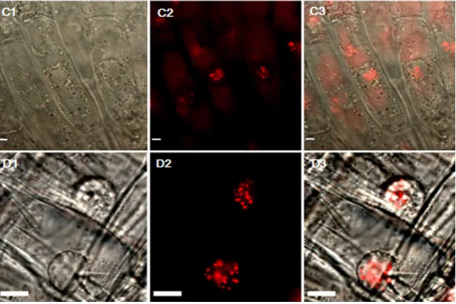
DOI: 10.4236/as.2019.107075 995 Agricultural Sciences maize somatic hybrids, cell or cell clusters were sampled 11 - 14 d after protop-last fusion and the localizations of wmCENH3-RFP and ZmCENH3-YFP were analyzed by immunostaining. Cultured cells in microplates were collected in a 2-ml tube by centrifugation at 2000 g for 5 min. After 30-min fixation in 4% (v/v) paraformaldehyde in fresh full-strength PBS, cells were collected by cen-trifugation at 2000 g for 5 min and washed with 1 × PBS containing 0.2% (v/v) Triton X-100. The fixed cells were digested in 20 μl of enzyme solution contain-ing 2% (w/v) Cellulase Onozuka R-10, 1% (w/v) Pectolyase Y-23 in 1 × PBS at 37˚C for 30 min. Digested cells were vortexed at low speed for about 20 s, cen-trifuged with a mini-centrifuge at 1500 g for 20 s, washed 3 times with 200 μl 1 × PBS and re-suspended in a final volume of 15 μl 1 × PBS. A total of 10 μl of di-gested cells was dropped onto a Poly-L-Lysine coated microscope slide and dried for 4 h in a water-wetted box. The wmCENH3-RFP and ZmCENH3-YFP pro-teins were detected with rabbit anti-RFP and mouse anti-GFP primary antibo-dies followed by sheep anti-rabbit Ig-Rhodamine (Millipore) secondary antibody (1:100 dilution in blocking solution) and FITC anti-mouse secondary antibody (Santa Cruz Biotechnology; 1:100 dilution in blocking solution), respectively. The slides were incubated for 2 h at room temperature, washed 3 times in 1 × PBS, mounted with a drop of Vectashield (Vector Laboratories) containing 1.5 μg/ml DAPI, and examined under the 100× oil immersion objective of Leica Leica DM5500B fluorescence microscope. Images were captured with a Leica DFC490 digital camera and processed with PHOTOSHOP CS software.
3. Results
3.1. Sequence Analysis of Wheat and Maize CENH3 and the Design
of a Synthetic wmCENH3
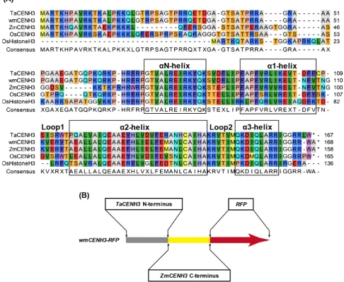
Sequence alignment of wheat, maize and rice CENH3 proteins were performed
(Figure 2(A)). The result showed conserved and diverged regions among the
CENH3 proteins. The N-terminal regions were highly diverged among the CENH3 proteins, whereas the HFDs including αN-helix, α1-helix, Loop 1, α2-helix, Loop2 and α3-helix were relatively conserved.
The large divergence between wheat and maize CENH3 proteins (74% simi-larity) could be a factor for the suppression of ZmCENH3 expression in trans-genic wheat [27]. To reduce the difference between transgenic CENH3 and the endogenous TaCENH3, a synthetic wmCENH3 gene was designed to have the N-terminus of TaCENH3 before the loop 1 domain, and the C-terminus of ZmCENH3 after loop 1 (Figure 2(B)). The synthetic wmCENH3 protein had 95% similarity to the TaCENH3. An RFP tag was fused in frame to the C-terminus of the synthetic wmCENH3 gene to trace the behavior of the wmCENH3 in transgenic plants.
DOI: 10.4236/as.2019.107075 996 Agricultural Sciences
Figure 2. Alignment of CENH3s from wheat (TaCENH3), maize (ZmCENH3), rice (OsCENH3), the synthetic wmCENH3, and a rice canonical histone H3 (OsHistoneH3) (A) and the schematic diagram of the synthetic wmCENH3-RFP fusion protein (B). The conserved αN, α1, α2 and α3 helix domains are boxed. The two loop regions (Loop 1 and Loop 2) are indicated. The synthetic wmCENH3-RFP is a fusion protein composed of N-terminal region of wheat CENH3 (TaCENH3) before Loop1, the C-terminal region of maize CENH3 (ZmCENH3) after Loop1, and a red fluorescence protein (RFP) tag fused in frame to the C-terminus of the wmCENH3 protein.
DOI: 10.4236/as.2019.107075 997 Agricultural Sciences
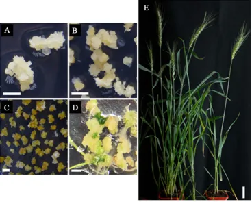
Figure 3. Genetic transformation and regeneration of transgenic wheat. (A) Wild type (WT) wheat calli induced from immature embryos; (B) Transformed wheat calli on Selection me-dium; (C) Resistant wheat calli on Regeneration meme-dium; (D) Plantlets regenerated from re-sistant calli; (E) wmCENH3-RFP transgenic wheat plants. Scale bars = 0.5 cm for ((A)-(D)) and 4 cm for (E).
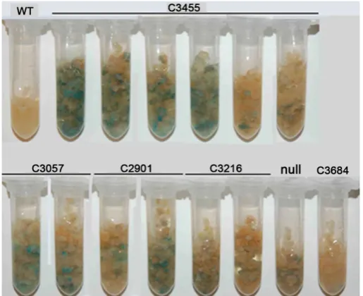
GUS staining was performed for fast growing callus lines C2901, C3057, C3216, C3455 and C3684 transformed with pCAMBIA3301-wmCENH3-RFP. The result suggested that not all cells in the calli expressed the transgene (Supplementary
Figure S2). Thus, PCR screenings with specific primers for wmCENH3-RFP,
[image:13.595.170.539.72.367.2]DOI: 10.4236/as.2019.107075 998 Agricultural Sciences
3.3. Southern Blot and RT-PCR Analysis for
Transgenic Wheat
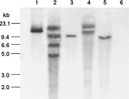
Southern blot with the DIG-labeled Bar gene was performed in T0 transgenic plants. The results showed wmCENH3-RFP construct was successfully inte-grated into the genome of T0 transgenic wheat lines of 11-1, C2901, C3057 and C3216. The copy numbers of the transgene were one for 11-1 and C3057 events, two for C2901 event, and four for C3216 event (Figure 4).
To analyze wmCNEH3-RFP gene expression in transgenic wheat, RT-PCR was performed with wild type wheat and transgenic events 11-1, C2901, C3057 and C3216. PCR primers were designed to detect a 399 bp product from the wmCENH3-RFP gene if it was expressed by RT-PCR. A 399 bp wmCENH3-RFP gene product was detected in the four transgenic wheat lines of 11-1, C2901, C3057 and C3216, but no product was detected in the WT wheat (Figure 5(A)).
3.4. Localization of Ectopically Expressed CENH3
The expression of fluorescence protein tagged CENH3 proteins were analyzed under a fluorescence microscope in root tips of regenerated transgenic plants. Dotted RFP signals were observed in the nuclei of root tip cells of wmCENH3-RFP transgenic plants (Supplementary Figure S3), suggesting that the wmCENH3-RFP was targeted to functional wheat centromeres. In contrast, fluorescence protein was not observed in the two transgenic plants with the ZmCENH3-YFP gene, being consistent with the previous report by Chen et al. [27]. This evidence in-dicated the suppression of ZmCENH3 gene expression in transgenic wheat.
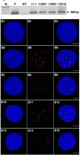
[image:14.595.261.486.462.635.2]The wmCENH3-RFP fusion protein was further detected by immunostaining with an antibody to the RFP tag at the C-terminus (Figure 5(B)). The expressed wmCENH3-RFP protein was detected in the nucleus of transgenic wheat in a
DOI: 10.4236/as.2019.107075 999 Agricultural Sciences
DOI: 10.4236/as.2019.107075 1000 Agricultural Sciences dotted pattern (Figure 5(B)), similar to the centromere localizations detected by direct observation of RFP fluorescence protein tag signals (Supplementary Fig-ure S3).
3.5. Symmetric Hybridization of Transgenic Wheat and Maize
Cell suspension cultures for WT wheat and wmCENH3-RFP transgenic wheat lines, 11-1, C2901, C3057, and C3216 were established. There was no significant difference in growth performance between WT and four wmCENH3-RFP trans-genic wheat cell suspension cultures (Supplementary Figure S4 and Supplemen-tary Figure S5). WT and one of the transgenic line C3216 cultured cells were used for protoplast isolation and somatic hybridization to maize protoplasts.Spherical protoplasts were isolated from WT and C3216 wheat suspension culture and maize young leaves (Supplementary Figure S6(A) and Figure
S6(B)). About equal amount of isolated wheat and maize protoplasts were mixed
and cell fusions were induced by PEG. Fused protoplasts were cultured in mi-croplates with P5 medium at a density of 1 - 5 × 10⁶ protoplasts per ml. The
oc-currence of hybridization was confirmed by checking the hybridization products with inverted microscope at 1 - 2 d after fusion (Supplementary Figure S6(C)). The growth of fused cells at early stage after fusion was observed by an inverted microscope (Supplementary Figure S6(D)).
3.6. Cytological Examinations of Somatic Hybrids
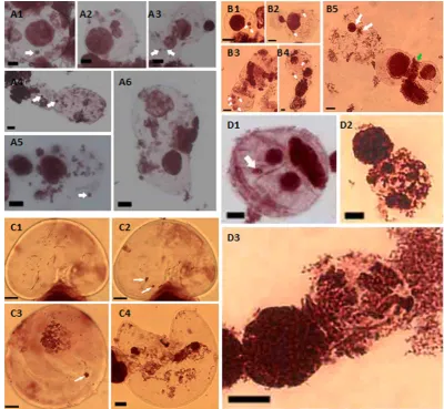
Aceto-orcein staining analysis was performed to investigate chromosome behaviors during mitosis in somatic hybrids. Abnormal chromosomes behaviors such as mi-cronuclear, asynchronous nucleus formation, lagging chromatin or chromosomes were observed in somatic hybrids from non-transgenic wheat/ZmCENH3-YFP transgenic maize and wmCENH3-RFP transgenic wheat/ZmCENH3-YFP trans-genic maize, respectively (Figure 6). In somatic hybrids of both hybridizations, micronuclear formation could be observed frequently in both single cells and di-viding cells. Micronuclei occurred during the whole process of mitosis, including interphase (Figure 6(A1) and Figure 6(B1)), telophase, and cytokinesis (Figures
6(A3)-(A5) and Figures 6(B2)-(B4)). Lagging chromosomes occurred at
ana-phase or lagging chromatin in the cleavage furrow (Figure 6(A5) and Figure
6(B5)), asynchronous nucleus formation as loose chromatin and condensed
chromatin (Figure 6(A6)), asynchronous cell cycle as metaphase and interphase occurred simultaneously (Figure 6(B5)) in the dividing cell, and also asyn-chronous mitosis (Figure 6(A2) and Figure 6(B2)). These results demonstrated that frequently abnormal chromosome behaviors might occur at early stage of somatic hybrid development and the synthetic wmCENH3 could not eliminate the abnormal chromosome behaviors in the somatic hybrid cells.
DOI: 10.4236/as.2019.107075 1001 Agricultural Sciences
Figure 6. Cytological analyses of somatic hybrids by Aceto-orcein staining. (A1)-(A6) Micronuclei (white arrows showed in (A1), (A3), (A4), and (A5)) and asynchronous nucleus formation ((A2), and (A6)) ob-served in the somatic hybrid of non-transgenic wheat × ZmCENH3YFP maize mesophyll protoplast. (B1)-(B5) Micronuclei (white arrows showed in B1-B4), asynchronous nucleus formation (white arrows showed in (B5)), and lagging chromatin (green arrow showed in (B5)) observed in the somatic hybrid of non-transgenic wheat × maize mesophyll protoplasts. (C1)-(C4) Micronuclei (white arrows showed in the same cell C1 and C2 at different focus levels) and also in (C3), and asynchronous nucleus formation (C4) observed in the fusion protoplasts of transgenic wheat C3057 and ZmCENH3YFP maize mesophyll. (D1)-(D3) Micronuclei (white arrows showed in D1) and asynchronous nucleus formation ((D2) and (D3)) observed in the fusion protoplasts of transgenic wheat C3216 and maize mesophyll protoplasts. Scale bars = 10 μm.
DOI: 10.4236/as.2019.107075 1002 Agricultural Sciences
DOI: 10.4236/as.2019.107075 1003 Agricultural Sciences could not be detected in the divided cells and microcell clusters (Figure 7(A),
Figure 7(B), Figure 7(F), Figure 7(G) & Figure 7(H)). The elimination of ma-ize chromosomes was observed in the second cell division of somatic hybrids (Figure 7(E)).
3.7. CENH3 Behavior in Somatic Hybrids
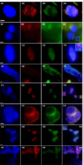
The wmCNEH3 and ZmCENH3 proteins in somatic hybrid cells were investi-gated by immunostaining analysis during 11 - 14 d after fusion. Both RFP and YFP signals were observed in the nuclei of somatic hybrid cells as diffused pat-terns (Figure 8), which was different from the dotted pattern observed before protoplast fusion (Figure 5(B) and Supplementary Figure S7). Fragments of chromatin were observed near the cell plate at anaphase of cell division, while the majority had moved to the polar regions. The loss of chromatin fragments was also reported in other somatic hybridization systems, which were considered as lost chromosome fragments [25] [46].
4. Discussions
4.1. Fragmentation of Transformed DNAs
Genetic transformation by particle bombardment usually produces complex trans-gene loci with multiple copies, truncated, rearranged sequences interspersed with genomic DNA fragments [47] [48]. In this research, we observed only 14 out of 109 transgenic wheat plants (12.84%) that had full length DNA fragments, and most of the transgenic events had fragmented transgenes, indicating the
DOI: 10.4236/as.2019.107075 1004 Agricultural Sciences fragmentation and rearrangement of transformed DNAs. The high frequent fragmentation of transgenes and nonrandomized transformation could be mainly caused by the following factors. First, plasmid DNAs might be vulnerable to shearing during gold particle coating. Plasmid DNAs were coated to golden par-ticles in biolistic transformation. The frequent vortexing, precipitation and cen-trifugation during coating may mechanically shear the plasmid DNA and pro-duce fragmented DNAs before transformation. Second, high velocity particle bombardment could be another factor to produce DNA fragmentation. High- speed gold particles during bombardment could create double strand breaks (DSBs) on genomic DNA that initiate the transgene integration, and at the same time, they could also create fragmentation of delivered DNAs on gold particle. Third, foreign DNA integration into the genome is an enzymatic reaction. For-eign DNAs are integrated as DSBs by endogenous DNA repair system, which re-lies mainly on the non-homologous end joining (NHEJ) system in plants. The binding of DSBs by KU70 - KU80 complex protects the broken DNA ends from degradation by exonuclease, but allows DNA end processing factors to modify the DSBs to produce DNA ends suitable for ligation by the DNA ligases [49]. Major or minor chromosome rearrangements could occur to separate or change the position of alien DNA elements [32] [47]. Southern blot and FISH analysis of transgene loci in transgenic hexaploid oat revealed that transgene integration was associated with chromosomal breakage and rearrangements [50], and more complex transgene integration loci and configuration could be induced through the minor rearrangements and undesirable plasmids backbone vector.
4.2. Biased Transformation and Recovery of Transgenic Wheat
with CENH3 Transgenes
DOI: 10.4236/as.2019.107075 1005 Agricultural Sciences wheat callus by PCR in T0 generation. But no YFP signals were observed in the root tips of these two events under fluorescence microscope. Similar results have been reported by Chen et al. [27], who observed the competition and suppres-sion of ZmCENH3 transcription by endogenous wheat TaCENH3. The trans-genic ZmCENH3 can be weakly transcribed and the ZmCENH3-YFP protein is not observed by both western and immunostaining analyses, whereas the expres-sion of endogenous TaCENH3 is up-regulated in transgenic wheat [27]. In con-trast, we observed higher transformation efficiency with the synthetic wmCENH3 gene (0.65%) than the ZmCENH3 construct (0.13%), and the expression of wmCENH3 was successful in transgenic wheat.
The difference between the two transgenes is the overall sequence similarity to wheat TaCENH3 and the N-terminal part of CENH3 proteins that is highly va-riable and critical for kinetichore assembly. In Arabidopsis, the N-terminal part of AtCENH3 is required for its loading to centromeres during meiosis and
mito-sis [51], and the divergence of the N-terminal tail could cause chromosome
mis-segregation and genome elimination in hybrids between wild type plants and engineered Arabidopsis that expressed a chimeric CENH3 [3]. In addition, high sequence similarity to endogenous CENH3 protein has been reported to be re-quired for heterologeous CENH3 recognition in A. thaliana centromeres [51], although there are other reports wherein the highly diverged maize ZmCENH3 localized in A. thaliana [3] [52]. The missegregation and chromosome elimina-tion are positively related to the degree of CENH3 divergence. Crosses between wild type and Arabidopsis cenh3 mutants that express the maize ZmCENH3 can produce more missegregation and haploids than those crosses with Arabidopsis cenh3 mutants that express CENH3 from a closely related L. oleraceum species
re-DOI: 10.4236/as.2019.107075 1006 Agricultural Sciences search.
4.3. Ectopically Expressed wmCENH3 in Wheat/Maize Somatic
Hybridization
Abnormal chromosome behaviors were observed frequently in the somatic hy-brids, indicating the occurrence of chromosome elimination. Cytological studies of these somatic hybrids revealed various abnormal chromosome behaviors such as micronuclei, asynchronous cell cycle, and chromosome lagging (Figure 6). Previous studies demonstrate that maize chromosomes without spindle micro-tubule attachment are stayed or delayed in the metaphase plate during first mi-tosis of zygotic hybrids of wheat/maize [26], and maize chromosomes could be gradually eliminated from the hybrids in several cell cycles [24].
Many evidences show that chromosome elimination is related to the loss or malfunction of CENH3 [2] [3] [4] [5] [52]. By analyzing CENH3 behavior in early somatic hybrids between transgenic wheat with a synthetic wmCENH3-RFP and transgenic maize with the ZmCENH3-YFP, we could detect the change of the CENH3 localization patterns from the normally dotted centromere localization to a diffused pattern in the whole nuclear chromatins (Figure 8).
Eukaryotic chromosomes can have three types of centromeres including point centromeres, monocentromeres and holocentromeres [53]. As a centromere marker, CNEH3 protein is mainly deposited to the active centromeres, and its distribution can vary depending on the type of centromeres. CENH3 on mono-centric or point centromeres is usually observed as dotted patterns. In contrast, CENH3 is almost evenly distributed on the whole holocentric chromosomes. Wheat and maize have monocentric chromosomes and thus their CENH3 loca-lization usually displays a dotted pattern in the nuclei. The heterogeneous ex-pression of wmCENH3-RFP in wheat also displayed a dotted pattern (Figure 5(B)), indicating that this heterogeneous CENH3 might be incorporated into wheat centromeres. Similar results have also been reported in heterogeneous ex-pressions of CENH3s from A. lyrata, A. arenosa, Capsella bursa-pastoris, Zea mays, Nicotiana tabacum, and L. oleraceum in A. thaliana [3] [51] [52] [54], as well as A. thaliana AtCENH3 in tobacco BY-2 cells [54]. Our observation of mislocalization of both ZmCENH3-YFP and wmCENH3-RFP in wheat/maize somatic hybrids may suggest a mechanism of centromere inactivation in somatic hybrids, which could be responsible for chromosome elimination partially.
Acknowledgements
DOI: 10.4236/as.2019.107075 1007 Agricultural Sciences and Research Grants Council (RGC) of Hong Kong (480209).
Author Contribution
X.Y, J.L, and W.Y designed the experiment and wrote the manuscript. X.Y and J.L performed the experiments.
Conflicts of Interest
The authors declare no conflicts of interest regarding the publication of this pa-per.
References
[1] Bloom, K. (2007) Centromere Dynamics. Current Opinion in Genetics & Develop-ment, 17, 151-156.https://doi.org/10.1016/j.gde.2007.02.009
[2] Ravi, M. and Chan, S.W.L. (2010) Haploid Plants Produced by Centromere-Media- ted Genome Elimination. Nature, 464, 615-618.
https://doi.org/10.1038/nature08842
[3] Maheshwari, S., Tan, E.H., West, A., Franklin, F.C., Comai, L. and Chan, S.W. (2015) Naturally Occurring Differences in CENH3 Affect Chromosome Segregation in Zygotic Mitosis of Hybrids. PLOS Genetics, 11, e1004970.
https://doi.org/10.1371/journal.pgen.1004970
[4] Sanei, M., Pickering, R., Kumke, K., Nasuda, S. and Houben, A. (2011) Loss of Cen-tromeric Histone H3 (CENH3) from Centromeres Precedes Uniparental Chromo-some Elimination in Interspecific Barley Hybrids. Proceedings of the National Academy of Sciences, 108, E498-E505.https://doi.org/10.1073/pnas.1103190108 [5] Jin, W., Melo, J.R., Nagaki, K., Talbert, P.B., Henikoff, S., Dawe, R.K. and Jiang, J.
(2004) Maize Centromeres: Organization and Functional Adaptation in the Genetic Background of Oat. Plant Cell, 16, 571-581.https://doi.org/10.1105/tpc.018937 [6] Malik, H.S. and Henikoff, S. (2002) Conflict Begets Complexity: The Evolution of
Centromeres. Current Opinion in Genetics & Development, 12, 711-718. https://doi.org/10.1016/S0959-437X(02)00351-9
[7] Talbert, P.B., Masuelli, R., Tyagi, A.P., Comai, L. and Henikoff, S. (2002) Centro-meric Localization and Adaptive Evolution of an Arabidopsis Histone H3 Variant. Plant Cell, 14, 1053-1066.https://doi.org/10.1105/tpc.010425
[8] Rieseberg, L.H. and Carney, S.E. (1998) Plant Hybridization. New Phytologist, 140, 599-624.https://doi.org/10.1046/j.1469-8137.1998.00315.x
[9] Brock, M. (2009) Prezygotic Barriers to Gene Flow between Taraxacum ceratopho-rum and the Invasive Taraxacum officinale (Asteraceae). Oecologia, 161, 241-251. https://doi.org/10.1007/s00442-009-1383-0
[10] Schiestl, F.P. and Schlüter, P.M. (2009) Floral Isolation, Specialized Pollination, and Pollinator Behavior in Orchids. Annual Review of Entomology, 54, 425-446. https://doi.org/10.1146/annurev.ento.54.110807.090603
[11] Swanson, R., Edlund, A.F. and Preuss, D. (2004) Species Specificity in Pollen-Pistil Interactions. Annual Review of Genetics, 38, 793-818.
https://doi.org/10.1146/annurev.genet.38.072902.092356
DOI: 10.4236/as.2019.107075 1008 Agricultural Sciences Reproduction, 21, 67-77.https://doi.org/10.1007/s00497-007-0060-x
[13] Stebbins, G.L. and Demerec, M. (1958) The Inviability, Weakness, and Sterility of Interspecific Hybrids. Advances in Genetics, 9, 147-215.
https://doi.org/10.1016/S0065-2660(08)60162-5
[14] Wu, C.I. (1992) A Note on Haldane’s Rule: Hybrid Inviability versus Hybrid Sterili-ty. Evolution, 46, 1584-1587.https://doi.org/10.1111/j.1558-5646.1992.tb01152.x [15] Johansen-Morris, A.D. and Latta, R.G. (2006) Fitness Consequences of
Hybridiza-tion between Ecotypes of Avena barbata: Hybrid Breakdown, Hybrid Vigor, and Transgressive Segregation. Evolution, 60, 1585-1595.
https://doi.org/10.1111/j.0014-3820.2006.tb00503.x
[16] Bomblies, K. and Weigel, D. (2007) Hybrid Necrosis: Autoimmunity as a Potential Gene-Flow Barrier in Plant Species. Nature Reviews Genetics, 8, 382-393.
https://doi.org/10.1038/nrg2082
[17] Gernand, D., Rutten, T., Varshney, A., Rubtsova, M., Prodanovic, S., Bruss, C., Kumlehn, J., Matzk, F. and Houben, A. (2005) Uniparental Chromosome Elimina-tion at Mitosis and Interphase in Wheat and Pearl Millet Crosses Involves Micro-nucleus Formation, Progressive Heterochromatinization, and DNA Fragmentation. Plant Cell, 17, 2431-2438.https://doi.org/10.1105/tpc.105.034249
[18] Gernand, D., Rutten, T., Pickering, R. and Houben, A. (2006) Elimination of Chromosomes in Hordeum vulgare × H. bulbosum Crosses at Mitosis and Inter-phase Involves Micronucleus Formation and Progressive Heterochromatinization. Cytogenetic and Genome Research, 114, 169-174.
https://doi.org/10.1159/000093334
[19] Baum, M., Lagudah, E.S. and Appels, R. (1992) Wide Crosses in Cereals. Annual Review of Plant Physiology and Plant Molecular Biology, 43, 117-143.
https://doi.org/10.1146/annurev.pp.43.060192.001001
[20] Sharma, D.R., Kaur, R. and Kumar, K. (1996) Embryo Rescue in Plants—A Review. Euphytica, 89, 325-337.
[21] Barclay, I.R. (1975) High Frequencies of Haploid Production in Wheat (Triticum aestivum) by Chromosome Elimination. Nature, 256, 410-411.
https://doi.org/10.1038/256410a0
[22] Laurie, D.A. and Bennett, M.D. (1986) Wheat × Maize Hybridization. Canadian Journal of Genetics and Cytology, 28, 313-316.https://doi.org/10.1139/g86-046 [23] Laurie, D.A. and Bennett, M.D. (1988) The Production of Haploid Wheat Plants
from Wheat × Maize Crosses. Theoretical and Applied Genetics, 76, 393-397. https://doi.org/10.1007/BF00265339
[24] Laurie, D.A. and Bennett, M.D. (1989) The Timing of Chromosome Elimination in Hexaploid Wheat × Maize Crosses. Genome, 32, 953-961.
https://doi.org/10.1139/g89-537
[25] Xu, C., Xia, G., Zhi, D., Xiang, F. and Chen, H. (2003) Integration of Maize Nuclear and Mitochondrial DNA into the Wheat Genome through Somatic Hybridization. Plant Science, 165, 1001-1008.https://doi.org/10.1016/S0168-9452(03)00287-5 [26] Mochida, K., Tsujimoto, H. and Sasakuma, T. (2004) Confocal Analysis of
Chro-mosome Behavior in Wheat × Maize Zygotes. Genome, 47, 199-205. https://doi.org/10.1139/g03-123
DOI: 10.4236/as.2019.107075 1009 Agricultural Sciences Genetics and Genomic, 42, 639-649.https://doi.org/10.1016/j.jgg.2015.05.006 [28] Christensen, A.H. and Quail, P.H. (1996) Ubiquitin Promoter-Based Vectors for
High-Level Expression of Selectable and/or Screenable Marker Genes in Monocoty-ledonous Plants. Transgenic Research, 5, 213-218.
https://doi.org/10.1007/BF01969712
[29] Redway, F.A., Vasil, V., Lu, D. and Vasil, I.K. (1990) Identification of Callus Types for Long-Term Maintenance and Regeneration from Commercial Cultivars of Wheat (Triticum aestivum L.). Theoretical and Applied Genetics, 79, 609-617.
https://doi.org/10.1007/BF00226873
[30] Jefferson, R.A., Kavanagh, T.A. and Bevan, M.W. (1987) GUS Fusions: Be-ta-Glucuronidase as a Sensitive and Versatile Gene Fusion Marker in Higher Plants. The EMBO Journal, 6, 3901.https://doi.org/10.1002/j.1460-2075.1987.tb02730.x [31] Bimboim, H.C. and Doly, J. (1979) A Rapid Alkaline Extraction Procedure for
Screening Recombinant Plasmid DNA. Nucleic Acids Research, 7, 1513-1523. https://doi.org/10.1093/nar/7.6.1513
[32] Altpeter, F., Vasil, V., Srivastava, V., Stöger, E. and Vasil, I.K. (1996) Accelerated Production of Transgenic Wheat (Triticum aestivum L.) Plants. Plant Cell Reports, 16, 12-17.https://doi.org/10.1007/BF01275440
[33] Vasil, I.K. and Vasil, V. (2006) Transformation of Wheat via Particle Bombard-ment. Methods in Molecular Biology, 318, 273.
https://doi.org/10.1385/1-59259-959-1:273
[34] Edwards, K., Johnstone, C. and Thompson, C. (1991) A Simple and Rapid Method for the Preparation of Plant Genomic DNA for PCR Analysis. Nucleic Acids Re-search, 19, 1349.https://doi.org/10.1093/nar/19.6.1349
[35] Kanai, R. and Edwards, G.E. (1973) Separation of Mesophyll Protoplasts and Bun-dle Sheath Cells from Maize Leaves for Photosynthetic Studies. Plant Physiology, 51, 1133-1137.https://doi.org/10.1104/pp.51.6.1133
[36] Armstrong, C.L., Petersen, W.L., Buchholz, W.G., Bowen, B.A. and Sulc, S.L. (1990) Factors Affecting PEG-Mediated Stable Transformation of Maize Protoplasts. Plant Cell Reports, 9, 335-339.https://doi.org/10.1007/BF00232864
[37] Xia, G. and Chen, H. (1996) Plant Regeneration from Intergeneric Somatic Hybri-dization between Triticum aestivum L. and Leymus chinensis (Trin.) Tzvel. Plant Science, 120, 197-203.https://doi.org/10.1016/S0168-9452(96)04492-5
[38] Grosser, J., Calović, M. and Louzada, E. (2010) Protoplast Fusion Technology-Somatic Hybridization and Cybridization. In: Plant Cell Culture, John Wiley & Sons, Hobo-ken, 175-198.https://doi.org/10.1002/9780470686522.ch10
[39] Ross, K., Fransz, P. and Jones, G. (1996) A Light Microscopic Atlas of Meiosis in Arabidopsis thaliana. Chromosome Research, 4, 507-516.
https://doi.org/10.1007/BF02261778
[40] Storey, W.B. and Mann, J.D. (1967) Chromosome Contraction by O-Isopropyl-N- Phenylcarbamate (IPC). Biotechnic & Histochemistry, 42, 15-18.
https://doi.org/10.3109/10520296709114976
[41] Alfenito, M.R. and Birchler, J.A. (1993) Molecular Characterization of a Maize B Chromosome Centric Sequence. Genetics, 135, 589-597.
[42] Ananiev, E.V., Phillips, R.L. and Rines, H.W. (1998) Chromosome-Specific Mole-cular Organization of Maize (Zea mays L.) Centromeric Regions. Proceedings of the National Academy of Sciences, 95, 13073-13078.
DOI: 10.4236/as.2019.107075 1010 Agricultural Sciences [43] Wiegant, J., Verwoerd, N., Mascheretti, S., Bolk, M., Tanke, H.J. and Raap, A.K.
(1996) An Evaluation of a New Series of Fluorescent dUTPs for Fluorescence in Situ Hybridization. Journal of Histochemistry & Cytochemistry, 44, 525-529.
https://doi.org/10.1177/44.5.8627009
[44] Kato, A., Lamb, J.C. and Birchler, J.A. (2004) Chromosome Painting Using Repeti-tive DNA Sequences as Probes for Somatic Chromosome Identification in Maize. Proceedings of the National Academy of Sciences, 101, 13554-13559.
https://doi.org/10.1073/pnas.0403659101
[45] Kato, A. (1999) Air Drying Method Using Nitrous Oxide for Chromosome Count-ing in Maize. Biotechnic & Histochemistry, 74, 160-166.
https://doi.org/10.3109/10520299909047968
[46] Cui, H., Sun, Y., Deng, J., Wang, M. and Xia, G. (2015) Chromosome Elimination and Introgression Following Somatic Hybridization between Bread Wheat and Other Grass Species. Plant Cell, Tissue and Organ Culture, 120, 203-210.
https://doi.org/10.1007/s11240-014-0594-1
[47] Kohli, A., Leech, M., Vain, P., Laurie, D.A. and Christou, P. (1998) Transgene Or-ganization in Rice Engineered through Direct DNA Transfer Supports a Two-Phase Integration Mechanism Mediated by the Establishment of Integration Hot Spots. Proceedings of the National Academy of Sciences, 95, 7203-7208.
https://doi.org/10.1073/pnas.95.12.7203
[48] Pawlowski, W.P. and Somers, D.A. (1998) Transgenic DNA Integrated into the Oat Genome Is Frequently Interspersed by Host DNA. Proceedings of the National Academy of Sciences, 95, 12106-12110.https://doi.org/10.1073/pnas.95.21.12106 [49] Waterworth, W.M., Drury, G.E., Bray, C.M. and West, C.E. (2011) Repairing Breaks
in the Plant Genome: The Importance of Keeping It Together. New Phytologist, 192, 805-822.https://doi.org/10.1111/j.1469-8137.2011.03926.x
[50] Svitashev, S., Ananiev, E., Pawlowski, W.P. and Somers, D.A. (2000) Association of Transgene Integration Sites with Chromosome Rearrangements in Hexaploid Oat. Theoretical and Applied Genetics, 100, 872-880.
https://doi.org/10.1007/s001220051364
[51] Moraes, I., Lermontova, I. and Schubert, I. (2011) Recognition of A. thaliana Cen-tromeres by Heterologous CENH3 Requires High Similarity to the Endogenous Protein. Plant Molecular Biology, 75, 253-261.
https://doi.org/10.1007/s11103-010-9723-3
[52] Maheshwari, S., Ishii, T., Brown, C.T., Houben, A. and Comai, L. (2017) Centro-mere Location in Arabidopsis Is Unaltered by Extreme Divergence in CENH3 Pro-tein Sequence. Genome Research, 27, 471-478.
https://doi.org/10.1101/gr.214619.116
[53] Heckmann, S., Macas, J., Kumke, K., Fuchs, J., Schubert, V., Ma, L., Novak, P., Neumann, P., Taudien, S., Platzer, M. and Houben, A. (2012) The Holocentric Spe-cies Luzula elegans Shows Interplay between Centromere and Large-Scale Genome Organization. The Plant Journal, 73, 555-565.https://doi.org/10.1111/tpj.12054 [54] Nagaki, K., Terada, K., Wakimoto, M., Kashihara, K. and Murata, M. (2010)
DOI: 10.4236/as.2019.107075 1011 Agricultural Sciences
[image:27.595.87.513.89.190.2]Supplementary Figures
Figure S1. FISH analysis of the maize B-repeat to chromosomes of an aneuploid wheat cell line. Maize B-repeat probe was labeled with Chroma Tide Alexa Fluor 594-5-dUTP (Invitrogen). White arrows point to hybridization signals in subtelomere regions of wheat chromosomes. Scale bars = 5 μm.
[image:27.595.170.428.245.455.2]DOI: 10.4236/as.2019.107075 1012 Agricultural Sciences
DOI: 10.4236/as.2019.107075 1013 Agricultural Sciences
Figure S4. Cell suspension cultures of wild type (WT) and transgenic wheat. (A1-A3), wild type, (B1-B3) transgenic wheat lines 11-1, (C1-C3), C2901, (D1-D3), C3057 and (E1-E3), C3216. The calli for initiating suspension cultures (A1-E1), suspension cul-tures (A2-E2), and cell clusters in suspension culcul-tures (A3-E3) were shown in the left, middle and right panels. WT culcul-tures were grown in wheat induction media, cultures of transgenic wheat were grown in selection media. Cell clusters (A3-E3) in suspension cultures were examined and images were captured under inverted microscope. Scale bar = 100 μm.
[image:29.595.151.448.237.649.2]DOI: 10.4236/as.2019.107075 1014 Agricultural Sciences
Figure S6. Wheat × maize somatic hybridization. (A) Protoplasts isolated from suspension cell of a wmCENH3-RFP transgenic wheat line C3216. (B) Protoplasts isolated from leaf mesophyll cells of ZmCENH3-YFP transgenic maize seedlings. (C) Fusion cells of wheat × maize observed at 1-2 d after fusion (DAF). (D) The growth of hybrid cells at 10 DAF (D1), 20 DAF (D2) and 30 DAF (D3). Scale bar = 20 μm.
[image:30.595.149.450.441.642.2]