University of Warwick institutional repository: http://go.warwick.ac.uk/wrap
This paper is made available online in accordance with publisher policies. Please scroll down to view the document itself. Please refer to the repository record for this item and our policy information available from the repository home page for further information.
To see the final version of this paper please visit the publisher’s website. Access to the published version may require a subscription.
Author(s): Zhe Liu, Abraha Habtemariam, Ana M. Pizarro, Guy J. Clarkson, and Peter J. Sadler
Article Title: Organometallic Iridium(III) Cyclopentadienyl Anticancer Complexes Containing C,N-Chelating Ligands
Year of publication: 2011
Link to published article: http://dx.doi.org/10.1021/om2005468 Publisher statement: This document is the Accepted Manuscript version of a Published Work that appeared in final form in
1 Organometallic Iridium(III) Cyclopentadienyl Anticancer Complexes
Containing C,N-Chelating Ligands
Zhe Liu, Abraha Habtemariam, Ana M. Pizarro, Guy J. Clarkson, and Peter J. Sadler*
Department of Chemistry, University of Warwick, Gibbet Hill Road, Coventry CV4
7AL, U.K.
Abstract: Organometallic Ir(III) cyclopentadienyl complexes [(η5-Cpx)Ir(C^N)Cl], Cpx = Cp*, C^N = 2-(p-tolyl)pyridine (1), 2-phenylquinoline (2), 2-(2,4-difluorophenyl)pyridine (3), Cpx = tetramethyl(phenyl)cyclopentadienyl (Cpxph), C^N = 2-phenylpyridine (4), and Cpx = tetramethyl(biphenyl)cyclopentadienyl (Cpxbiph), C^N = 2-phenylpyridine (5), have been synthesized and characterized. The X-ray crystal structures of 2 and 5 have been determined and show typical “piano-stool” geometry. All the complexes hydrolyzed
rapidly in aqueous solution (<5 min) even at 278 K. The pKa values of the aqua
adducts 1A−5A are in the range of 8.31−8.87, and follow the order 1A > 2A > 4A >
5A ≈ 3A. Hydroxo-bridged dimers {[(η5-Cpx)Ir]2(μ-OD)3}+ (Cpx = Cp*, 6; Cpxph, 7;
Cpxbiph, 8) are readily formed during pH titrations at ca. pH 8.7. Complexes 1 and 3−5
bind strongly to 9-ethylguanine (9-EtG), moderately strongly to 9-methyladenine (9-MeA), and hence preferentially to 9-EtG when in competition with 9-MeA. The extent of guanine and adenine binding to complex 2 was significantly lower for both purines due to steric hindrance from the chelating ligand. All complexes showed potent cytotoxicity, with IC50 values ranging from 6.5 μM to 0.7 μM towards A2780
human ovarian cancer cells. Potency toward these cancer cells increased with additional phenyl substitution on Cp*: Cpxbiph > Cpxph > Cp*. Cpxbiph, with complex 5
2 of the chelating and cyclopentadienyl ligands.
3
Introduction
Organometallic complexes offer enormous scope for the design of anticancer candidates due to their versatile structures, potential redox features and wide range of ligand substitution rates.1 For examples, titanocenyl, ferrocenyl and RuII arene anticancer complexes are attracting current attention.2 However, there are only a limited number of reported studies on the anticancer activity of organometallic iridium complexes.3 In general iridium(III) complexes are usually thought to be relatively inert.4
The negatively-charged pentamethylcyclopentadienyl ligand (Cp*) is often an excellent stabilizing ligand for organometallic iridium(III) complexes. However, a number of Cp* IrIII complexes of the type [(η5-C5Me5)Ir(XY)Cl]0/+, where XY =
N,N-bound 1,3,5-triaza-7-phosphatricyclo-[3.3.1.1]decane),3i ethylenediamine (en), 2,2′-bipyridine (bpy), 1,10-phenanthroline (phen), or N,O-bound picolinate (pico),3a have been reported to be inactive towards A2780 human ovarian cancer cells. However, the electronic and steric properties of the ligands can have a major effect on the chemical and biological activities of transition metal complexes.5 Recently we have demonstrated that introduction of phenyl or biphenyl substituents on Cp* can improve cancer cell cytotoxicity of N,N-chelated IrIII complexes significantly.3a In addition, we have found that replacement of the neutral N,N-bound chelating ligand (bpy) by the negatively-charged C,N-bound 2-phenylpyridine (phpy) ligand in a Cp* IrIII chlorido complex can switch on biological activity.3b This finding has led us to make detailed investigations of Cp* IrIII complexes with C,N-bound ligands and to study the influence of phenyl- or biphenyl-substituted Cp* on their chemical and biological properties.
4 electron-withdrawing substituents, and with Cp* and substituted Cp* ligands, Chart 1. The results suggest that this new class of organometalic Ir(III) complexes is well suited for development as anticancer agents.
Chart 1. Iridium Cyclopentadienyl Complexes Studied in This Work
N
N
N N
F F
phpy tpy phq dfphpy
Cpxbiph Cp* Cpxph
Ir Cpx Z C N 0/+ Cpx C^N
Z=Cl Z=D2O Z=9-EtG Z=9-MeA Cp x
C^N
1 1A 1G 1Ad Cp* tpy
2 2A 2G 2Ad Cp* phq
3 3A 3G 3Ad Cp* dfphpy
4 4A 4G 4Ad Cpxph phpy
5 5A 5G 5Ad Cpxbiph phpy
Experimental Section
Materials. 2-Phenylpyridine, 2-(2,4-difluorophenyl)pyridine, 2-(p-tolyl)pyridine,
2-phenylquinoline, 9-ethylguanine, and 9-methyladenine were purchased from Sigma-Aldrich. Methanol was distilled over magnesium/iodine prior to use. Dimers
Cpx Ir D O D O O D Ir Cpx dimer Cpx
5 [(η5-C5Me5)IrCl2]2,6 [(η5-C5Me4C6H5)IrCl2]2,3a and [(η5-C5Me4C6H4C6H5)IrCl2]2,3a
were prepared according to reported methods.
Syntheses.
[(η5-C5Me5)Ir(tpy)Cl] (1). A solution of [(η5-C5Me5)IrCl2]2 (48 mg, 0.06 mmol),
2-(p-tolyl)pyridine (20 mg, 0.12 mmol) and sodium acetate (20 mg, 0.24 mmol) in CH2Cl2 (15 mL) was stirred for 2 h at ambient temperature. The solution was filtered
through celite. The filtrate was evaporated to dryness on a rotary evaporator, and washed with diethyl ether. The product was recrystallized from CHCl3/hexane. Yield:
45 mg (70%). 1H NMR (CDCl3): δ = 8.65 (d, 1H, J = 5.7 Hz), 7.75 (d, 1H, J = 8.3
Hz), 7.62 (m, 2H), 7.57 (d, 1H, J = 8.0 Hz), 7.03 (t, 1H, J = 6.3 Hz), 6.86 (d, 1H, J = 7.8 Hz), 1.68 (s, 15H). 13C NMR (CDCl3): δ = 151.29, 140.79, 136.74, 128.35,
123.27, 121.65, 118.43, 88.41, 77.36, 8.83. Anal. Calcd. for C22H25ClNIr (531.14): C,
49.75; H, 4.74; N, 2.64. Found: C, 49.66; H, 4.65; N, 2.68. MS: m/z 496 [M − Cl]+.
[(η5-C5Me5)Ir(phq)Cl] (2). The synthesis was performed as for 1 using
[(η5-C5Me5)IrCl2]2 (48 mg, 0.06 mmol), 2-phenylquinoline (25 mg, 0.12 mmol), and
sodium acetate (20 mg, 0.24 mmol). Yield: 43 mg (75%). 1H NMR (CDCl3): δ = 8.71
(d, 1H, J = 8.8 Hz), 8.02 (d, 1H, J = 8.7 Hz), 7.93 (d, 2H, J = 8.8 Hz), 7.77 (m, 2H), 7.69 (t, 1H, J = 8.1 Hz), 7.53 (t, 1H, J = 6.7 Hz), 7.24 (t, 1H, J = 7.8 Hz), 7.07 (t, 1H,
J = 7.7 Hz), 1.57 (s, 15H). 13C NMR (CDCl3): δ = 156.22, 137.76, 136.49, 131.36,
130.87, 130.33, 127.97, 126.34, 125.30, 122.11, 116.56, 89.11, 77.35, 76.71, 9.26.
Anal. Calcd. for C25H25ClNIr (567.13): C, 52.94; H, 4.44; N, 2.47. Found: C, 53.06;
H, 4.41; N, 2.42. MS: m/z 531 [M − Cl]+. Crystals suitable for X-ray diffraction were obtained by slow evaporation of a methanol/diethyl ether solution at ambient temperature.
[(η5-C5Me5)Ir(dfphpy)Cl] (3). The synthesis was performed as for 1 using
[(η5-C5Me5)IrCl2]2 (48 mg, 0.06 mmol), 2-(2,4-difluorophenyl)pyridine (23 mg, 0.12
6 Hz), 7.31 (d, 1H, J = 8.8 Hz), 7.10 (t, 1H, J = 6.3 Hz), 6.49 (t, 1H, J = 9.5 Hz), 1.67 (s, 15H). 13C NMR (CDCl3): δ = 151.54, 143.53, 137.52, 122.7, 117.28, 98.20. 89.02,
77.35, 8.78. Anal. Calcd. for C21H21ClF2NIr (553.10): C, 45.60; H, 3.83; N, 2.53.
Found: C, 45.76; H, 3.71; N, 2.46. MS: m/z 517 [M − Cl]+.
[(η5-C5Me4C6H5)Ir(phpy)Cl] (4). A solution of [(η5-C5Me4C6H5)IrCl2]2 (46 mg,
0.05 mmol), 2-phenylpyridine (15 mg, 0.10 mmol) and sodium acetate (16 mg, 0.20 mmol) in CH2Cl2 (15 mL) was heated under reflux in an N2 atmosphere for 24 h. The
solution was filtered through celite. The filtrate was evaporated to dryness on a rotary evaporator and washed with diethyl ether. The product was recrystallized from CHCl3/hexane. Yield: 37 mg (57%). 1H NMR (MeOD-d4): δ = 8.60 (d, 1H, J = 5.3
Hz), 8.04 (d, 1H, J = 8.3 Hz), 7.84 (m, 2H), 7.65 (d, 1H, J = 7.8 Hz), 7.38 (m, 3H), 7.33 (m, 2H), 7.16 (t, 1H, J = 6.1 Hz), 7.13 (t, 1H, J = 7.2 Hz), 7.09 (t, 1H, J = 7.3 Hz), 1.85 (s, 3H), 1.74 (s, 3H),1.72 (s, 3H), 1.56 (s, 3H). 13C NMR (CDCl3): δ =
151.53, 137.13, 135.65, 131.14, 128.77, 127.33, 123.90, 122.20, 118.96, 77.35, 9.66.
Anal. Calcd. for C26H25ClNIr (579.16): C, 53.92; H, 4.35; N, 2.42. Found: C, 53.77;
H, 4.31; N, 2.41. MS: m/z 543 [M − Cl]+.
[(η5-C5Me4C6H4C6H5)Ir(phpy)Cl] (5). Thesynthesis was performed as for 4 using
[(η5-C5Me4C6H4C6H5)IrCl2]2 (53 mg, 0.05 mmol), 2-phenylpyridine (15 mg, 0.10
mmol) and sodium acetate (16 mg, 0.20 mmol). Yield: 37 mg (57%). 1H NMR (CDCl3): δ = 8.51 (d, 1H, J = 5.3 Hz), 7.81 (d, 1H, J = 7.3 Hz), 7.72 (m, 2H), 7.64 (m,
5H), 7.51 (m, 4H), 7.37 (d, 1H, J = 7.6 Hz), 7.16 (t, 1H, J = 7.3 Hz), 7.05 (t, 1H, J = 6.0 Hz), 6.94 (t, 1H, J = 7.3 Hz), 1.92 (s, 3H), 1.82 (s, 3H),1.79 (s, 3H), 1.67 (s, 3H).
13
C NMR (CDCl3): δ = 151.45, 139.90, 136.98, 135.57, 131.05, 129.01, 127.22,
123.93, 122.51, 118.94, 77.34, 9.88. Anal. Calcd. for C32H29ClNIr (655.25): C, 58.66;
H, 4.46; N, 2.14. Found: C, 58.46; H, 4.35; N, 2.18. MS: m/z 619 [M − Cl]+.Crystals suitable for X-ray diffraction were obtained by slow evaporation of a methanol/diethyl ether solution at ambient temperature.
7
X-ray crystallography. All diffraction data were obtained on an Oxford
Diffraction Gemini four-circle system with a Ruby CCD area detector using Mo Kα radiation. Absorption corrections were applied using ABSPACK.7 The crystals were mounted in oil and held at 100(2) K with the Oxford Cryosystem Cobra. The structures were solved by direct methods using SHELXS (TREF)8 with additional light atoms found by Fourier methods. Complexes 2 and 5 were refined against F2
using SHELXL9, and hydrogen atoms were added at calculated positions and refined riding on their parent atoms.
X-ray crystallographic data for complexes 2 and 5 have been deposited in the Cambridge Crystallographic Data Centre under the accession numbers CCDC 829525 and 829524, respectively.
NMR Spectroscopy.1H NMR spectra were acquired in 5 mm NMR tubes at 298 K
(unless stated otherwise) on either Bruker DPX 400 (1H = 400.03 MHz) or AVA 600 (1H = 600.13 MHz) spectrometers. 1H NMR chemical shifts were internally referenced to CHCl3 (7.26 ppm) for chloroform-d1, CHD2OD (3.33 ppm) for
methanol-d4 or to 1,4-dioxane (3.75 ppm) for aqueous solutions. All data processing was carried out using XWIN-NMR version 3.6 (Bruker UK Ltd.).
Mass Spectrometry. Electrospray ionization mass spectra (ESI-MS) were obtained
on a Bruker Esquire 2000 Ion Trap Spectrometer. Samples were prepared in 50% CH3CN and 50% H2O (v/v). The mass spectra were recorded with a scan range of m/z
50–1000 for positive ions.
Elemental Analysis. CHN elemental analyses were carried out on a CE-440
elemental analyzer by Exeter Analytical (UK) Ltd.
pH Measurement. pH* values (pH meter reading without correction for the effect
of deuterium on the glass electrode) of NMR samples in D2O were measured at ca.
8
Determination of pKa Values. To generate the aqua complexes, chlorido
complexes were dissolved in D2O and 0.98 mol equiv of AgNO3 were added. The
solution was stirred for 24 h at 298 K, and AgCl was removed by filtration. For determinations of pKa* values (pKa values for solutions in D2O), the pH* values of
solutions of the aqua complexes in this studywere varied from ca. pH* 2 to 10 by the addition of dilute NaOD and DClO4, and 1H NMR spectra were recorded. The
chemical shifts of the chelating ligand protons and/or of the methyl protons of Cpx were plotted against pH*. The pH* titration curves were fitted to the Henderson-Hasselbalch equation, with the assumption that the observed chemical shifts are weighted averages according to the populations of the protonated and deprotonated species. These pKa* values can be converted to pKa values by use of the
equation pKa = 0.929pKa* + 0.42 as suggested by Krezel and Bal10 for comparison
with related values in the literature.
Interactions with Nucleobases. The reaction of complexes 1–5 (ca. 1 mM) with nucleobases typically involved addition of a solution containing 1 mol equiv of nucleobase in D2O to an equilibrium solution of complexes 1–5 in 20% MeOD-d4/80%
D2O (v/v). 1H NMR spectra of these solutions were recorded at 310 K after various
time intervals.
Cytotoxicity. The A2780 human ovarian cancer cell line was obtained from the
ECACC (European Collection of Animal Cell Cultures, Salisbury, UK). The cells were maintained in RPMI 1640 media (supplemented with 10% fetal calf serum, 1% L-glutamine, and 1% penicillin/streptomycin). All cells were grown at 310 K in an humidified atmosphere containing 5% CO2. Stock solutions of the IrIII complexes
9 grow for three doubling times (72 h). Protein content (proportional to cell survival) was then measured using the sulforhodamine B (SRB) assay.11 The standard errors are based on two independent experiments carried out in triplicate.
Results
Five IrIII half-sandwich complexes of the type [(η5-Cpx)Ir(C^N)Cl], where Cpx is pentamethylcyclopentadienyl Cp*, or its phenyl (Cpxph) or biphenyl (Cpxbiph) derivatives, and the C,N-chelating ligands are 2-(p-tolyl)pyridine (tpy, 1), 2-phenylquinoline (phq, 2), 2-(2,4-difluorophenyl)pyridine (dfphpy, 3), or 2-phenylpyridine (phpy, 4 and 5), were synthesized in good yields by reaction of the chelating ligand with the appropriate dimer [(η5-Cpx)IrCl2]2 in CH2Cl2. All the
synthesized complexes were fully characterized by 1H NMR and 13C NMR spectroscopy, ESI-MS and CHN elemental analysis. All the complexes in this study
are chiral, but no attempt was made to resolve them and racemates are used in the
following studies.
X-ray Crystal Structures. The X-ray crystal structures of [(η5-C5Me5)Ir(phq)Cl]
(2) and [(η5-C5Me4C6H4C6H5)Ir(phpy)Cl] (5) were determined. The structures and
atom numbering schemes are shown in Figure 1. Crystallographic data are shown in Table 1, and selected bond lengths and angles are listed in Table 2. Complexes 2 and
5 adopt the expected half-sandwich pseudo-octahedral “three-leg piano-stool” geometry with the iridium bound to a η5-cyclopentadienyl ligand (Ir to ring centroid distances of 1.829 and 1.825 Å, respectively). The Ir−Cl bond distances are 2.3989(16) and 2.3886(8) Å for 2 and 5, respectively. The Ir−C(chelating ligand) bond lengths in
10 that between the bound ring and the terminal phenyl ring is 28.50°. The phenyl rings are twisted by 21.55°. No intermolecular π-ring stacking in the unit cell is observed in the two crystal structures.
Figure 1. X-ray crystal structures and atom numbering schemes for complexes
[(η5-C5Me5)Ir(phq)Cl] (2) and [(η5-C5Me4C6H4C6H5)Ir(2-phpy)Cl] (5).
Table 1. Crystallographic Data for [(η5-C5Me5)Ir(phq)Cl] (2) and
[(η5-C5Me4C6H4C6H5)Ir(2-phpy)Cl] (5)
2 5
formula C25H25ClIrN C32H29ClIrN
MW 567.11 655.21
cryst color orange block orange block
cryst size (mm) 0.40 × 0.40 × 0.10 0.22 × 0.18 × 0.12
λ (Å) 0.71073 0.71073
temp (K) 100 100
cryst syst orthorhombic monoclinic
space group P2(1)2(1)2(1) P2(1)/n
a (Å) 8.05090(13) 10.0094(4)
b (Å) 15.9169(3) 22.9497(6)
c (Å) 16.0586(3) 11.2128(4)
α (°) 90 90
β (°) 90 103.473(3)
γ (°) 90 90
[image:11.595.91.514.177.310.2] [image:11.595.87.486.413.741.2]11
Z 4 4
density(calc) (Mg·m−3) 1.830 1.737
abs coeff (mm−1) 6.628 5.459
F(000) 1104 1288
θ range (deg) 3.11 to 30.45 3.10 to 29.33
index ranges −11 ≤ h ≤ 6, −20 ≤ k ≤ 22,
−22 ≤ l ≤ 11
−13 ≤ h ≤ 12, −31 ≤ k ≤ 31,
−14 ≤ l ≤ 15
reflections collected 10123 23877
independent reflections 5496 [R(int) = 0.0266] 6211 [R(int) = 0.0542]
data/restraints/params 5496/0/253 6211/0/320
final R indices [I > 2σ(I)] R1 = 0.0368, wR2 = 0.0856 R1 = 0.0293, wR2 = 0.0680
R indices (all data) R1 = 0.0407, wR2 = 0.0886 R1 = 0.0377, wR2 = 0.0699
GOF 1.053 0.999
largest diff peak and hole
(e Å−3)
[image:12.595.84.477.67.326.2]6.037 and −1.728 1.754 and −2.149
Table 2. Selected Bond Lengths (Å) and Angles (deg) for [(η5-C5Me5)Ir(phq)Cl] (2)
and [(η5-C5Me4C6H4C6H5)Ir(2-phpy)Cl] (5)
Bond(s) 2 5
Ir−C(Cpx) 2.139(6) 2.151(3) 2.157(6) 2.163(3) 2.167(5) 2.183(3) 2.243(6) 2.240(3) 2.304(6) 2.243(3) Ir−C(centroid) 1.829 1.825 Ir−C 2.045(6) 2.057(3) Ir−N 2.128(5) 2.080(3) Ir−Cl
Ir−Ir
2.3989(16) 2.3886(8) C−Ir−N 77.4(2) 78.27(13) C−Ir−Cl 89.66(17) 88.20(9) N−Ir−Cl 87.23(13) 86.34(8)
Hydrolysis Studies. The hydrolysis of complexes 1–5 in 20% MeOD-d4/80% D2O
12 acquired (~5 min) even at 278 K. At equilibrium 20%–50% of complex 1–5 was in the hydrolyzed form, based on 1H NMR peak integrals.
To confirm the hydrolysis of the complexes, NaCl was added to equilibrium solutions containing the chlorido complexes and their aqua adducts to final concentrations of 4, 23 and 104 mM NaCl, mimicking the chloride concentrations in cell nucleus, cell cytoplasm and blood plasma, respectively.12 1H NMR spectra were then recorded within 10 min of the Cl− additions at 298 K. Upon addition of NaCl, the
1
H NMR peaks corresponding to the chlorido complexes increased in intensity whilst peaks for the aqua adducts decreased, Figures 2 and S1. These data confirm the formation of the aqua adducts and the reversibility of the process. On the basis of 1H NMR data, anation of aqua complexes 1A−5A was almost complete in 104 mM [Cl−] or in 23 mM [Cl−], and ca. 5% of aqua complex was observed at 4 mM [Cl−] after 10 min with no further change after 24 h.
Figure 2. Confirmation of hydrolysis of IrIII complex [(η5-C5Me5)Ir(tpy)Cl] (1). (A) 1
H NMR spectrum of an equilibrium solution of 1 (1 mM) in 20% MeOD-d4/80%
D2O (v/v) at 298 K. (B) 1H NMR spectrum recorded 10 min after addition of NaCl
(final concentration, 4 mM) to the equilibrium solution of 1. Complex 1A corresponds to the aqua complex [(η5-C5Me5)Ir(tpy)(D2O)] +. The peaks for the chlorido complex 1
[image:13.595.98.440.423.592.2]13 remaining) upon addition of NaCl.
pKa* Determination. Changes in the 1H NMR chemical shifts of the methyl
protons of Cp* or protons of the coordinated chelating ligands in aqua complexes
1A−5A and [(η5-C5Me5)Ir(phpy)(D2O)]+ (for comparison), were followed with
change in pH* over a range of 2−10 (Figure S2). 1H NMR peaks assigned to aqua complexes gradually shifted to high field with increase in pH*. The resulting pH titration curves were fitted to the Henderson-Hasselbalch equation, from which the pKa* values of the coordinated water were determined. This gave pKa values between
8.31 and 8.87 (Table 3), with the Cpxbiph aqua complex 5A and fluoro-substituted phenylpyridine Cp* complex 3A having the lowest pKa values (8.31 and 8.32,
respectively) and the methyl-substituted phenylpyridine complex 1A having the highest (8.87).
Table 3. pKa* and pKa Valuesa for the Deprotonation of the Coordinated D2O in
Complexes 1A−5A and [(η5-C5Me5)Ir(phpy)(D2O)]+
Aqua Complex pKa* pKa
[(η5-C5Me5)Ir(tpy)(D2O)]+ (1A) 9.10 8.87
[(η5-C5Me5)Ir(phq)(D2O)]+ (2A) 8.86 8.65
[(η5-C5Me5)Ir(dfphpy)(D2O)]+ (3A) 8.51 8.32
[(η5-C5Me4C6H5)Ir(phpy)(D2O)]+ (4A) 8.64 8.45
[(η5-C5Me4C6H4C6H5)Ir(phpy)(D2O)]+(5A) 8.50 8.31
[(η5-C5Me5)Ir(phpy)(D2O)]+ 8.97 8.75
a
pKa values calculated from pKa* according to Krezel and Bal.10
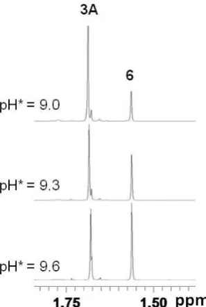
During the pH titrations for aqua complexes 1A−5A, the appearance of a new set of peaks was detected with increasing pH* (>8.7). The new peaks are attributable to the free C,N-chelating ligands and to the hydroxo-bridged dimers {[(η5-Cpx)Ir]2(μ-OD)3}+
14 [(η5-C5Me5)Ir(dfphpy)(D2O)]+ (3A), the amount of dimer 6 increased from 23% at
pH*9.0 to 50% at pH* 9.6, Figure 3. ESI-MS studies on the diluted sample (0.2 mM) gave a major peak at m/z 709.2, consistent with the presence of {[(η5-C5Me5)Ir]2(μ-OD)3}+ (calcd m/z 710.2).
Figure 3. Methyl region of 1H NMR spectra from the pH titration of the aqua complex [(η5-C5Me5)Ir(dfphpy)(D2O)]+ (3A), showing an increase in intensity of the
peak for the hydroxo-bridged dimer {[(η5-C5Me5)Ir]2(μ-OD)3}+ (6) with increase in
pH*.
Interactions with Nucleobases. Since DNA is a potential target for transition
metal anticancer drugs,13 nucleobase binding reactions of complexes [(η5-C5Me5)Ir(tpy)Cl] (1), [(η5-C5Me5)Ir(phq)Cl] (2), [(η5-C5Me5)Ir(dfphpy)Cl] (3),
[(η5-C5Me4C6H5)Ir(phpy)Cl] (4) and [(η5-C5Me4C6H4C6H5)Ir(phpy)Cl] (5), with
9-ethylguanine (9-EtG) and 9-methyladenine (9-MeA) were investigated. Solutions of
[image:15.595.99.248.206.427.2]15 D2O (v/v) were prepared, and 1H NMR spectra were recorded at different time
[image:16.595.98.328.196.336.2]intervals at 310 K. The percentages of nucleobase adducts formed by the complexes after 24 h reaction, based on 1H NMR peak integrals, are shown in Table S1 and Figure 4.
Figure 4. Bar chart showing the extent of binding of complexes 1−5 (ca. 1 mM in 20%
MeOD-d4/80% D2O) to the nucleobases 9-EtG and 9-MeA at equilibrium (24 h),
based on 1H NMR peak integrals.
In the 1H NMR spectrum of a solution containing 4 (0.8 mM) and 1 mol equiv 9-ethylguanine (20% MeOD-d4/80% D2O, pH* 7.4, 310 K), one set of new peaks
assignable to the 9-EtG adduct 4G appeared, showing that 100% of 4 had reacted after 10 min (Figure S3). A significant change in chemical shift from 8.62 ppm for the chlorido complex 4 to 9.28 ppm for 9-EtG adduct 4G for the CH=N (phpy ligand) proton was observed. A new 9-EtG H8 peak appeared at 7.44 ppm (singlet), shifted by 0.34 ppm tohigh field relative to that of free 9-EtG. After 24 h, no further change was observed. The ESI-MS of an equilibrium solution contained a major peak at m/z
723.2, confirming the formation of the 9-EtG adduct 4G, [(η5-C5Me4C6H5)Ir(phpy)(9-EtG)]+ (calcd m/z 722.9). Similarly, complexes 1, 3 and 5
also formed 9-EtG adducts to the extent of 100% after 24 h. Only complex 2
16 Complexes 3−5 formed moderately strong 9-MeA adducts (35−63% at equilibrium after 24 h, Table S1). Only complex 1 [(η5-C5Me5)Ir(tpy)Cl] showed an exceptionally
high affinity for 9-MeA, with 90% adduct formation after 24 h. Complex 2 containing 2-phenylquinoline formed almost no 9-MeA adduct (<5%). Except for 2, two adenine nucleobase adducts were formed in the reactions between complexes 1, 3−5 with 9-MeA, most likely through iridium binding to N1 or N7 of adenine in a ratio typically of 1:5.
Competition reactions between equi-molar amounts of 9-EtG and 9-MeA and complexes 1, and 3−5 (ca. 1 mM) in 20% MeOD-d4/80% D2O (v/v, pH* ca. 7.4) at
310 K were investigated. In the competitive experiment, 9-EtG adducts 3G, 4G or 5G
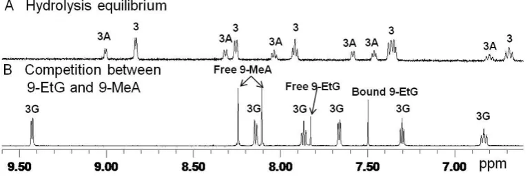
were observed as the only product for complexes 3−5, and as 80% 9-EtG adduct for complex 1 (20% 9-MeA adduct), Table S1. The 1H NMR spectra for the competition reaction for 3 are shown in Figure 5.
Figure 5. Low field region of the 1H NMR spectra for the competitive reaction between 9-EtG and 9-MeA and complex 3 [(η5-C5Me5)Ir(dfphpy)Cl].(A) Equilibrium
solution of 3 (1.0 mM) in 20% MeOD-d4/80% D2O (v/v, pH* 7.4) at 310 K,
[image:17.595.91.474.418.548.2]17
Figure 6. Low field region of the 1H NMR spectra for the reaction between 9-EtG and
complex 4 [(η5-C5Me4C6H5)Ir(phpy)Cl] at 310 K.(A) Equilibrium solution of 4 (0.8
mM) in 20% MeOD-d4/80% D2O (v/v, pH* 7.4), containing both the chlorido
complex 4 and its aqua adduct 4A. (B) 10 min after addition of NaCl (final concentration, 4 mM) to the equilibrium solution. (C) The complete formation of 9-EtG adduct 4G after addition of 1 mol equiv 9-EtG.
Reactions of complexes [(η5-C5Me4C6H5)Ir(phpy)Cl] (4) and
[(η5-C5Me4C6H4C6H5)Ir(phpy)Cl] (5) with 9-EtG were also investigated in the
presence of 4 mM NaCl, Figure 6. A solution of 4 (ca. 0.8 mM) in 20% MeOD-d4/80%
D2O (v/v) containing an equilibrium mixture of 4 and aqua adduct 4A was prepared,
and its 1H NMR spectrum was recorded, Figure 6A. Then NaCl was added to give a 4 mM [Cl−] solution. The aqua adduct 4A was suppressed to ca. 5%, Figure 6B. Addition of ca. 1 mol equivalent of 9-EtG resulted in complete formation of 9-EtG adduct after 24 h at 310 K. Complex 5 also formed a guanine adduct quantitatively in the presence of 4 mM [Cl−].
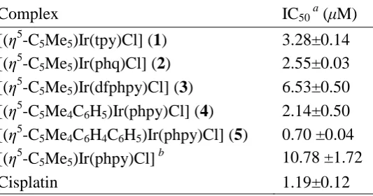
Cytotoxicity. The cytotoxicity of complexes 1−5 towards A2780 human ovarian
cancer cells was investigated, Table 4. The IC50 value (concentration at which 50% of
18 cisplatin. Complexes 4 and 5 containing Cpxph or Cpxbiph were even more potent, especially complex 5 with an IC50 value of 0.7 μM (ca. twice as active as cisplatin).
[image:19.595.92.359.241.381.2]Overall, the cytotoxic potency increases with phenyl substitution on Cp*: Cpxbiph > Cpxph > Cp*, Table 4 and Figure S4.
Table 4. Inhibition of Growth of A2780 Human Ovarian Cancer Cells by Complexes
1−5 and Comparison withCisplatin
Complex IC50 a (μM)
[(η5-C5Me5)Ir(tpy)Cl] (1) 3.28±0.14
[(η5-C5Me5)Ir(phq)Cl] (2) 2.55±0.03
[(η5-C5Me5)Ir(dfphpy)Cl] (3) 6.53±0.50
[(η5-C5Me4C6H5)Ir(phpy)Cl] (4) 2.14±0.50
[(η5-C5Me4C6H4C6H5)Ir(phpy)Cl] (5) 0.70 ±0.04
[(η5-C5Me5)Ir(phpy)Cl] b 10.78 ±1.72
Cisplatin 1.19±0.12
a
Drug-treatment period was 24 h. bRef 3b.
Discussion
X-ray Crystal Structures. A search of the Cambridge Crystallographic Database
showed that the crystal structure of complex 5 is only the second example to be reported of a metal complex containing the Cpxbiph ligand. The only other example is the bipyridine complex [(η5-C5Me4C6H4C6H5)Ir(bpy)Cl]PF6 (where bpy =
bipyridine).3a The two complexes are structurally very similar. In complex 5, the chelating ligand is closer to the IrIII center than in [(η5-C5Me4C6H4C6H5)Ir(bpy)Cl]PF6
19 [(η5-C5Me4C6H4C6H5)Ir(bpy)Cl]PF6. The Ir–Cl bond lengths are similar in the two
complexes, with distances of 2.3886(8) Å (Table 2) and 2.3840(14) Å,3a respectively. This behavior is similar to that observed for the C,N-chelated complex [(η5-C5Me5)Ir(phpy)Cl]14 when compared to the N,N-chelated complex
[(η5-C5Me5)Ir(bpy)Cl]Cl.3b
The bond lengths and bond angles in complexes [(η5-C5Me5)Ir(phq)Cl] (2) and
[(η5-C5Me5)Ir(phpy)Cl]14 are similar, except that the Ir−N bond for 2 [2.128(5) Å] is
longer than that of [(η5-C5Me5)Ir(phpy)Cl] [2.080(2) Å], implying weaker σ donation
from N to the iridium center due to the electron-withdrawing phenyl ring in the quinoline moiety.
Hydrolysis and pKa of Aqua Adducts. Since M–OH2 (M = metal) aqua
complexes are often more reactive than the equivalent chlorido complexes,3a,12,15 hydrolysis of the M–Cl bonds can represent an activation step for transition metal anticancer complexes.16 There are only a few previous studies of the aquation (substitution of Cl by H2O) of organometallic IrIII complexes.3a,4a,17 In the work
reported here all the [(η5-Cpx)Ir(C^N)Cl] complexes 1−5 hydrolyzed too rapidly for determination of their hydrolysis rates by NMR, even for the biphenyl substituted Cpxbiph complex 5 [(η5-C5Me4C6H4C6H5)Ir(2-phpy)Cl]. We have previously reported
half-lives for the hydrolysis of some Cpxph or Cpxbiph IrIII complexes containing N,N-bound 2,2′-bipyridine (bpy) or 1,10-phenanthroline (phen) chelating ligands of ca. 4 min at 310 K.3a The electron-donor methyl groups on the Cp ring and the negatively-charged C,N-chelating ligands may together contribute to the fast hydrolysis observed for the complexes reported here since the increased effective charge on the Ir center may facilitate chloride loss. Fast hydrolysis rates also have been reported for some hexamethylbenzene RuII complexes,18 acetylacetonate RuII and OsII complexes,19 and picolinate IrIII complexes.3a These results illustrate that IrIII complexes are not always inert and that Ir−Cl bonds can be labile.
20 (23 mM), complexes 1−5 were found to be almost all present in solution as the relatively unreactive chlorido species, and only about 5% of complexes 1−5 was present as the reactive aqua species at a chloride concentration of 4 mM close to that of the cell nucleus. These data suggest that aquation is suppressed almost totally at the saline concentration in blood. However, complexes 1−5 might be activated by aquation in the cell nucleus. Another possibility is that the complexes might react by direct substitution of chloride by nucleobases (DNA).
The aqua complexes [(η5-C5Me5)Ir(tpy)(D2O)]+ (1A), [(η5-C5Me5)Ir(phq)(D2O)]+
(2A), [(η5-C5Me5)Ir(dfphpy)(D2O)]+ (3A), [(η5-C5Me4C6H5)Ir(phpy)(D2O)]+ (4A),
[(η5-C5Me4C6H4C6H5)Ir(phpy)(D2O)]+ (5A), and [(η5-C5Me5)Ir(phpy)(D2O)]+, have
similar pKa values, ranging from 8.31 to 8.87 (Table 3). Although the substituents on
the Cp* ring and the 2-phenylpyridine chelating ligand do not significantly affect the acidity of the bound water, the observed trend caused by these substituents is clear, following the order 1A > 2A > 4A > 5A ≈ 3A. The presence of phenyl and biphenyl substituents on the Cp* ring lower the pKa value by ca. 0.3 to 0.4 units, consistent
with the electron-withdrawing properties of these groups. Substitution of cyclometalated 2-phenylpyridine by the fluorinated chelating ligand 2-(2,4-difluorophenyl)pyridine leads to a decrease in pKa by 0.4 units. However,
replacing cyclometalated 2-phenylpyridine by 2-(p-tolyl)pyridine or by the more π-delocalized 2-phenylquinoline, has little effect on the pKa value (ca. 0.1 unit)
However, the aqua complexes [(η5-C5Me5)Ir(phpy)(D2O)]+,
[(η5-C5Me4C6H5)Ir(phpy)(D2O)]+ (4A) and [(η5-C5Me4C6H4C6H5)Ir(phpy)(D2O)]+ (5A)
containing the C,N-chelated 2-phenylpyridine ligand, have significantly higher pKa
21 complexes would be present in the active aqua form at physiological pH.
The pH titration also showed that additional species were formed for all complexes studied here above pH 8.7 (Figure 3), indicating the formation of the hydroxo-bridged dimers {[(η5-Cpx)Ir]2(μ-OD)3}+ (6−8, Chart 1). However, this does not occur when the
chelating ligand is N,N-chelated bipyridine ligand.3a Clearly the Ir−C bond shows less stability with respect to the formation of dimers 6−8 than the Ir−N bond. It seems likely that the mechanism of formation of 6−8 involves initial cleavage of the Ir−C bond, followed by Ir−N bond breakage. Some OsII and RuII complexes containing N,O-bound or O,O-bound ligands have been reported to form hydroxo-bridged dimers readily during their hydrolysis, resulting in their inactivity toward cancer cell lines.20 In this work, no hydroxo-bridged dimers of 6−8 were observed during hydrolysis, and all the complexes are active toward A2780 human ovarian cancer cells. This indicates that the C,N-chelated IrIII complexes are stable in aqueous solution and the dimers
6−8 formed only appear at high pH and have no negative effect on their cytotoxicity at physiological pH.
Interactions with Nucleobases. Interaction with DNA is often associated with the
cytotoxicity of metal anticancer drugs.13 In this study, the interactions with model nucloebases, 9-EtG and 9-MeA, and with complexes 1−5 were investigated (Figure 4 and Table S1). All the C,N-chelated IrIII complexes, except 2, showed an exceptionally high nucleobase affinity with 100% guanine adduct formation for 9-EtG, which may contribute to their high cytotoxicity. Complexes [(η5-C5Me4C6H5)Ir(phpy)Cl] (4) and [(η5-C5Me4C6H4C6H5)Ir(phpy)Cl] (5) still bind
22 to be present as the reactive aqua form compared to the N,N-chelated complexes.
Complexes 1−5 bind more weakly to adenine compared to guanine. The competition between 9-EtG and 9-MeA for the C,N-bound IrIII complexes give rise to 9-EtG adducts as the only product for 3−5 and as major product for 1 (Figure 4 and Table S1), confirming that binding to guanine is stronger than to adenine. This may due to the steric hindrance caused by the NH2 group at the 6-position of the adenine
ring. In addition, guanine is usually considered to be a stronger electron donor than adenine.21 The widely used anticancer drug in clinical, cisplatin, also prefers guanine over adenine.22 Organometallic RuII, OsII and IrIII complexes containing N,O-chelating ligands or O,O-chelating ligands such as picolinate and acetylacetonate, which possess oxygen as an H-bond acceptor for adenine C6NH2, also bind to both
guanine and adenine residues.3a,19a,20b,23
IrIII cyclopentadienyl complexes containing a neutral N,N-chelating ligand (phen, bpy, or ethylenediamine) bind selectively to 9-EtG, but not to adenine.3a The formation of adenine adducts in this work may be due to the interaction between NH2
of 9-MeA and negatively-charged carbons on the C,N-chelating ligand.3b Thus complexes 1−3 containing different substituents on the phenylpyridine bind to 9-MeA differently. The electron donating methyl group on the phenyl ring in complex [(η5-C5Me5)Ir(tpy)Cl] (1) increases electron density and may facilitate the interaction
with 9-MeA (>90%). In contrast, only 35% of complex [(η5-C5Me5)Ir(dfphpy)Cl] (3)
formed 9-MeA adducts which may be due to the presence of the electron-withdrawing fluoro group. Complex 2 [(η5-C5Me5)Ir(phq)Cl] has the lowest affinity for both model
nucleobases among the five complexes, with 45% and <5% binding to 9-EtG and 9-MeA, respectively. The weaker binding is most likely due to the steric hindrance caused by the quinoline ligand.
Cytotoxicity. We have reported that some Cp* IrIII complexes containing N,N-, or
23 and [(η5-C5Me5)Ir(dfphpy)Cl] (3) studied here showed promising activity toward the
human ovarian A2780 cancer cell line with IC50 values ranging from 2.5−6.5 μM
(Table 4), close to the value we reported recently for [(η5-C5Me5)Ir(phpy)Cl].3b Thus
the introduction of C,N-chelating ligands is an effective method for switching on the cancer cell cytotoxicity of Cp* IrIII complexes. The strong binding of Ir to nucleobases, especially to guanine bases, may provide an important contribution towards the cytotoxicity. Also the neutral C,N- complexes display a more hydrophobic character than the positively-charged N,N- complexes3b and therefore possess enhanced cellular uptake which may also contribute to the cytotoxicity. The introduction of substituents on the phenylpyridine ring enhanced the cytotoxicity of [(η5-C5Me5)Ir(phpy)Cl], Table 4. Particularly active is the complex
[(η5-C5Me5)Ir(phq)Cl] (2), which is 4 times as potent as [(η5-C5Me5)Ir(phpy)Cl]. The
ability of the phq ligand to intercalate into DNA24 may contribute to this enhanced potency.
The introduction of phenyl or biphenyl substituents onto the tetramethylcyclopentadienyl ring to give complexes [(η5-C5Me4C6H5)Ir(phpy)Cl] (4)
and [(η5-C5Me4C6H4C6H5)Ir(phpy)Cl] (5), results in a dramatic increase in
cytotoxicity compared to the parent Cp* complex [(η5-C5Me5)Ir(phpy)Cl], Table 4
24 action from that of cisplatin.
Conclusions
Iridium-based anticancer agents, including organometallic iridium complexes, are currently attracting attention as potential anticancer agents with novel mechanisms of action. We have studied here the effects of changing the Cpx ligand and negatively-charged C,N-chelating ligand of IrIII cyclopentadienyl complexes of the type [(η5-Cpx)Ir(C^N)Cl] on the hydrolysis of the chlorido complex, acidity of the aqua adduct, nucleobase binding, and cancer cell cytotoxicity.
All the complexes undergo rapid hydrolysis (<5 min at 278 K) due to the strongly electron-donating methyl group and negatively-charged C,N-chelating ligand. However the complexes are likely to be present in their unhydrolyzed forms in the extracellular fluid and cell cytoplasm (typically 104 and 23 mM [Cl−]), whereas they are likely to be activated by hydrolysis in the cell nucleus ([Cl−] ca. 4 mM). Generally, the aqua adducts of the C,N- complexes studied here possess low acidity, with pKa
values 1.9 units higher than N,N- analogues, which ensures that the active aqua adduct is the major form after hydrolysis at physiological pH, and may contribute to the strong binding to guanine. The substituents on both the Cp* ring and on 2-phenylpyridine can fine-tune the acidity of aqua adduts according to their electronic effects. Hydroxo-bridged dimers {[(η5-Cpx)Ir]2(μ-OD)3}+ (6–8) are observed at high
pH (>8.7), which suggests the stability of C,N- complexes is not as high as N,N- analogues under strongly basic conditions.
25 All the C,N- complexes showed very promising anticancer activity toward A2780 human ovarian cancer cells. The Cp* complexes possess activity comparable to cisplatin. The introduction of phenyl or biphenyl substituent significantly improved their cytotoxicity, especially for the Cpxbiph complex 5 which has submicromolar activity against A2780 cancer cells. The strong binding to guanine bases and hydrophobicity may contribute to their high activity. This study shows that desirable features can be introduced into this class of complexes to optimize their design as anticancer agents.
Acknowledgements.
Z.L. was supported by a University of Warwick Research Scholarship (WPRS). We thank the ERC (grant no. 247450 for P.J.S.), EPSRC, ORSAS, ERDF and AWM for Science City funding, and members of COST Action D39 for stimulating discussions.
Supporting Information Available: Details of the extent of nucleobase binding
(Table S1), aqueous chemistry (Figures S1 and S2), nucleobase studies (Figure S3), IC50 comparison (Figure S4). This material is available free of charge via the Internet
at http://pubs.acs.org. X-ray crystallographic data in CIF format are available from the Cambridge Crystallographic Data Centre (http://www.ccdc.cam.ac.uk).
Notes and References
26 Hubbard, C. D., Eds.; Academic Press: 2009; Vol. 61, pp 1-62.
(2) (a) Allen, O. R.; Gott, A. L.; Hartley, J. A.; Hartley, J. M.; Knox, R. J.; McGowan, P. C. Dalton Trans.2007, 5082-5090. (b) Tan, Y. L. K.; Pigeon, P.; Hillard, E. A.; Top, S.; Plamont, M.-A.; Vessieres, A.; McGlinchey, M. J.; Muller-Bunz, H.; Jaouen, G.
Dalton Trans.2009, 10871-10881. (c) van Rijt, S. H.; Sadler, P. J. Drug Discov. Today 2009, 14, 1089-1097. (d) Strohfeldt, K.; Tacke, M. Chem. Soc. Rev. 2008, 37, 1174-1187. (e) Eger, S.; Immel, T. A.; Claffey, J.; Muller-Bunz, H.; Tacke, M.; Groth, U.; Huhn, T. Inorg. Chem. 2010, 49, 1292-1294. (f) Vessières, A.; Top, S.; Pigeon, P.; Hillard, E.; Boubeker, L.; Spera, D.; Jaouen, G. J. Med. Chem.2005, 48, 3937-3940. (g) Chatterjee, S.; Biondi, I.; Dyson, P.; Bhattacharyya, A. J. Biol. Inorg. Chem.2011, 1-10. (h) Hanif, M.; Schaaf, P.; Kandioller, W.; Hejl, M.; Jakupec, M. A.; Roller, A.; Keppler, B. K.; Hartinger, C. G. Aust. J. Chem.2010, 63, 1521-1528.
(3) (a) Liu, Z.; Habtemariam, A.; Pizarro, A. M.; Fletcher, S. A.; Kisova, A.; Vrana, O.; Salassa, L.; Bruijnincx, P. C. A.; Clarkson, G. J.; Brabec, V.; Sadler, P. J. J. Med. Chem. 2011, 54, 3011-3026. (b) Liu, Z.; Salassa, L.; Habtemariam, A.; Pizarro, A. M.; Clarkson, G. J.; Sadler, P. J. Inorg. Chem. 2011, 50, 5777-5783. (c) Schäfer, S.; Sheldrick, W. S. J. Organomet. Chem. 2007, 692, 1300-1309. (d) Sliwinska, U.; Pruchnik, F. P.; Ulaszewski, S.; Latocha, M.; Nawrocka-Musial, D. Polyhedron2010,
27
Chem., Int. Ed.2010, 49, 8304-8305.
(4) (a) Helm, L.; Merbach, A. E. Coord. Chem. Rev. 1999, 187, 151-181. (b) Wilbuer, A.; Vlecken, D. H.; Schmitz, D. J.; Kräling, K.; Harms, K.; Bagowski, C. P.; Meggers, E. Angew. Chem., Int. Ed.2010, 49, 3839-3842.
(5) (a) van Rijt, S. H.; Peacock, A. F. A.; Johnstone, R. D. L.; Parsons, S.; Sadler, P. J.
Inorg. Chem. 2009, 48, 1753-1762. (b) Kostrhunova, H.; Florian, J.; Novakova, O.; Peacock, A. F. A.; Sadler, P. J.; Brabec, V. J. Med. Chem.2008, 51, 3635-3643.
(6) White, C. Y., A.; Maitlis, P. M. Inorg. Chem.1992, 29, 228-234.
(7) CrysAlis PRO; Oxford Diffraction Ltd.: Abington, Oxfordshire, U. K., 2007. (8) Sheldrick, G. M. Acta Crystallogr.1990, A46, 467-473.
(9) Sheldrick, G. M. SHELXL-97; University of Göttingen: Göttingen, Germany, 1997.
(10) Krezel, A.; Bal, W. J. Inorg. Biochem.2004, 98, 161-166.
(11) Skehan, P.; Storeng, R.; Scudiero, D.; Monks, A.; McMahon, J.; Vistica, D.; Warren, J. T.; Bokesch, H.; Kenney, S.; Boyd, M. R. J. Natl. Cancer Inst. 1990, 82, 1107-1112.
(12) Martin, R. B. In Cisplatin: Chemistry and Biochemistry of a Leading Anticancer Drug; Lippert, B., Ed.; VHCA & Wiley-VCH: Zürich, Switzerland, 1999, pp 181-205. (13) (a) Zhang, C. X.; Lippard, S. J. Curr. Opin. Chem. Biol. 2003, 7, 481-489. (b) Deubel, D. V.; Lau, J. K.-C. Chem. Commun.2006, 2451-2453.
(14) Li, L.; Brennessel, W. W.; Jones, W. D. J. Am. Chem. Soc. 2008, 130, 12414-12419.
(15) Hohmann, H.; Hellquist, B.; Van Eldik, R. Inorg. Chem.1992, 31, 345-351. (16) Pizarro, A. M.; Habtemariam, A.; Sadler, P. J. In Medicinal Organometallic Chemistry (Topics in Organometallic Chemistry), 1st ed.; Jaouen, G., Metzler-Nolte, N., Ed.; Springer-Verlag: Heidelberg, Germany, 2010; 32, pp 21-56.
(17) Koelle, U. Coord. Chem. Rev.1994, 135-136, 623-650.
28 Deeth, R. J.; Aird, R.; Guichard, S.; Fabbiani, F. P. A.; Lozano-Casal, P.; Oswald, I. D. H.; Jodrell, D. I.; Parsons, S.; Sadler, P. J. Proc. Natl. Acad. Sci. U.S.A. 2005, 102, 18269-18274.
(19) (a) Fernández, R.; Melchart, M.; Habtemariam, A.; Parsons, S.; Sadler, P. J.
Chem.-Eur. J.2004, 10, 5173-5179. (b) Peacock, A. F. A.; Melchart, M.; Deeth, R. J.; Habtemariam, A.; Parsons, S.; Sadler, P. J. Chem.-Eur. J.2007, 13, 2601-2613.
(20) (a) Peacock, A. F. A.; Habtemariam, A.; Fernández, R.; Walland, V.; Fabbiani, F. P. A.; Parsons, S.; Aird, R. E.; Jodrell, D. I.; Sadler, P. J. J. Am. Chem. Soc.2006, 128, 1739-1748. (b) Peacock, A. F. A.; Parsons, S.; Sadler, P. J. J. Am. Chem. Soc.2007,
129, 3348-3357. (c) Kljun, J.; Bytzek, A. K.; Kandioller, W.; Bartel, C.; Jakupec, M. A.; Hartinger, C. G.; Keppler, B. K.; Turel, I. Organometallics2011, 30, 2506-2512. (21) Pullman, B.; Pullman, A. Biochimica et Biophysica Acta1959, 36, 343-350. (22) Baik, M.-H.; Friesner, R. A.; Lippard, S. J. J. Am. Chem. Soc. 2003, 125, 14082-14092.
(23) Melchart, M.; Habtemariam, A.; Parsons, S.; Sadler, P. J. Inorg. Biochem. 2007,
101, 1903-1912.
(24) (a) Atwell, G. J.; Bos, C. D.; Baguley, B. C.; Denny, W. A. J. Med. Chem.1988,
31, 1048-1052. (b) Atwell, G. J.; Baguley, B. C.; Denny, W. A. J. Med. Chem.1989,
32, 396-401.

![Table 2. Selected Bond Lengths (Å) and Angles (deg) for [(η5-C5Me5)Ir(phq)Cl] (2)](https://thumb-us.123doks.com/thumbv2/123dok_us/9665531.468471/12.595.84.477.67.326/table-selected-bond-lengths-a-angles-deg-me.webp)
![Figure 2. Confirmation of hydrolysis of IrIII complex [(η5-C5Me5)Ir(tpy)Cl] (1). (A)](https://thumb-us.123doks.com/thumbv2/123dok_us/9665531.468471/13.595.98.440.423.592/figure-confirmation-hydrolysis-iriii-complex-me-ir-tpy.webp)