ORIGINAL RESEARCH
ADULT BRAIN
Multimodal CT Imaging: Time to Treatment and Outcomes in
the IMS III Trial
XA. Vagal, XL.D. Foster, XB. Menon, XA. Livorine, XJ. Shi, XE. Qazi, XS.D. Yeatts, XA.M. Demchuk, XM.D. Hill, XT.A. Tomsick, and XM. Goyal
ABSTRACT
BACKGROUND AND PURPOSE: The importance of time in acute stroke is well-established. Using the Interventional Management of Stroke III trial data, we explored the effect of multimodal imaging (CT perfusion and/or CT angiography) versus noncontrast CT alone on time to treatment and outcomes.
MATERIALS AND METHODS: We examined 3 groups: 1) subjects with baseline CTP and CTA (CTP⫹CTA), 2) subjects with baseline CTA without CTP (CTA), and 3) subjects with noncontrast head CT alone. The demographics, treatment time intervals, and clinical outcomes in these groups were studied.
RESULTS:Of 656 subjects enrolled in the Interventional Management of Stroke III trial, 90 (13.7%) received CTP and CTA, 216 (32.9%) received CTA (without CTP), and 342 (52.1%) received NCCT alone. Median times for the CTP⫹CTA, CTA, and NCCT groups were as follows: stroke onset to IV tPA (120.5 versus 117.5 versus 120 minutes;P⫽.5762), IV tPA to groin puncture (77.5 versus 81 versus 91 minutes;P⫽.0043), groin puncture to endovascular therapy start (30 versus 38 versus 44 minutes;P⫽.0001), and endovascular therapy start to end (63 versus 46 versus 74 minutes;P⬍.0001). Compared with NCCT, the CTA group had better outcomes in the endovascular arm (OR, 2.12; 95% CI, 1.36 –3.31; adjusted for age, NIHSS score, and time from onset to IV tPA). The CTP⫹CTA group did not have better outcomes compared with the NCCT group.
CONCLUSIONS: Use of CTA with or without CTP did not delay IV tPA or endovascular therapy compared with NCCT in the Interventional Management of Stroke III trial.
ABBREVIATIONS:ED⫽emergency department; EVT⫽endovascular therapy; IMS III⫽Interventional Management of Stroke III trial
T
he importance of time is well-established in acute ischemic stroke. Ample evidence suggests that the benefit of treatment decreases with increasing time to treatment in intravenous andendovascular therapy (EVT).1-3It is critical to minimize time
de-lays in stroke treatment workflow components, including imag-ing time delays. However, imagimag-ing protocols vary by institution, and there is no nationally or internationally recognized standard imaging protocol for evaluating patients with acute stroke.
Multimodal imaging, including CT angiography, CT perfusion, or MR angiography and MR perfusion may provide useful infor-mation on arterial occlusion, collateral flow, and tissue viability. On the contrary, these imaging techniques may also delay initia-tion of revascularizainitia-tion therapies. Recent endovascular stroke trials (Multicenter Randomized CLinical trial of Endovascular treatment for Acute ischemic stroke in the Netherlands [MR CLEAN],4Endovascular Treatment for Small Core and Proximal
Occlusion Ischemic Stroke [ESCAPE],5Extending the Time for
Thrombolysis in Emergency Neurological Deficits–Intra-Arterial [EXTEND-IA],6Solitaire With the Intention For Thrombectomy
as Primary Endovascular Treatment [SWIFT PRIME],7
Endovas-cular RevasEndovas-cularization With Solitaire Device Versus Best Medical Therapy in Anterior Circulation Stroke Within 8 Hours [REVAS-Received October 14, 2015; accepted after revision January 22, 2016.
From the Department of Radiology (A.V., A.L., J.S., T.A.T.), University of Cincinnati Medical Center, Cincinnati, Ohio; Department of Public Health Sciences (L.D.F., S.D.Y.), Medical University of South Carolina, Charleston, South Carolina; and Departments of Neurology (A.M.D., M.D.H.) and Radiology (B.M, E.Q., M.G.), University of Calgary, Calgary Alberta, Canada.
A.V. was funded by the Clinical and Translational Science Awards 8 UL1
TR000077-05 CT2 Award, the Imaging Core Lab, A Study of the Efficacy and Safety of Activase (Alteplase) in Patients With Mild Stroke (PRISMS) Trial, and Genentech. S.D.Y. was funded by the National Institutes of Health/National Institute of Neu-rological Disorders and Stroke (U01 NS052220). B.M. was funded by the Canadian Institute of Health Research, Heart and Stroke Foundation, and Alberta Innovates Health Solutions.
Paper previously presented at: International Stroke Conference, February 10 –13, 2015; Nashville, Tennessee.
Please address correspondence to Achala Vagal, MD, MS, Department of Radiology, University of Cincinnati Medical Center, 234 Goodman St, Cincinnati, OH 45267; e-mail: Achala.Vagal@uchealth.com
Indicates open access to non-subscribers at www.ajnr.org
CAT],8Assess the Penumbra System in the Treatment of Acute
Stroke [THERAPY],9and Trial and Cost Effectiveness Evaluation
[image:2.594.54.377.56.194.2]of Intra-arterial Thrombectomy in Acute Ischemic Stroke [THRACE]10) demonstrating the benefit of endovascular therapy
have used supplementary imaging, in addition to noncontrast CT, for patient selection. It is therefore imperative to understand whether additional imaging causes delays in the initiation of treat-ment of patients with acute ischemic stroke.
The Interventional Management of Stroke III (IMS III) trial was a phase III randomized trial of endovascular treatment after intravenous tPA versus IV tPA alone.11Multimodal imaging was
available in a subgroup of patients at the discretion of the enroll-ing site. Our objective was to explore the effect of multimodal imaging: CTP⫹CTA or CTA (without CTP) versus NCCT on time to treatment and outcomes in the IMS III trial.
MATERIALS AND METHODS
Study Population
IMS III was a multicenter, phase III, randomized, open-label clin-ical trial testing the approach of IV tPA followed by endovascular treatment, compared with standard IV tPA alone, determined by the primary outcome of a modified Rankin Scale score of 0 –2 at 90 days. Details of the design, methodology, and outcomes of the IMS III trial have been published.11
A noncontrast head CT was the only required baseline im-aging. Additional imaging, including CTP and/or CTA, was not a prerequisite for entry or patient selection in the IMS III trial. However CTA or CTP was performed at a few study sites at their discretion.
Imaging Subgroups
The analysis consisted of 3 subgroups based on baseline imaging: 1) subjects with baseline CTP and CTA (CTP⫹CTA), 2) subjects with CTA (without a concurrent CTP study), and 3) subjects without CTA or CTP (noncontrast head CT alone). The baseline demographics, time intervals, and outcomes in these 3 groups were studied.
Time Intervals
The following intervals were calculated for all subjects: 1) stroke onset to emergency department (ED) arrival, 2) ED arrival to start of imaging, and 3) start of imaging to IV tPA initiation.
Additional time intervals were calcu-lated for the endovascular treatment arm: 1) IV tPA initiation to groin puncture, 2) groin puncture to the start of EVT, and 3) the duration of EVT (for subjects who received endo-vascular treatment).
Baseline imaging time was defined as the time of the first imaging performed. EVT initiation was defined as the start of intra-arterial tPA/balloon occlusion/ thrombus aspiration or the first deploy-ment of the device, depending on the type of endovascular therapy adminis-tered. Similarly, the EVT end time was defined as the time of the last deploy-ment, time of last thrombus aspiration attempt, or time of the end of intra-arterial infusion.
Statistical Analysis
Because the time intervals described above did not follow a normal distribution, descriptive statistics are reported as median (interquartile range). Normality was assessed graphically via histogram and quantile-quantile plot. The Hodges-Lehmann esti-mator of location shift was used to calculate differences, with confi-dence intervals, in time intervals between groups. To compare the 3 groups (CTP⫹CTA versus CTA versus NCCT), we used the Kruskal-Wallis test for continuous variables and the2test for categoric
vari-ables. The primary clinical outcome (mRS 0 –2 at 90 days) was ana-lyzed via a logistic regression model to estimate adjusted odds ratios. The linearity in the log assumption was confirmed for continuous covariates. All tests were conducted at the .05 level of signifi-cance. If there was a significant association, pair-wise compar-ison was also performed between the CTP⫹CTA and CTA groups. Because this analysis is exploratory,Pvalues were not adjusted for multiple testing. Because the endovascular group was of particular interest in light of recent trial results, separate models were created to estimate odds ratios by treatment. Sta-tistical analysis was performed in SAS, Version 9.3T (SAS In-stitute, Cary, North Carolina).
RESULTS
Population Demographics
Of 656 subjects enrolled in the IMS III trial, 90 (13.7%) received CTP and CTA, 216 (32.9%) received CTA (without CTP), and 342 (52.1%) did not receive CTP or CTA (NCCT). Eight subjects with CTP performed without CTA were excluded from the anal-ysis to increase the homogeneity within groups. Demographic characteristics are provided inTable 1.
Time Intervals
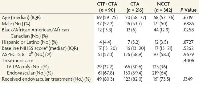
The time intervals for the 3 imaging groups are provided in Table 2. The use of CTP or CTA imaging did not cause delays in IV tPA (Fig 1) or endovascular treatment (Fig 2). Time from the start of imaging to IV tPA initiation was shorter in the CTP⫹CTA and CTA groups, compared with NCCT (6 minutes [95% CI, 1–11 minutes] and 8 minutes [95% CI, 4 –12 minutes] shorter, respec-Table 1: Demographic characteristics
CTP+CTA (n= 90)
CTA (n= 216)
NCCT
(n= 342) PValue Age (median) (IQR) 69 (59–75) 70 (58–77) 68 (57–76) .6719
Male (No.) (%) 47 (52.2) 116 (53.7) 171 (50) .6885
Black/African American/African Canadian (No.) (%)
12 (13.3) 13 (6) 44 (12.9) .0258
Hispanic or Latino (No.) (%) 4 (4.4) 7 (3.2) 12 (3.5) .8727 Baseline NIHSS scorea(median) (IQR) 17 (13–20) 16 (13–20) 17 (13–21) .5262 ASPECTS 8–10b(No.) (%) 51 (57.3) 126 (58.9) 197 (58.3) .9679
Treatment arm .4006
IV tPA only (No.) (%) 29 (32.2) 66 (30.6) 123 (36) Endovascular (No.) (%) 61 (67.8) 150 (69.4) 219 (64)
Received endovascular treatment (No.) (%) 49 (80.3) 123 (82.0) 161 (73.5) .1349 Note:—IQR indicates interquartile range.
a
Two subjects have missing data (CTP⫹CTA,n⫽1; CTA,n⫽1).
b
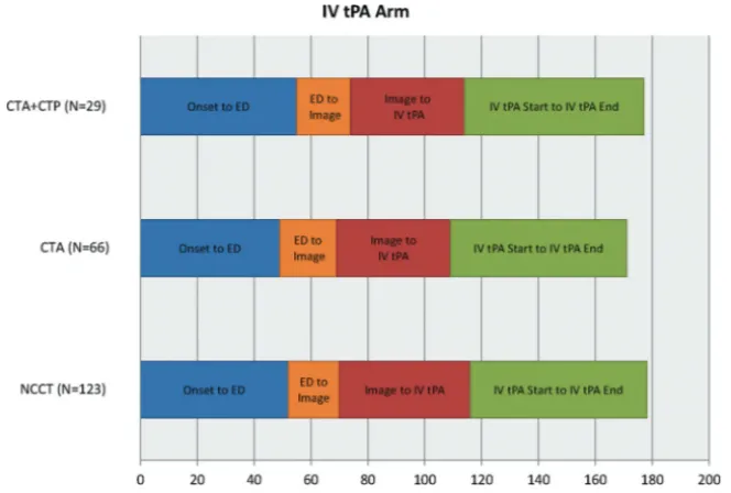
tively). Door to IV tPA initiation was not significantly different among groups. Time from baseline imaging to puncture was shorter in the CTP⫹CTA and CTA groups compared with NCCT (17 utes [95% CI, 4 –29 minutes] and 22 minutes [95% CI, 14 –30 min-utes] shorter, respectively). Groin puncture to EVT start was faster in the CTP⫹CTA and CTA groups, compared with the NCCT group (12 minutes [95% CI, 7–18 minutes] and 7 minutes [95% CI, 2–12 minutes] faster, respectively). EVT duration was 26 minutes (95% CI 14 –37) faster in the CTA compared with the NCCT group, though the CTP⫹CTA and NCCT groups were not significantly different. Total ED arrival to puncture time was shorter in the CTP⫹CTA and CTA groups compared with the NCCT group (14 minutes [95% CI, 0 –27 minutes] and 17 minutes [95% CI, 8–26 minutes] shorter, respectively).
There were no significant pair-wise time interval differences between the CTP⫹CTA and CTA groups. The rate of mother ship enrollment (direct admit of patient to tertiary stroke center) differed by subgroup: 97% in CTP⫹CTA group, 94% in CTA group, and 77% in NCCT group. When transfer subjects were removed, no sta-tistical difference remained among groups for the time from onset to
IV tPA bolus (P⫽.5568). Times from im-aging to groin puncture and groin punc-ture to the start of EVT were still shorter in the CTP⫹CTA and CTA populations (P⫽.0088 andP⫽.0006, respectively).
The high-enrolling sites (defined as sites that enrolled⬎6 subjects) were iden-tified because these are usually high-vol-ume, experienced stroke centers and tend to have established systems of care with streamlined workflows. There was no dif-ference in the proportion of subjects from high-enrolling sites by imaging subgroups (85% CTP⫹CTA, 88% CTA, 86% NCCT; 2,P⫽.7947). The sites were grouped by
the ability to perform CTP, defined as hav-ing at least 1 subject in the CTP⫹CTA group, and we compared time to treat-ment and outcome. There were no signif-icant differences between CTP sites and non-CTP sites in time from ED to IV tPA initiation, time from ED to groin punc-ture, or 90-day mRS of 0 –2 (Table 3).
Outcomes
In the IV tPA treatment arm, there was not a significant difference in good clin-ical outcome in the 3 imaging subgroups (34.5% in CTP⫹CTA versus 40.9% in CTA versus 38.2% in NCCT;P⫽.8338). In the endovascular arm, more good outcomes were observed with CTA (39.3% in CTP⫹CTA versus 52.0% in CTA versus 33.8% in NCCT;P⫽.0022). In the endovascular arm, subjects with CTA had about twice the odds of good clinical outcome compared with sub-jects with NCCT, after adjustment for age, NIHSS strata, and time from onset to IV tPA initiation (ad-justed OR, 2.12; 95% CI, 1.36 –3.31;Table 4).
DISCUSSION
The principal finding of our study is that additional imaging, in-cluding CT angiography with or without CT perfusion, did not delay treatment times compared with noncontrast CT in the IMS III trial. In fact, the imaging algorithms of NCCT⫹CTP⫹CTA or NCCT⫹CTA are possible with faster treatment times than NCCT alone.
The guiding principle of stroke therapy is rapid treatment be-cause potential benefits and less adverse events are greatest with earlier reperfusion. Recently, multiple acute stroke trials have demonstrated that endovascular therapy improves functional outcomes in subjects with major arterial occlusions compared with standard therapy, including IV tPA.4-10These trials included
[image:3.594.53.381.55.172.2]CTA and/or CTP imaging techniques to identify patients who would be likely to benefit from endovascular therapy. The MR CLEAN and REVASCAT trials used CTA to identify proximal vessel occlusion; ESCAPE evaluated vessel occlusion and collat-FIG 1. Time intervals of 3 imaging subgroups in the IV tPA treatment arm.
Table 2: Time intervals for the 3 imaging groups
Time (min) (Median) (IQR) CTP+CTA (n= 90) CTA (n= 216) NCCT (n= 342) PValue Onset to ED arrival 55.5a(41–80) 51 (38–69) 48 (33–67) .0216 ED arrival to start of imaging 19.5 (14–26) 21a(14–28) 18 (10–25) .0016 Imaging start to IV tPA initiation 40a(29–54) 38 (24–51) 47 (33–64) .0001 Onset to IV tPA initiation 120.5 (100–160) 117.5 (96–145) 120 (97–145) .5762 ED to IV tPA initiation 62 (50–75) 59.5 (47–77) 65.5 (49–86) .1554 IV tPA start to groin punctureb 77.5 (58.5–105.5) 81a(65–96) 91 (66–115) .0043 Imaging to groin punctureb 124a(97–155.5) 119a(95–140) 142 (115–170.5) ⬍.0001 ED to groin punctureb 146.5 (118.5–182.5) 144a(120–167) 160 (132–191) .0008 Groin puncture to EVT startc 30a(26–45) 38a(25–56) 44 (34–60) .0001 EVT start to EVT finishc 63 (20–115) 46a(14–83) 74 (42–119) ⬍.0001 Note:—IQR indicates interquartile range.
a
Significant pair-wise difference between the imaging subgroup and the NCCT group.
b
Applicable only to subjects with groin puncture: CTP⫹CTA (n⫽60), CTA (n⫽150), NCCT (n⫽210).
c
[image:3.594.53.385.239.463.2]eral circulation by using multiphase CTA; EXTEND-IA used CTA and CTP with automated perfusion processing; and the SWIFT PRIME study started with CTA/CTP or MRA/MR perfusion but amended the imaging protocol to a CT/CTA/MRA strategy to accommodate more sites. Multimodal imaging selection in these trials may lead to a paradigm shift in the use of imaging in acute ischemic stroke work-up. Investigation of workflow while using these imaging algorithms is even more important in the current era.
The strength of our study is that we have analyzed time to treatment with 3 different imaging paradigms in the setting of a large, randomized multicenter acute stroke trial (IMS III). The recent endovascular stroke trials selected patients on the basis of additional imaging, including CTA, multiphase CTA, or CTP. These trials do not have a control NCCT-alone group for similar workflow comparisons, as we have in our study. Our results
sup-port the findings of these trials by sug-gesting benefits with the use of CTA with or without CTP in the work-up of pa-tients with ischemic stroke.
We found that the CTA group of subjects was associated with better clin-ical outcome in the endovascular arm, likely a marker of faster treatment times among more advanced stroke centers. In a previous analysis by using data from the IMS III trial, we found that the odds of favorable outcome in subjects with baseline CTA were higher.12,13In this
study, we have further explored this group by adding CTP to the imaging analysis. In a prior study involving a multicenter, prospective, single-arm thrombectomy trial, our group demon-strated that multimodal CT-based im-aging did not affect workflow.3Our
re-sults are consistent with a single-center experience in which change in acute stroke imaging protocol to include mul-timodal CT did not delay administra-tion of IV tPA.14Our findings differed
from those in a retrospective, multi-in-stitutional study, in which advanced im-aging was associated with delays in en-dovascular treatment without added benefit in outcomes.15However, there
are considerable differences between the 2 studies. The study by Sheth et al15was
a retrospective analysis of both CT and MR imaging, which included only sub-jects with endovascular treatment; per-fusion imaging was used for decision-making, and the study did not address the vascular imaging times and time to IV tPA.
Most surprising, in our study, the NCCT group had longer treatment times, including imaging to IV tPA ini-tiation and imaging to endovascular treatment times. This may be multifactorial. A CTA study can be advantageous for endovascu-lar therapy planning, adding valuable information regarding the presence or absence of large-vessel occlusion, the exact site of the occlusion, anatomic details of the vessels, and the presence of atherosclerotic plaque, and as a result, CTA can potentially reduce procedural time.13In addition, institution-specific practice and
workflow patterns may translate into faster treatments. Experi-enced, high-volume stroke centers may have parallel processes in place, whereby the treatment decisions are made in the CT con-sole, including administration of IV tPA (if appropriate) after NCCT.16A workflow that offers IV tPA administration in parallel
to imaging and rapid activation of the interventional team has the potential to reduce treatment times.
[image:4.594.53.376.45.280.2]Perfusion may provide information regarding tissue viability. However, in our analysis the addition of CTP did not add an FIG 2. Time intervals of 3 imaging subgroups in the endovascular treatment arm.
Table 3: Time intervals and outcome for CTP and non-CTP sites
CTP Sitesa (20 Sites/266 Subjects)
Non-CTP Sites (38 Sites/382 Subjects) ED to IV tPA initiationb(median) (IQR) 63 (47.5–76) 63 (49–87) ED to groin puncturec(median) 145 (122.5–176) 157 (128–185)
90-Day mRS 0–2 (No.) (%) 114 (42.9%) 146 (38.2%)
Note:—IQR indicates interquartile range.
aSites that performed at least 1 baseline CTP. b
Two subjects were missing from the CTP sites; 4 subjects were missing from the non-CTP sites.
cApplicable only to subjects with groin puncture: 172 subjects from the CTP sites, 249 subjects from the non-CTP sites.
Table 4: Logistic regression for outcome (mRS 0 –2 at 90 days) by treatment category
Unadjusted OR (95% CI) AdjustedaOR (95% CI) Endovascular
CTP⫹CTA vs NCCT 1.27 (0.71–2.28);P⫽.4218 1.32 (0.71–2.44);P⫽.3765 CTA vs NCCT 2.12 (1.39–3.25);P⫽.0005 2.12 (1.36–3.31);P⫽.0010 CTA vs CTP⫹CTA 1.67 (0.91–3.06);P⫽.0968 1.60 (0.85–3.03);P⫽.1444 IV tPA only
[image:4.594.54.378.317.374.2] [image:4.594.53.374.430.525.2]outcome benefit compared with CTA alone. Potential reasons for this observation include the following: 1) CTP was not always used for treatment decisions; 2) there is marked variability in CTP postprocessing, and its ability to reliably identify nonviable tissue likely varies substantially across centers; and 3) there is a high potential for selection bias, and patients who underwent addi-tional CTP imaging may have had different baseline characteris-tics from those who did not.
Limitations
Our study has several important limitations, including the heter-ogeneity of imaging protocols in the IMS III centers. Our data did not address the institutional variables affecting workflow, which is critical in understanding the differences in time parameters. In-stitutional practice variability due not only to the large number of participating IMS III centers (58 centers) but also changes in prac-tice during the trial (2006 –2012) confound analysis of center-specific practice patterns. Institution-center-specific data on workflow details around IV tPA administration, neurointervention team activation, and time taken for study consent were not available. We also acknowledge that other unmeasured variables, including general-versus-local anesthesia and complete-versus-focused an-giograms, can influence the time intervals.
It is likely that multimodal imaging (CTA and/or CTP) did not necessarily lead to faster treatment but was simply performed in centers that were generally faster. High-volume stroke centers that perform CTA and CTP routinely may have expedited work-flows leading to shorter times to revascularization. The recent ESCAPE trial had aggressive time targets (median time from im-aging to first reperfusion of 84 minutes), and most of subjects were enrolled at endovascular centers that successfully imple-mented well-organized workflow and imaging processes.5
Although CTA and CTP can be performed quickly with the new-generation scanners (median time to perform CTP⫹CTA and CTA was 9 and 5 minutes, respectively, in our study), data postprocessing and interpretation and decision-making to acti-vate endovascular teams can cause time delays, and we could not analyze this step of the workflow. Ideally, the image processing and interpretation should be performed in parallel with other workflow steps rather than being a sequential flow, to minimize delays.
Another limitation of our study is that it is unclear whether CTA/CTP imaging was used for treatment decisions with poten-tial impact on outcomes, particularly in the endovascular arm, though as per the study protocol, advanced imaging was not used for treatment decisions. We acknowledge that there are con-straints in the data that particularly limit what can be said about the role and importance of CTP. The key ones are the relatively small number of patients who underwent CTP and the lack of information about how the CTP data were processed or used in the study.
We did not analyze the MR imaging– based paradigms due to a very small number of subjects who underwent baseline MR im-aging in IMS III. Although MR imim-aging has obvious diagnostic advantages, it can cause delays in treatment compared with CT-based paradigms.3,17,18 Finally, this is a post hoc analysis and
additional confounding factors and baseline differences may
account for the differences in outcome, thus limiting the general-izability of our results. The outcome differences should be viewed with caution and in the context of limitations of a post hoc analysis.
CONCLUSIONS
Use of CTA with or without CTP did not delay IV tPA throm-bolysis or endovascular therapy compared with NCCT in the IMS III trial. Efficient institutional protocols and expedited workflows to allow rapid neuroimaging for patient selection without delaying treatment are very relevant in the current landscape.
Disclosures: Achala Vagal—RELATED: Grant: Clinical and Translational Science Awards 8 UL1 TR000077-05,* Genentech,*Comments: research grant: Clinical and Translational Science Awards KL2 Career Award; research grant: Imaging Core Lab, PRISMS trial, Genentech. Lydia D. Foster—RELATED:Grant: National Institute of Neurological Disorders and Stroke (IMS U01 NS077304).* Sharon D. Yeatts— RELATED:Grant: National Institutes of Health/National Institute of Neurological Disorders and Stroke (U01 NS052220),*Comments: Statistics and Data Coordination Center for IMS III: unblinded statistician;UNRELATED:Consultancy: Genentech, Comments: PRISMS Trial Steering Committee member. Andrew M. Demchuk— RELATED:Grant: National Institutes of Health/National Institute of Neurological Disorders and Stroke,*Comments: National Institutes of Health funded the Calgary CT Core Laboratory for the IMS-III trial;UNRELATED:Payment for Lectures (includ-ing service on Speakers Bureaus): Medtronic,Comments: honorarium for Continuing Medical Education lectures. Michael D. Hill—RELATED:Grant: National Institutes of Health/National Institute of Neurological Disorders and Stroke*;UNRELATED: Con-sultancy: Merck,Comments: paid Adjudication Committee for outcomes assess-ment in clinical trials;Grants/Grants Pending: Covidien,*Comments: grant for the ESCAPE trial;Stock/Stock Options: Calgary Scientific (stock ownership, imaging company). Thomas A. Tomsick—RELATED:Grant: National Institute of Neurological Disorders and Stroke,*Comments: Angiography Core Lab. Interventional Co-Princi-pal Investigator;UNRELATED:Expert Testimony: Multiple legal entities. Mayank Goyal—RELATED:Grant: Covidien,*Comments: partial funding of ESCAPE trial through an unrestricted grant to University of Calgary;Consulting Fee or Honorar-ium: Covidien, Stryker,Comments: speaking engagement on acute stroke, design and conduct of SWIFT PRIME trial;Other: GE Healthcare,Comments: licensing agree-ment regarding further developagree-ment of systems of stroke diagnosis. *Money paid to the institution.
REFERENCES
1. Khatri P, Yeatts SD, Mazighi M, et al; IMS III Trialists.Time to
an-giographic reperfusion and clinical outcome after acute ischaemic stroke: an analysis of data from the Interventional Management of Stroke (IMS III) phase 3 trial. Lancet Neurol 2014;13:567–74 CrossRef Medline
2. Lees KR, Bluhmki E, von Kummer R, et al.Time to treatment with
intravenous alteplase and outcome in stroke: an updated pooled analysis of ECASS, ATLANTIS, NINDS, and EPITHET trials.Lancet
2010;375:1695–703CrossRef Medline
3. Menon BK, Almekhlafi MA, Pereira VM, et al; STAR Study
Investi-gators.Optimal workflow and process-based performance
mea-sures for endovascular therapy in acute ischemic stroke: analysis of the Solitaire FR thrombectomy for acute revascularization study. Stroke2014;45:2024 –29CrossRef Medline
4. Berkhemer OA, Fransen PS, Beumer D, et al.A randomized trial of
intraarterial treatment for acute ischemic stroke.N Engl J Med2015;
372:11–20CrossRef Medline
5. Goyal M, Demchuk AM, Menon BK, et al; ESCAPE Trial
Investi-gators.Randomized assessment of rapid endovascular treatment
of ischemic stroke.N Engl J Med 2015;372:1019 –30CrossRef Medline
6. Campbell BC, Mitchell PJ, Kleinig TJ, et al; EXTEND-IA
Investi-gators.Endovascular therapy for ischemic stroke with
perfu-sion-imaging selection.N Engl J Med2015;372:1009 –18CrossRef Medline
after intravenous tPA vs. tPA alone in stroke.N Engl J Med2015;372:
2285–95CrossRef Medline
8. Jovin TG, Chamorro A, Cobo E, et al; REVASCAT Trial Investigators.
Thrombectomy within 8 hours after symptom onset in ischemic stroke.N Engl J Med2015;372:2296 –306CrossRef Medline
9. Mocco J, Zaidat O, von Kummer R, et al.Results of the THEPAPY
trial.In:Proceedings of the European Stroke Organisation, Glasglow, UK. April 17–19, 2015
10. Bracard S GF, Ducrocq X.THRACE study: intermediate analysis
results.In:Proceedings of the European Stroke Organisation, Glasglow, UK. April 17–19, 2015
11. Broderick JP, Palesch YY, Demchuk AM, et al; Interventional
Man-agement of Stroke (IMS) III Investigators.Endovascular therapy
af-ter intravenous tPA versus tPA alone for stroke.N Engl J Med2013;
368:893–903CrossRef Medline
12. Demchuk AM, Goyal M, Yeatts SD, et al; IMS III Investigators.
Re-canalization and clinical outcome of occlusion sites at baseline CT angiography in the Interventional Management of Stroke III trial. Radiology2014;273:202–10CrossRef Medline
13. Goyal M, Almekhlafi MA, Fan L, et al.Evaluation of interval times
from onset to reperfusion in patients undergoing endovascular
therapy in the Interventional Management of Stroke III trial. Cir-culation2014;130:265–72CrossRef Medline
14. Salottolo KM, Fanale CV, Leonard KA, et al.Multimodal imaging
does not delay intravenous thrombolytic therapy in acute stroke.AJNR Am J Neuroradiol2011;32:864 – 68CrossRef Medline
15. Sheth KN, Terry JB, Nogueira RG, et al.Advanced modality
im-aging evaluation in acute ischemic stroke may lead to delayed endovascular reperfusion therapy without improvement in clinical outcomes. J Neurointerv Surg 2013;5(suppl 1):i62– 65 CrossRef Medline
16. Goyal M, Menon BK, Hill MD, et al.Consistently achieving
com-puted tomography to endovascular recanalization<90 minutes: solutions and innovations. Stroke 2014;45:e252–56 CrossRef Medline
17. Yoo SH, Kwon SU, Lee DH, et al.Comparison between MRI
screen-ing and CT-plus-MRI screenscreen-ing for thrombolysis within 3 h of isch-emic stroke.J Neurol Sci2010;294:119 –23CrossRef Medline
18. Chalela JA, Kidwell CS, Nentwich LM, et al.Magnetic resonance