Original Article
Combined detection of homocysteine
and hypersensitive c-reactive protein in
the risk assessment of coronary heart disease
Zhen Liu1, Chunhua Gu3, Huiling Liu2
Departments of 1Emergency, 2Medical, Shenzhen Bao’an Hospital of Traditional Chinese Medicine (Group),
Shen-zhen, Guangdong Province, China; 3Department of Emergency, Shenzhen Bao’an People’s Hospital (Group), The
First People’s Hospital, Shenzhen, Guangdong Province, China
Received April 22, 2019; Accepted July 4, 2019; Epub August 15, 2019; Published August 30, 2019
Abstract: Objective: The goal of this study was to determine the clinical value of combined detection of homocys-teine (Hcy) and hypersensitive C-reactive protein (hs-CRP) in risk assessment of coronary heart disease (CHD). Methods: A total of 182 patients with CHD were enrolled as the CHD group, and 213 healthy people who came to Shenzhen Bao’an Hospital of Traditional Chinese Medicine (Group) for physical examination during the same period were selected as the control group. Baseline data for both groups were analyzed retrospectively, including gender, age, body mass index, systolic blood pressure, diastolic blood pressure, triglyceride, total cholesterol, blood glucose, smoking history, and drinking history. The venous plasma parameters of the two groups such as red blood cells, white blood cells, platelets, low density lipoprotein, uric acid, Hcy and hs-CRP were compared. ROC curve and logis-tic regression analysis were used to evaluate the results. Results: The baseline data of the two groups were equally comparable (P > 0.05). Compared with the control group, the levels of white blood cells, low density lipoprotein, uric acid, Hcy and hs-CRP in the CHD group were significantly increased (all P < 0.05). Multivariate logistic regression analysis indicated that low density lipoprotein, Hcy and hs-CRP were independent risk factors for CHD (P < 0.05). The area under the ROC curve of the serum Hcy level was 0.791 while the hs-CRP was 0.850, and the combined detection of the two was 0.946. The specificity (85%) and sensitivity (97%) of Hcy combined with hs-CRP detec-tion were better than the single detecdetec-tion, and the differences were statistically significant (P < 0.05). Conclusion: Serum Hcy and hs-CRP levels are closely related to CHD. Combined detection of Hcy and hs-CRP can be used as an important indicator for clinical risk assessment of patients with CHD, thereby providing a basis for clinical early diagnosis and contributing to the improvement of the prevention, treatment and prognosis of CHD.
Keywords: Homocysteine, hypersensitive C-reactive protein, coronary heart disease, combined detection
Introduction
Coronary atherosclerotic heart disease is a series of clinical syndromes caused by athero-sclerotic lesions in the coronary arteries, lead-ing to cardiovascular stenosis or obstruction, and myocardial hypoxia, ischemia and even necrosis [1-3]. Coronary heart disease (CHD) is one of the most common heart diseases. Ac- cording to the National Health Service Survey data, compared with 4.6‰ in 2003, the preva-lence rate of ischemic heart disease in China was 10.2‰ in 2015, which was increased by more than two times [4, 5]. In 2009, the mortal-ity rate of CHD in Chinese urban residents was about 94.96/100,000, showing an increasing trend year by year [6].
Clinical manifestations of CHD mainly depend on the ischemia degree of the affected myocar-dium. In mild cases, symptoms such as chest distress, shortness of breath, palpitation and angina pectoris may occur. In severe cases, myocardial infarction, arrhythmia, shock and even sudden death may appear, which serious-ly endangers the life safety of patients [7, 8]. However, CHD is always too insidious to be dis-covered. It is less likely to cause the patient’s attention until it develops to angina pectoris. At this time, the disease often has developed to a
more serious stage, which is difficult to control
At present, coronary angiography is commonly used as the gold standard for the diagnosis of CHD in clinical practice, but it is complex and invasive with certain operation taboos and adverse reactions. Therefore, it is not suitable for promotion as a routine examination method
for CHD, and cannot reflect the development
and change of the disease in real time [9, 10]. Hypersensitive C-reactive protein (hs-CRP) is
an inflammatory response marker. Su et al.
showed that the level of hs-CRP was positively correlated with the degree and scope of
coro-nary artery lesions [11]. Ridker et al. confirmed that inflammatory response was involved in the
occurrence and development of CHD, indicat-ing that hs-CRP was a risk factor and predictor of CHD [12]. Furthermore, abnormal lipid meta- bolism is one of the important causes of CHD. Studies have shown that low density lipoprotein (LDL) level is not only a key factor in the forma-tion of atherosclerotic plaque, but also one of the crucial indicators to guide the tertiary pre-vention and treatment of patients with CHD [13, 14]. Currently, detection of serum lipids and hypersensitive C-reactive protein is gener-ally carried out for the diagnosis of CHD in clini-cal practice. However, in recent years, hyperho-mocysteinemia has been shown to be closely related to the severity of CHD. Liu et al. found that the risk of sudden acute myocardial
infarc-tion increased significantly in patients with hy-perhomocysteinemia (OR (95% CI): 6.38
(1.18-34.46)) [15].
Homocysteine (Hcy) is an important risk factor for the occurrence of cardiovascular disease. The higher the concentration of Hcy is, the greater the risk is. In healthy people, if the bl- ood Hcy concentration continued to rise, CHD would be more likely to occur [16]. Therefore, Hcy has great potential and application value in predicting and assessing the risk of CHD. However, the diagnostic value of single detec-tion for CHD was always limited (sensitivity was
81.1%, specificity was 54.3%), and the com -bined detection of multiple indicators was more in line with the development direction of clinical needs [17].
In this study, single or combined detection of LDL, Hcy and hs-CRP were used to explore the application value of them in the screening, pre-vention, diagnosis and risk assessment of CHD. This research aims to improve the detection ra-
te of high-risk patients with CHD, reduce the incidence of cardiovascular accidents, and im- prove the therapeutic effect and prognosis of patients with CHD. The results are reported below.
Materials and methods
The general information
A total of 182 patients with CHD in Shenzhen Bao’an Hospital of Traditional Chinese Medici- ne (Group) from June 2017 to June 2018 we- re enrolled as the CHD group, and 213 healthy people who came to the hospital for physical examination during the same period were se- lected as the control group. All included re- search subjects signed the informed consent, and this study was reviewed and approved by the Ethics Committee of Shenzhen Bao’an Hospital of Traditional Chinese Medicine (Gr- oup).
Inclusion criteria: All cases met the diagnostic criteria of ischemic heart disease of the Ame- rican College of Cardiology and the World He-
alth Organization in 2014, and were confirmed
by electrocardiogram, echocardiography, coro-nary angiography, myocardial enzyme and oth- er examination indicators combined with clini-cal manifestations [18]. Exclusion criteria: Pa- tients with anemia, infection, tumor, liver and kidney dysfunction, abnormal lipid metabolism and congenital heart disease; the patient who took folic acid and vitamin B12, non-steroidal
anti-inflammatory drugs, hormones and other
drugs in the past 3 months. Methods
All research subjects in both groups received detection of plasma parameters such as red blood cells, white blood cells, platelets, LDL,
uric acid, Hcy and hs-CRP. To be specific, 2 mL
of fasting venous blood was extracted from the patients in two groups through the elbow vein in the early morning. Red blood cells, white blood cells, and platelets were measured us- ing a Countess II FL automatic cell counter
(American Thermo Fisher Scientific, Inc.). LDL
zyme-linked immunosorbent assay kit (XY-E10836, Shanghai Xinyu Biotechnology Co., Ltd, China). The level of hs-CRP was also deter-mined by the enzyme-linked immunosorbent assay kit (XY-E11183, Shanghai Xinyu Biote- chnology Co., Ltd, China). All operations were carried out in strict accordance with the rea- gent instructions, and the obtained data were recorded in detail.
sure, triglyceride, total cholesterol, blood glu-cose, smoking history and drinking history between the control group and the CHD group (all P > 0.05). Thus, they were comparable as shown in Table 1.
The comparison of plasma parameters
[image:3.612.89.406.87.402.2]As shown in Table 2, the levels of white blood cells, LDL, uric acid, Hcy and hs-CRP in the CHD Table 1. Comparison of baseline data (_x ± sd, n, %)
Related factors CHD group (n = 182) Control group (n = 213) t/χ2 P
Gender 0.127 0.722
Male 101 (55.5%) 122 (57.3%)
Female 81 (44.5%) 91 (42.7%)
Age (year) 1.335 0.248
Range 56-72 51-77
Average 60.9 ± 6.0 59.7 ± 6.3
BMI 26.00 ± 0.82 25.99 ± 0.82 0.034 0.983
Systolic blood pressure (mmHg) 0.014 0.905
Range 108-124 103-126
Average 109.3 ± 6.0 110.8 ± 5.9
Diastolic blood pressure (mmHg) 0.941 0.332
Range 72-91 75-88
Average 74.6 ± 3.2 78.7 ± 3.1
Triglyceride (mmol/L) 1.47 ± 0.29 1.51 ± 0.30 -1.242 0.215 Total cholesterol (mmol/L) 4.07 ± 0.30 4.11 ± 0.28 -1.303 0.193 Blood glucose (mmol/L) 6.04 ± 0.29 5.99 ± 0.28 1.516 0.130
Smoking history 0.041 0.839
Yes 95 (52.2%) 109 (51.2%)
No 87 (47.8%) 104 (48.8%)
Drinking history 0.096 0.757
Yes 78 (42.9%) 88 (41.3%)
No 104 (57.1%) 125 (58.7%)
Note: CHD, coronary heart disease; BMI, body mass index.
Statistical analysis
The data were ana-lyzed with SPSS22.0. The measurement da- ta are expressed as mean ± standard devi-ation (_x ± sd) and t test was used for in- tragroup comparison. In addition, the count data were expressed as cases/percentage
(n/%) and evaluated by the χ2 test. Correlation
factors were analyzed using logistic regressi- on analysis, with ven- ous plasma paramet- ers as the independ- ent variables and the result of whether or not suffering from CHD as a dependent vari-able. The ROC curve was used to analyze and compare the area under the curve and the performance pa- rameters of single or combined detection. P
< 0.05 was statistical-ly significant.
Results
The comparison of general information
There were no signifi -cant differences in the gender composition, age, body mass index, systolic blood pressu- re, diastolic blood pres- Table 2. Comparison of plasma parameters (_x ± sd)
Parameters CHD group (n = 182) Control group (n = 213) t P Red blood cells (*106/mL) 4.94 ± 0.60 4.98 ± 0.59 -0.643 0.520
White blood cells (*103/mL) 11.77 ± 1.20 6.38 ± 1.25 43.584 < 0.001
Platelets (*103/mL) 174.73 ± 14.63 173.66 ± 13.70 0.750 0.454
LDL (mmol/L) 2.51 ± 0.61 2.11 ± 0.62 6.524 < 0.001 Uric acid (μmol/L) 363.26 ± 29.84 346.4 ± 27.78 5.793 < 0.001 Hcy (μmol/L) 17.77 ± 3.54 12.31 ± 4.84 12.897 < 0.001 Hs-CRP (mg/L) 13.1 ± 3.05 7.06 ± 3.56 17.705 < 0.001
[image:3.612.90.405.452.567.2]group were significantly higher than the control group (all P < 0.05). However, there was no sta
-tistically significant difference in the levels of
red blood cells and platelets between the two groups (P > 0.05).
The univariate logistic repression analysis of CHD plasma parameters
According to the univariate logistic regression analysis, white blood cells, LDL, uric acid, Hcy and hs-CRP were used as independent
vari-possible risk factors, the variables with
statisti-cal significance in the univariate analysis were
introduced into the multivariate logistic model. LDL, uric acid, Hcy and hs-CRP were taken as independent variables, and whether or not suf-fering from CHD was used as a dependent vari-able. The screening was performed at the level
of α = 0.05. The results show that LDL, Hcy and
hs-CRP were risk factors for the occurrence of
CHD (P < 0.05), among which LDL was the most dangerous factor, with OR (95% CI) being 3.804 (1.759, 8.227) (P < 0.05) as shown in Table 4. The single detection performance of LDL, Hcy and hs-CRP
Taking coronary angiography results as the gold standard, the results of ROC curve showed the value of Hcy, hs-CRP and LDL for risk prediction in patients with CHD under the detection of
single parameter (P < 0.05), as shown in Fi- gure 1. The areas under the curve of Hcy, hs-CRP and LDL were 0.791 (0.735, 0.848), 0.850 (0.801, 0.899) and 0.690 (0.640, 0.759), re-
spectively, and the detection efficiency of hs-CRP was the highest (P < 0.05). The sensitivity and specificity of the three parameters were
compared when the Youden index (sensitivity +
specificity-1) was the maximum. The sensitivity
of hs-CRP detection was the highest (0.84),
and the specificity of Hcy detection was the
Table 3. Univariate logistic repression analysis of CHD plasma parameters
Parameters χ2 P OR (95% CI)
White blood cells (*103/mL) < 0.001 0.984
LDL (mmol/L) 25.007 < 0.001 2.879 (1.902, 4.359) Uric acid (μmol/L) 20.547 < 0.001 1.020 (1.011, 1.029) Hcy (μmol/L) 61.608 < 0.001 1.372 (1.268, 1.485) Hs-CRP (mg/L) 74.102 < 0.001 1.870 (1.621, 2.156)
Note: CHD, coronary heart disease; LDL, low density lipoprotein; Hcy,
homocys-teine; Hs-CRP, hypersensitive C-reactive protein; OR, odds ratio; CI, confidence
[image:4.612.91.359.96.178.2]interval.
Table 4. Multivariate logistic repression analysis of CHD plasma parameters
Parameters β χ2 P OR 95% CI
LDL (mmol/L) 1.336 11.525 < 0.001 3.804 (1.759, 8.227) Uric acid (μmol/L) 0.322 0.637 0.425 1.380 (0.626, 3.040) Hcy (μmol/L) 0.310 26.971 < 0.001 1.364 (1.213, 1.533) Hs-CRP (mg/L) 0.556 52.964 < 0.001 1.743 (1.501, 2.025)
Note: CHD, coronary heart disease; LDL, low density lipoprotein; Hcy,
homocys-teine; Hs-CRP, hypersensitive C-reactive protein; OR, odds ratio; CI, confidence
[image:4.612.90.360.251.321.2]interval.
Figure 1. ROC curve for single detection of LDL, Hcy and hs-CRP. LDL: low density lipoprotein; Hcy: homo-cysteine; hs-CRP: hypersensitive C-reactive protein.
ables and whether or not suf- fering from CHD was used as a dependent variable. The results showed that LDL, uric acid, Hcy and hs-CRP were closely related to CHD, and the risk degree was ranked as LDL > hs-CRP > Hcy >
uric acid (P < 0.05), as shown in
Table 3. LDL was the most dan-gerous factor in the occurrence
of CHD, and the OR (95% CI) was 2.879 (1.902, 4.359) (P < 0.05). Moreover, the OR (95% CI)
of the other three parameters were as follow: hs-CRP: 1.870 (1.621, 2.156), Hcy: 1.372 (1.268, 1.485), uric acid: 1.020
(1.011, 1.029) (P < 0.05).
The multivariate logistic repres-sion analysis of CHD plasma parameters
[image:4.612.92.288.372.533.2]best (0.79). In addition, the sensitivity and
specificity of LDL detection were the lowest
(0.56 and 0.69, respectively) as shown in Table 5.
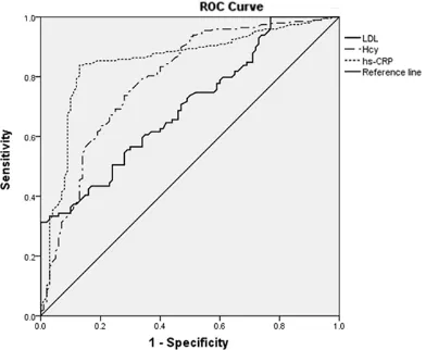
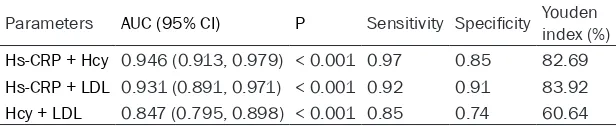
The combined detection performance of LDL, Hcy and hs-CRP
The ROC curves of three combined detection methods including Hcy + LDL, hs-CRP + LDL and hs-CRP + Hcy were calculated, and by
which the sensitivity and specificity of com -bined detections in the diagnosis of CHD were evaluated with the results of coronary angiog-raphy as the gold standard, as shown in Figure 2 and Table 6. The results showed that the three combined detection methods had risk
prediction value for CHD (P < 0.05). The area
under the curves of Hcy + LDL, hs-CRP + LDL and hs-CRP + Hcy were 0.847 (0.795, 0.898), 0.931 (0.891, 0.971) and 0.946 (0.913, 0.979),
respectively, and the detection efficiency of hs-CRP combined with Hcy was the highest (P < 0.05). The sensitivity and specificity of the
th-ree combined detection methods were
com-ed with Hcy had higher area under the ROC
curve, sensitivity and specificity o than the sin -gle detection of hs-CRP or Hcy separately, whi- ch had a better risk assessment value for the diagnosis of CHD.
Discussion
A study showed that high Hcy was an indepen-dent risk factor for coronary atherosclerotic heart disease, and elevated serum Hcy level
was significantly associated with an increased risk of CHD death (OR (95% CI): 1.66
(1.12-2.47)) [19]. Hcy cannot be synthesized in vivo and can only be generated by the hydrolysis of methionine [20]. Excessive intake of high
ani-mal protein diet or deficiency of methionine
metabolic cofactors such as folic acid, vitamin B6 and B12 can cause the metabolism disor-der of methionine and further lead to hyperho-mocysteinemia [21].
On the one hand, the increased Hcy in the blood had vascular toxicity. It produced a lot of free radicals through oxidative stress, which damaged vascular endothelial cells, activated platelets and promoted blood coagulation. On the other hand, Hcy could also inhibit the syn-thesis of nitric oxide and promote its degrada-tion, resulting in decreased endogenous vaso-active substances and abnormal vasodilatory function, thus promoting thrombosis and
lead-ing to inflammation and the generation of vas -cular wall plaques [22].
Hs-CRP was a kind of reactive protein in the
acute phase of inflammation and was one of
the risk predictors of cardiovascular events [23]. Hs-CRP can bind to lipoprotein, activate
the complement system, promote the infiltra
-tion of inflammatory cells and have other
im-mune regulatory effects, so as to produce a
large number of inflammatory mediators,
[image:5.612.90.403.85.149.2]rele-ase oxygen free radicals, and cause vascular in- jury and vasospasm, plaque formation, lumen Table 5. Single detection performance of LDL, Hcy and hs-CRP
Parameters AUC (95% CI) P Sensitivity Specificity index (%)Youden LDL (mmol/L) 0.690 (0.640, 0.759) < 0.001 0.56 0.69 25.06 Hcy (μmol/L) 0.791 (0.735, 0.848) < 0.001 0.76 0.79 55.76 Hs-CRP (mg/L) 0.850 (0.801, 0.899) < 0.001 0.84 0.70 54.84
Note: LDL, low density lipoprotein; Hcy, homocysteine, Hs-CRP, hypersensitive C-reactive
protein; AUC, area under the curve; CI, confidence interval.
Figure 2. ROC curve for combined detection of LDL, Hcy and hs-CRP. LDL: low density lipoprotein; Hcy: homocysteine; hs-CRP: hypersensitive C-reactive protein.
pared when the Youden index (sensitivity + spe-
cificity-1) was the maxi -mum. The sensitivity of hs-CRP combined with Hcy was the highest
[image:5.612.93.288.193.352.2]stenosis, and the occurrence of cardiovascular and cerebrovascular events [24].
Low density lipoprotein (LDL) is a transport car-rier for endogenous cholesterol in the blood [25]. LDL level was obviously positively
corre-lated with the incidence of CHD (OR (95% CI):
1.95 (1.31-2.90)), which may be due to the fact
that LDL can release various pro-inflammatory factors, recruit inflammatory response cells
and damage arterial endothelium [26]. Further- more, LDL can also promote excessive accumu-lation of cholesterol in the blood vessel wall, leading to decreased endothelial function and the formation of atherosclerotic plaques [27]. This study shows that the Hcy level of patients in the CHD group was markedly higher than
that in the healthy group, which confirmed the
close association between hyperhomocystein-emia and CHD. This result was basically consis-tent with the report of Yeh et al. on the risk ratio of CHD in patients with elevated Hcy level (OR
(95% CI): 1.29 (1.02-1.64)) [28]. Furthermore, the hs-CRP level of CHD group was significantly
higher than that of the healthy group, indicating that the test of hs-CRP level can provide labora-tory basis for the detection and prevention of CHD in high-risk patients, which was the same as Held et al. [29]. The LDL level of patients in the CHD group was clearly higher than that in the healthy group, suggesting that the detec-tion of LDL had certain diagnostic value for CHD, which was in line with the report of Puri et al. on the risk ratio of CHD in patients with
ele-vated LDL level (OR (95% CI): 1.30 (0.93-1.84))
[30].
From the perspective of the sensitivity of single detection, hs-CRP (0.84) > Hcy (0.76) > LDL (0.56). The sensitivity of hs-CRP to the risk as- sessment of CHD was much higher than that of other parameters, which probably because
C-reactive protein was a non-specific indica-
tor reflecting acute inflammatory reaction and
which may be inferred although the hs-CRP can
reflect the inflammatory response of the whole body, it lacks specificity and directivity.
Po-isoning, infection, autoimmune reaction, and other factors can cause the rise of hs-CRP
level. The specificity of Hcy (0.79) for the risk assessment of CHD was significantly higher
than that of other parameters, which may be related to the toxic effect of hyperhomocyste-ine on cardiovascular and cerebrovascular di- seases.
This study confirmed that the combined detec -tion of the two parameters was more valuable for the risk assessment of CHD, among which the detection of hs-CRP combined with Hcy had
the highest diagnostic benefit (ROC curve AUC
was 0.946 (0.913, 0.979)). From the perspec-tive of sensitivity, hs-CRP + Hcy (0.97) > hs-CRP + LDL (0.92) > Hcy + LDL (0.85). From the
per-spective of specificity, hs-CRP + LDL (0.91) >
hs-CRP + Hcy (0.85) > Hcy + LDL (0.74). Con-
sidering the sensitivity and specificity of the
detection parameters comprehensively, detec-tion of hs-CRP combined with Hcy has a better reference value for screening, diagnosis, pre-vention and risk assessment of CHD.
This study still has the following shortcomings. First, this study was a retrospective analysis, and there may be some bias in the inclusion of study subjects and the collection of clinical data. Second, the detection of venous plasma parameters was based on the kit provided by the biological company, and there were some technical problems and possible errors. In
addi-tion, this research lacked a stratified study on
[image:6.612.90.398.86.149.2]the subtype and severity of CHD, which will also be the focus of further studies. In future clinical observation, simultaneously combined detec-tion of hs-CRP and Hcy together with coronary angiography will be performed, combined with the comprehensive analysis of clinical charac-teristics of the patients, and conduct follow-up Table 6. Combined detection performance of LDL, Hcy and hs-CRP
Parameters AUC (95% CI) P Sensitivity Specificity Youden index (%) Hs-CRP + Hcy 0.946 (0.913, 0.979) < 0.001 0.97 0.85 82.69 Hs-CRP + LDL 0.931 (0.891, 0.971) < 0.001 0.92 0.91 83.92 Hcy + LDL 0.847 (0.795, 0.898) < 0.001 0.85 0.74 60.64
Note: LDL, low density lipoprotein; Hcy, homocysteine; Hs-CRP, hypersensitive C-reactive
protein; AUC, area under the curve; CI, confidence interval.
closely related to the occurrence, developm- ent and deterioration of CHD. From the perspe-
ctive of the specificity
to provide more technical support for the diag-nosis, treatment and prevention of CHD. In conclusion, LDL, Hcy and hs-CRP are related to CHD, and all of them can be used as predic-tors for patients with CHD. In risk assessment of CHD, compared with single parameter de- tection, combined detection of Hcy and hs-CRP
has good specificity and sensitivity, which is
more advantageous and worth promoting its use in clinical practice.
Disclosure of conflict of interest
None.
Address correspondence to: Zhen Liu, Department of Emergency, Shenzhen Bao’an Hospital of Tradi- tional Chinese Medicine (Group), No. 25 Yu’an Se- cond Road, Bao’an District, Shenzhen 518133, Guangdong Province, China. Tel: +86-0755-2962- 9333-8131; E-mail: liuzhen071@126.com
References
[1] Chapman AR, Adamson PD and Mills NL. As-sessment and classification of patients with myocardial injury and infarction in clinical practice. Heart 2017; 103: 10-18.
[2] White H, Thygesen K, Alpert JS and Jaffe A. Uni-versal MI definition update for cardiovascular disease. Curr Cardiol Rep 2014; 16: 492. [3] Moussa ID, Klein LW, Shah B, Mehran R, Mack
MJ, Brilakis ES, Reilly JP, Zoghbi G, Holper E, Stone GW; Society for Cardiovascular Angiogra-phy and Interventions. Consideration of a new definition of clinically relevant myocardial in-farction after coronary revascularization: an expert consensus document from the Society for Cardiovascular Angiography and Interven-tions (SCAI). Catheter Cardiovasc Interv 2014; 83: 27-36.
[4] Han C, Liu F, Yang X, Chen J, Li J, Cao J, Li Y, Shen C, Yu L, Liu Z, Wu X, Zhao L, Hu D, Lu X and Gu D. Ideal cardiovascular health and inci-dence of atherosclerotic cardiovascular dis-ease among Chinese adults: the China-PAR project. Sci China Life Sci 2018; 61: 504-514. [5] Younus A, Aneni EC, Spatz ES, Osondu CU,
Roberson L, Ogunmoroti O, Malik R, Ali SS, Aziz M, Feldman T, Virani SS, Maziak W, Agatston AS, Veledar E and Nasir K. A systematic review of the prevalence and outcomes of ideal car-diovascular health in US and non-US popula-tions. Mayo Clin Proc 2016; 91: 649-670. [6] Guo L and Zhang S. Association between ideal
cardiovascular health metrics and risk of car-diovascular events or mortality: a
meta-analy-sis of prospective studies. Clin Cardiol 2017; 40: 1339-1346.
[7] Guimaraes PO, Granger CB, Stebbins A, Chiswell K, Held C, Hochman JS, Krug-Gourley S, Lonn E, Lopes RD, Stewart RAH, Vinereanu D, Wallentin L, White HD, Hagstrom E and Danchin N. Sex differences in clinical charac-teristics, psychosocial factors, and outcomes among patients with stable coronary heart dis-ease: insights from the STABILITY (stabilization of atherosclerotic plaque by initiation of darap-ladib therapy) trial. J Am Heart Assoc 2017; 6. [8] Moore B, Yu C, Kotchetkova I, Cordina R and
Celermajer DS. Incidence and clinical charac-teristics of sudden cardiac death in adult con-genital heart disease. Int J Cardiol 2018; 254: 101-106.
[9] Thygesen K, Alpert JS, Jaffe AS and White HD. Diagnostic application of the universal defini-tion of myocardial infarcdefini-tion in the intensive care unit. Curr Opin Crit Care 2008; 14: 543-548.
[10] Vafaie M. State-of-the-art diagnosis of myocar-dial infarction. Diagnosis (Berl) 2016; 3: 137-142.
[11] Su H, Pei Y, Tian C, Zhang Q, Liu L, Meng G, Yao Z, Wu H, Xia Y, Bao X, Gu Y, Sun S, Wang X, Zhou M, Jia Q, Song K, Sun Z and Niu K. Rela-tionship between high-sensitivity C-reactive protein and subclinical carotid atherosclerosis stratified by glucose metabolic status in Chi-nese adults. Clin Cardiol 2019; 42: 39-46. [12] Ridker PM and Morrow DA. C-reactive protein,
inflammation, and coronary risk. Cardiol Clin 2003; 21: 315-325.
[13] Gao S, Zhao D, Wang M, Zhao F, Han X, Qi Y and Liu J. Association between circulating oxi-dized LDL and atherosclerotic cardiovascular disease: a meta-analysis of observational stu- dies. Can J Cardiol 2017; 33: 1624-1632. [14] Gao S and Liu J. Association between
circulat-ing oxidized low-density lipoprotein and athero-sclerotic cardiovascular disease. Chronic Dis Transl Med 2017; 3: 89-94.
[15] Liu C, Yang Y, Peng D, Chen L and Luo J. Hyper-homocysteinemia as a metabolic disorder pa-rameter is independently associated with the severity of coronary heart disease. Saudi Med J 2015; 36: 839-846.
[16] Di Minno MN, Tremoli E, Coppola A, Lupoli R and Di Minno G. Homocysteine and arterial thrombosis: challenge and opportunity. Th- romb Haemost 2010; 103: 942-961.
[17] Sun Y, Chien KL, Hsu HC, Su TC, Chen MF and Lee YT. Use of serum homocysteine to predict stroke, coronary heart disease and death in ethnic Chinese. 12-year prospective cohort study. Circ J 2009; 73: 1423-1430.
Rosenbaum L, Shaw LJ, Stainback RF and Al-len JM. ACCF/AHA/ASE/ASNC/HFSA/HRS/SC- AI/SCCT/SCMR/STS 2013 multimodality ap-propriate use criteria for the detection and risk assessment of stable ischemic heart disease: a report of the American college of cardiology foundation appropriate use criteria task force, American heart association, American society of echocardiography, American society of nu-clear cardiology, heart failure society of Ameri-ca, heart rhythm society, society for cardiovas-cular angiography and interventions, society of cardiovascular computed tomography, society for cardiovascular magnetic resonance, and society of thoracic surgeons. J Am Coll Cardiol 2014; 63: 380-406.
[19] Peng HY, Man CF, Xu J and Fan Y. Elevated ho-mocysteine levels and risk of cardiovascular and all-cause mortality: a meta-analysis of pro-spective studies. J Zhejiang Univ Sci B 2015; 16: 78-86.
[20] Chen KJ, Pan WH, Yang FL, Wei IL, Shaw NS and Lin BF. Association of B vitamins status and homocysteine levels in elderly Taiwanese. Asia Pac J Clin Nutr 2005; 14: 250-255. [21] Debreceni B and Debreceni L. The role of
ho-mocysteine-lowering B-vitamins in the primary prevention of cardiovascular disease. Cardio-vasc Ther 2014; 32: 130-138.
[22] Katsiki N, Perez-Martinez P and Mikhailidis DP. Homocysteine and non-cardiac vascular dis-ease. Curr Pharm Des 2017; 23: 3224-3232. [23] Yahagi K, Kolodgie FD, Lutter C, Mori H,
Rome-ro ME, Finn AV and Virmani R. Pathology of hu-man coronary and carotid artery atherosclero-sis and vascular calcification in diabetes me- llitus. Arterioscler Thromb Vasc Biol 2017; 37: 191-204.
[24] Koenig W. High-sensitivity C-reactive protein and atherosclerotic disease: from improved risk prediction to risk-guided therapy. Int J Car-diol 2013; 168: 5126-5134.
[25] Yoshida H. Front line of oxidized lipoproteins: role of oxidized lipoproteins in atherogenesis and cardiovascular disease risk. Rinsho Byori 2010; 58: 622-630.
[26] Simons LA, Simons J, Friedlander Y and McCal-lum J. LDL-cholesterol predicts a first CHD event in senior citizens, especially so in those with elevated lipoprotein (a): dubbo study of the elderly. Heart Lung Circ 2018; 27: 386-389.
[27] Dong Y, Fernandes C, Liu Y, Wu Y, Wu H, Brophy ML, Deng L, Song K, Wen A, Wong S, Yan D, Towner R and Chen H. Role of endoplasmic re-ticulum stress signalling in diabetic endotheli-al dysfunction and atherosclerosis. Diab Vasc Dis Res 2017; 14: 14-23.
[28] Yeh JK, Chen CC, Hsieh MJ, Tsai ML, Yang CH, Chen DY, Chang SH, Wang CY, Lee CH and Hsieh IC. Impact of homocysteine level on long-term cardiovascular outcomes in patients after coronary artery stenting. J Atheroscler Thromb 2017; 24: 696-705.
[29] Held C, White HD, Stewart RAH, Budaj A, Can-non CP, Hochman JS, Koenig W, Siegbahn A, Steg PG, Soffer J, Weaver WD, Ostlund O and Wallentin L. Inflammatory biomarkers interleu-kin-6 and C-reactive protein and outcomes in stable coronary heart disease: experiences from the STABILITY (stabilization of atheroscle-rotic plaque by initiation of darapladib therapy) trial. J Am Heart Assoc 2017; 6.