Original Article
miR-486 inhibits Gli1 expression and attenuates
epithelial mesenchymal transition
and invasion of prostate cancer
Derong Zhou, Hao Lin, Xiaohong Chen, Junhong Zheng
Department of Urology, The Second Affiliated Hospital of Shantou University Medical College, Shantou 515000, Guangdong, China
Received April 29, 2019; Accepted July 12, 2019; Epub September 15, 2019; Published September 30, 2019
Abstract: Abnormal expression of glioma-associated oncogene protein 1 (Gli1) is associated with various tumors. Studies have found that decreased miR-486 expression is associated with prostate cancer. Bioinformatics analysis revealed that miR-486 has a complementary binding site in the 3’-UTR region of Gli1 mRNA. This study investigated whether miR-486 plays a role in regulating Gli1 expression and affects epithelial mesenchymal transition (EMT) and invasion of prostate cancer cells. The dual luciferase reporter gene assay validated the targeted regulation between miR-486 and Gli1. qRT-PCR was used to measure the expression of miR-486 and Gli1 mRNA. The Gli1 protein expression was detected by western blot. PC3M cells were cultured in vitro and divided into two groups: miR-NC group and miR-486 mimic group. The expressions of Gli1, E-cadherin and N-cadherin were detected, and the cell invasion ability was detected by Transwell assay. There was a targeted regulatory relationship between miR-486
and Gli1 mRNA. Compared with RWPE-1 cells, miR-486 expression was significantly decreased and Gli1 expres
-sion was significantly increased in PC3 and PC3M cells, and both of them were increased in high metastatic PC3M cells. Transfection of miR-486 mimic significantly down-regulated the expression of Gli1 and N-cadherin in PC3M
cells, which increased E-cadherin expression, inhibited EMT and decreased the invasive ability. The decreased expression of miR-486 plays a role in up-regulating Gli1 expression, promotes EMT and invasion of prostate cancer cells. Increasing miR-486 expression can inhibit Gli1 expression and attenuate the EMT process and the invasion of prostate cancer cells.
Keywords: miR-486, Gli1, prostate cancer, EMT, invasion
Introduction
Prostate cancer (PC) refers to epithelial malig-nant tumors that occur in the prostate. In recent years, the incidence of prostate cancer in China has been increasing year by year and has ranked sixth in the incidence of male malig-nant tumors, and third place in male urinary tumors [1, 2].
The Hedgehog signaling pathway is widely expressed in various tissues and cells and is involved in the regulation of various biological processes such as cell proliferation, apoptosis, migration and invasion [3-5]. Glioma-associated oncogene protein 1 (Gli1) is an important trans-regulatory factor in the Hedgehog signaling pathway, which can regulate the expression of a variety of genes, thereby affecting cell
prolif-eration, differentiation, migration, etc., and plays a promoting role in the occurrence of tumors [13] or the role of the tumor suppressor genes [14]. In recent years, studies have found that abnormal expression of miR-486 is associ-ated with prostate cancer [15]. Bioinformatics analysis showed that miR-486 has a target-complement relationship with Gli1’s 3’-UTR. This study investigated whether miR-486 regu-lates Gli1 expression and affects the EMT pro-cess and invasion ability of prostate cancer cells.
Materials and methods
Main reagents and materials
Highly metastatic human prostate cancer cells PC3M, low metastatic prostate cancer cells PC3 were purchased from Nanjing Kezhen cell bank; HEK293T cells were purchased from Wuhan Punosi organism; human normal pros-tate epithelial RWPE-1 cells were purchased from Hunan Fenghui organism; DMEM culture Base, fetal bovine serum (FBS), Opti-MEM and penicillin were purchased from Gibco (USA); Lip 2000 was purchased from Invitrogen (USA);
RNA extraction reagent EasyPure RNA Kit, fluo -rescent quantitative PCR reagent TransScript Green One-Step qRT-PCR SuperMix was pur-chased In Beijing, the whole-type golden biolo-gy; miR-NC, miR-486 mimic was designed and synthesized by Guangzhou Ruibo Bio; rabbit
anti-E-cadherin, N-cadherin, Gli1, β-actin poly -clonal antibody were purchased from American Abcam; HRP coupled two The anti-purchase was purchased from Shanghai Biotech; the Transwell cell was purchased from Corning (USA); the Matrigel gel was purchased from BD biosciences; the Dual-Luciferase Reporter Assay System, and the pGL3 plasmid was pur-chased from Promega (USA); the recombinant
TGF-β1 protein was purchased from Peprotech
(USA).
Cell culture and EMT induction
PC3M, PC3 and RWPE-1 cells were cultured in DMEM medium containing 10% FBS in an incu-bator containing 5% CO2 at 37°C, subcultured at a ratio of 1:4, and harvested in logarithmic phase for the experiment.
In the EMT induction experiment, PC3M cells were inoculated into 6-well plates at a density
of 3 × 10⁴. After adherence for 24 h, TGF-β1 at
a final concentration of 10 ng/mL was added
to the medium for 48 h to induce EMT. Conventional cultured cells induced without the
addition of TGF-β1 were used as controls.
Dual luciferase gene reporter assay
Using the PC3M cell genome as a template, the full-length 3’-UTR fragment of Gli1 gene was
amplified, and the PCR product was digested. The amplified product was ligated into the lucif -erase reporter vector pGL3 and transformed
into DH5α competent cells followed by screen -ing positive clones by colony PCR to select the correct sequencing plasmid for transfection cells and subsequent experiments, which were named as pGL3-Gli1-WT, pGL3-Gli1-MUT. pGL3-Gli1-WT (or pGL3-Gli1-MUT) was trans-fected into HEK293T cells with miR-486 mimic (or miR-NC) using Lipo2000. After 48 h of cul-ture, dual luciferase assay was performed fol-lowing the instructions of Dual-Glo Luciferase Assay System Kit.
Cell transfection and grouping
PC3M cells were cultured in vitro and divided into two groups: miR-NC transfection group and miR-486 mimic transfection group. The general procedure for transfection was as follows: 10
μL of Lip 2000, 50 nmol miR-NC, 50 were dilut
-ed with 100 μL Opti-MEM. Nmol miR-486 mimic
incubated for 5 min at room temperature, mixed Opti-MEM with Lip 2000, NC or miR-486 mimic, incubated for 20 min at room tem-perature, added the transfectant mixture to the cell culture medium, continued to culture 72 hours followed by collection of cells for experiments.
qRT-PCR detection of gene expression
The RNA extracted using the EasyPure RNA Kit was subjected to one-step qRT-PCR to detect the relative expression of the gene using TransScript Green One-Step qRT-PCR SuperMix
40 cycles, detection of gene expression on Bio-Rad CFX96 real-time PCR instrument. The prim-er sequence was: miR-486-Forward-5’-TGGG- ATCCATGAGGAAGGGACATGAAGA-3’; miR-486- Reverse-5’-ACCGAAGCTTAAAAAAGCTCGGTCCC AGAGTCAG-3’; U6-Forward-5’-CTCGCTTCGGC- AGCACA-3’; U6-Reverse-5’-AACGCTTCACGAAT- TTGCGT-3’; GLI-1 (forward) 5’-CTCCCGAAGG- ACAGGTATGTAAC-3’ and (reverse) 5’-CCCTAC- TCTTTAGGCACTAGAGTTG-3’; GAPDH (forward) 5’-CACCACCAACTGCTTAG-3’ and (reverse) 5’- CTTCACCACCTTCTTGATG-3’; E-cadherin (For- ward) 5’-CAGAAAGTTTTCCACCAAAG-3’ and (reverse) 5’-ACTGAACCTGACCGTACAAAATGTG- AGCAATTCTGCTT-3’.
Western blot
The total protein was extracted using RIPA lysis buffer, and the mass concentration was deter-mined by BCA method according to the kit instructions. After adding 3 times the volume of 4 × protein loading buffer, it was boiled for 5
min, and 40 μg was loaded, and separated on
8-10% SDS-PAGE followed by being transferred to PVDF membrane (300 mA, 90 min), blocked with 5% skim milk powder for 60 min at room temperature, and incubated with the primary
antibody at 4°C overnight (E-cadherin, N- The
dilution ratios of cadherin, Gli1, and β-actin
were 1:2000, 1:2000, 1:800, 1:5000, respec-tively). After that, the membrane was washed 3 times with PBST, and then HRP-conjugated sec-ondary antibody (1:10000 ratio dilution) was added and incubated for 60 min at room tem-perature. After washing the membrane 3 times with PBST, added ECL luminescence solution
for 2~3 min, exposure, developed, fixed in the dark room, scaned the film and saved the data.
Transwell assay analysis of cell invasion
100 μL of Matrigel gel was placed on the upper surface of the Transwell chamber filter and
incubated in a 37°C cell incubator for 30 min to allow complete polymerization. We added 500
μL of complete medium containing 10% FBS to
a 24-well plate, placed the Transwell chamber
in a 24-well plate, and added 200 μL of PC3M
cells resuspended in serum-free DMEM medi-um to the upper chamber to continue the cul-ture. After 48 h, the medium in the upper cham-ber of Transwell was discarded, and the cells that failed to pass were wiped off with a sterile
cotton swab. After fixation with methanol and
crystal violet, the microscope was inverted (manufactured by Olympus, model: IX51) and counted. The number of perforated cells in
each field of view was calculated and the aver -age value was calculated.
Statistical analysis
Statistical analysis was performed using SPSS 18.0. The measurement data were expressed as mean ± standard deviation (SD). The com-parison between the measurement data of the groups was analyzed by student t test. P < 0.05
was considered statistically significant. Results
A targeted regulation relationship between miR-486 and Gli1
Bioinformatics analysis revealed a complemen-tary binding site between miR-486 and the 3’-UTR of Gli1 mRNA (Figure 1A). The dual lucif-erase gene reporter assay showed that
trans-fection of miR-486 mimic significantly reduced
the relative luciferase activity in pGL3-Gli1-WT-transfected HEK293T cells, while transfection of miR-NC or miR-486 mimic did not have a sig-Figure 1. Dual luciferase reporter gene assay
analy-sis of the targeted regulation relationship between miR-486 and Gli1. A. Schematic diagram of a target-ed binding site between miR-486 and Gli1. B. Dual luciferase reporter gene assay. *P < 0.05 compared
[image:3.612.88.286.78.301.2]nificant effect on the relative luciferase activity
in HEK293T cells transfected with pGL3-Gli1-MUT (Figure 1B), indicating a targeted regula-tory relationship between miR-486 and Gli1 mRNA.
Abnormal expression of Gli1 and miR-486 in prostate cancer cells
The results of qRT-PCR showed that the expres-sion of Gli1 mNRA in prostate cancer PC3 and
PC3M cells was significantly higher than that in
human normal prostate epithelial RWPE-1 cells
EMT process and invasion of PC3M cells
The qRT-PCR results showed that the
expres-sion of miR-486 was significantly increased in
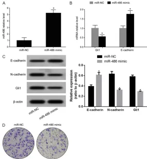
PC3M cells in the miR-486 mimic group com-pared with the miR-NC group (Figure 4A), and
the expression of Gli1 mRNA was significantly
decreased (Figure 4B). The expression of
E-cadherin mRNA was significantly increased
(Figure 4B). Western blot analysis showed that compared with NC, transfection of
miR-486 mimic significantly decreased the expres -sion of Gli1 and N-cadherin proteins in PC3M Figure 2. Abnormal expression of Gli1 and miR-486 in prostate cancer cells,
and is related to cell transfer ability. A. qRT-PCR detection of intracellular Gli1 mRNA expression. B. qRT-PCR detection of intracellular miR-486 ex-pression. C. Western blot analysis of intracellular Gli1 protein exex-pression.
*P < 0.05 compared with RWPE-1 cells; #P < 0.05 compared to PC3 cells.
Figure 3. Abnormal changes in the expression of miR-486 and Gli dur-ing EMT in PC3M cells. A. qRT-PCR detection of E-cadherin mRNA ex-pression in PC3M cells. B. qRT-PCR detection of miR-486 expression in PC3M cells. C. qRT-PCR detection of Gli1 mRNA expression in PC3M cells. *P < 0.05 compared to the
Control group.
(Figure 2A), while the
expres-sion of miR-486 was signifi -cantly decreased (Figure 2B). The expression of Gli1 mNRA in high metastatic PC3M cells was higher than that in low metastatic PC3 cells, while the expression of miR-486 was lower than that in low metastatic PC3 cells. Western blot analysis showed that the expression of Gli1 protein in human normal prostate epi-thelial RWPE-1 cells, prostate cancer PC3 and PC3M cells
was significantly decreased,
and the expression of Gli1 pro-tein in PC3M cells was lower than that in PC3 cells (Figure 2C).
Abnormal expression of miR-486 and Gli in EMT of PC3M cells
The results of qRT-PCR sh- owed that the expression of E-cadherin mRNA was
sig-nificantly down-regulated dur -ing the EMT of PC-3M cells
induced by TGF-β1 treatment,
indicating that the epithelial properties were reduced (Fi- gure 3A). Meanwhile, the ex- pression of miR-486 was
sig-nificantly decreased during
the EMT of PC-3M cells in-
duced by TGF-β1 treatment
(Figure 3B), and the expres-sion of Gli1 mRNA (Figure 3C)
was significantly increased.
cells, and significantly increased the expres -sion of E-cadherin protein (Figure 4C). Transwell assays showed that the invasive ability of PC3M cells in the miR-486 mimic transfection group
was significantly reduced compared to the
miR-NC group (Figure 4D).
Discussion
Prostate cancer has the characteristics of high malignancy, rapid progress and poor therapeu-tic effect, low survival rate and unsatisfactory prognosis. Therefore, studying the abnormal changes in the pathogenesis of prostate
can-cer is of great significance in improving the
diagnosis, the therapeutic effect, the survival rate as well as the prognosis.
Under physiological conditions, the Hedgehog signaling pathway is usually in an inactive form or has low functional activity. When the
[image:5.612.87.373.71.381.2]abolishes the inhibitory effect of Ptch on Smo, which in turn invalidates the degradation of Gli and enters the nucleus in full length to promote the transcription and expression of target genes, thereby promoting cell proliferation, migration, malignant transformation, leading to tumors [20]. The abnormal expression and function of Gli1 is associated with abnormal Hedgehog signaling pathway, and plays an important role in the occurrence and progres-sion of several tumors such as breast cancer [9], cervical cancer [10], and lung cancer [11]. Studies have found that elevated expression and functional activity of Gli1 is associated with prostate cancer [12]. A number of studies have shown that the expression of miR-486 is decreased in multiple tumors such as non-small cell lung cancer [21], colorectal cancer [22], cervical cancer [23]. In recent years, stud-ies have found that abnormal expression of miR-486 is associated with prostate cancer Figure 4. Elevated miR-486 reduces Gli1 expression and inhibits EMT
pro-cess and invasion of PC3M cells. A. qRT-PCR detection of miR-486 expres-sion in PC3M cells. B. qRT-PCR was used to detect the expresexpres-sion of Gli1 and E-cadherin mRNA in PC3M cells. C. Western blot detection of protein expres-sion. D. Transwell assay to detect cell invasion ability. *P < 0.05 compared to
their corresponding miR-NC.
[15]. This study investigated whether miR-486 plays a role in regulating Gli1 expression and affecting the EMT process and invasion ability of prostate cancer cells.
In this study, the dual luciferase gene reporter assay showed that transfection of miR-486
mimic significantly reduced the relative lucifer -ase activity in pGL3-Gli1-WT-transfected
HEK293T cells, but did not have significant
effect on the relative luciferase activity in pGL3-Gli1-MUT-transfected HEK293T cells,
confirming the targeted regulatory relationship
between miR-486 and Gli1. The results of this study showed that compared with human nor-mal prostate epithelial RWPE-1 cells, the expression of miR-486 in prostate cancer PC3
and PC3M cells was significantly decreased, while the expression of Gli1 was significantly
increased. The abnormal expression of
miR-486 and Gli1 was significantly higher in highly
metastatic PC3M cells than that in low meta-static PC3 cells. The results showed that the decreased expression of miR-486 may play a role in up-regulating Gli1 expression and pros-tate pathogenesis, and this regulation is involved in the regulation of prostate cancer metastasis. Epithelial mesenchymal transition (EMT) is a biological process in which epithelial cells are transformed into mesenchymal cells, which mediate tight junctions between cell-cells and cell-cells-extracellular matrix (E-cadherin). Decreased expression of cadherin is an impor-tant marker of EMT process. The EMT process of tumor cells is closely related to tumor pro-gression, metastasis, recurrence and poor
prognosis [24, 25]. In this study, TGF-β1 was
administered to PC3M cells to induce EMT in PC3M cells, and the expression of miR-486
was significantly decreased in EMT, while the expression of Gli1 was significantly increased, further confirming the targeted regulation
between miR-486 and Gli1 is involved in the regulation of EMT processes in prostate cancer PC3M cells. In the study of the relationship between miR-486 and prostate cancer, the sequencing results of Song et al [26] showed that the expression of miR-486 in prostate
can-cer patients was significantly lower than that of
benign prostatic hyperplasia, and the results of
qRT-PCR further confirmed the sequencing test
results. The results of Zhang et al [15] showed that compared with non-metastatic prostate cancer LNCaP cells, the expression of miR-486
in metastatic prostate cancer C4-2 cells was
significantly reduced. Compared with in situ prostate cancer tumor tissue, the expression of miR-486 in the metastatic prostate cancer
tis-sue was significantly reduced. In this study, the
decreased expression of miR-486 is associat-ed with prostate cancer and high metastatic characteristics, consistent with the results of Song et al [26], and Zhang et al [15].
In this study, miR-486 mimic was further trans-fected into prostate cancer PC3M cells to observe changes in cell EMT and invasion abil-ity. The results showed that transfection of miR-486 mimic in prostate cancer PC3M cells
significantly decreased the expression of Gli,
increased the expression of E-cadherin, decre- ased the expression of N-cadherin, and inhibit-ed the cell EMT process. The ability of cells to
invade was also significantly inhibited. Zhang et
al [15] showed that the expression of miR-486 in high-metastasis prostate cancer C4-2 cells can inhibit the expression of target gene Snail and inhibit the EMT process of C4-2 cells. The
ability to migrate and invade was significantly
weakened. In this study, the effect of miR-486 expression in prostate cancer cells was attenu-ated by decreasing the expression of miR-486 in prostate cancer cells, consistent with the results of Zhang et al [15]. This study combines the targeted regulatory relationship between miR-486 and Gli1, revealing that decreased expression of miR-486 plays a role in up-regu-lating Gli1 expression and promoting EMT and invasion of prostate cancer cells, while increas-ing miR-486. The expression can inhibit the expression of Gli1 by targeting, and attenuate the EMT process and invasion ability of pros-tate cancer cells, which has not been reported in previous studies, and is the innovation of this study. Whether MiR-486 regulates Gli1 is relat-ed to prostate cancer invasion in human body is unclear and requires further investigations in the future.
Conclusion
Disclosure of conflict of interest
None.
Address correspondence to: Dr. Derong Zhou, De-
partment of Urology, The Second Affiliated
Hospi-tal of Shantou University Medical College, Dongxia North Road, Shantou 515000, Guangdong, China. Tel: +86-0754-88915666; Fax: +86-0754-88346- 543; E-mail: [email protected]
References
[1] Fenner A. Prostate cancer: ERSPC calculator recalibrated for China. Nat Rev Urol 2017; 14: 66.
[2] Ren SC, Chen R and Sun YH. Prostate cancer research in China. Asian J Androl 2013; 15: 350-353.
[3] Liu Y, Gao S, Zhu J, Zheng Y, Zhang H and Sun H. Dihydroartemisinin induces apoptosis and inhibits proliferation, migration, and invasion in epithelial ovarian cancer via inhibition of the hedgehog signaling pathway. Cancer Med 2018; 7: 5704-5715.
[4] Xu Z, Huang C and Hao D. MicroRNA-1271 in-hibits proliferation and promotes apoptosis of multiple myeloma cells through inhibiting smoothened-mediated Hedgehog signaling pathway. Oncol Rep 2017; 37: 1261-1269. [5] Bai Y, Chen B, Hong W, Liang Y, Zhou M and
Zhou L. Sedum sarmentosum Bunge extract induces apoptosis and inhibits proliferation in pancreatic cancer cells via the hedgehog sig-naling pathway. Oncol Rep 2016; 35: 2775-2784.
[6] Wang K, Chen D, Qian Z, Cui D, Gao L and Lou M. Hedgehog/Gli1 signaling pathway regulates MGMT expression and chemoresistance to te-mozolomide in human glioblastoma. Cancer Cell Int 2017; 17: 117.
[7] Lin Z, Sheng H, You C, Cai M, Zhang Y, Yu LS, Yu X, Lin J and Zhang N. Inhibition of the CyclinD1 promoter in response to sonic hedgehog sig-naling pathway transduction is mediated by Gli1. Exp Ther Med 2017; 13: 307-314. [8] Cai X, Yu K, Zhang L, Li Y, Li Q, Yang Z, Shen T,
Duan L, Xiong W and Wang W. Synergistic inhi-bition of colon carcinoma cell growth by hedge-hog-Gli1 inhibitor arsenic trioxide and pho- sphoinositide 3-kinase inhibitor LY294002. Onco Targets Ther 2015; 8: 877-883.
[9] Villegas VE, Rondon-Lagos M, Annaratone L, Castellano I, Grismaldo A, Sapino A and Zaphi-ropoulos PG. Tamoxifen treatment of breast cancer cells: impact on hedgehog/GLI1 signal-ing. Int J Mol Sci 2016; 17: 308.
[10] Nayak A, Satapathy SR, Das D, Siddharth S, Tripathi N, Bharatam PV and Kundu C.
Nano-quinacrine induced apoptosis in cervical can-cer stem cells through the inhibition of hedge-hog-GLI1 cascade: role of GLI-1. Sci Rep 2016; 6: 20600.
[11] Li X, Zhou X, Fan Y, Zhang Y, Zu L, Yao F and Zhou Q. WW45, a Gli1 binding protein, nega-tively regulated hedgehog signaling in lung cancer. Oncotarget 2016; 7: 68966-68975. [12] Zhang L, Li L, Jiao M, Wu D, Wu K, Li X, Zhu G,
Yang L, Wang X, Hsieh JT and He D. Genistein inhibits the stemness properties of prostate cancer cells through targeting hedgehog-Gli1 pathway. Cancer Lett 2012; 323: 48-57. [13] Li C, Deng L, Zhi Q, Meng Q, Qian A, Sang H, Li
X and Xia J. MicroRNA-183 functions as an on-cogene by regulating PDCD4 in gastric cancer. Anticancer Agents Med Chem 2016; 16: 447-455.
[14] Qiu ZA and He GP. MicroRNA-134 functions as a tumor suppressor gene in gastric cancer. Am J Transl Res 2016; 8: 4320-4328.
[15] Zhang X, Zhang T, Yang K, Zhang M and Wang K. miR-486-5p suppresses prostate cancer metastasis by targeting snail and regulating epithelial-mesenchymal transition. Onco Tar-gets Ther 2016; 9: 6909-6914.
[16] Chakrabarti J, Holokai L, Syu L, Steele NG, Chang J, Wang J, Ahmed S, Dlugosz A and Zav-ros Y. Hedgehog signaling induces PD-L1 ex-pression and tumor cell proliferation in gastric cancer. Oncotarget 2018; 9: 37439-37457. [17] Nedjadi T, Salem N, Khayyat D, Sayyad A,
Al-Ammari A and Al-Maghrabi J. Sonic hedgehog expression is associated with lymph node inva-sion in urothelial bladder cancer. Pathol Oncol Res 2019; 25: 1067-1073.
[18] Liu X, Liu J, Zhang X, Tong Y and Gan X. MiR-520b promotes the progression of non-small cell lung cancer through activating hedgehog pathway. J Cell Mol Med 2019; 23: 205-215. [19] Yu Y, Cheng L, Yan B, Zhou C, Qian W, Xiao Y,
Qin T, Cao J, Han L, Ma Q and Ma J. Overex-pression of Gremlin 1 by sonic hedgehog sig-naling promotes pancreatic cancer progres-sion. Int J Oncol 2018; 53: 2445-2457. [20] Gu D, Schlotman KE and Xie J. Deciphering the
role of hedgehog signaling in pancreatic can-cer. J Biomed Res 2016; 30: 353-360. [21] Yu S, Geng S and Hu Y. miR-486-5p inhibits cell
proliferation and invasion through repressing GAB2 in non-small cell lung cancer. Oncol Lett 2018; 16: 3525-3530.
[22] Zhang Y, Fu J, Zhang Z and Qin H. miR-486-5p regulates the migration and invasion of colorectal cancer cells through targeting PI-K3R1. Oncol Lett 2018; 15: 7243-7248. [23] Ye H, Yu X, Xia J, Tang X, Tang L and Chen F.
[24] Matysiak M, Kapka-Skrzypczak L, Jodlowska-Jedrych B and Kruszewski M. EMT promoting transcription factors as prognostic markers in human breast cancer. Arch Gynecol Obstet 2017; 295: 817-825.
[25] Zhou P, Li B, Liu F, Zhang M, Wang Q, Liu Y, Yao Y and Li D. The epithelial to mesenchymal tran-sition (EMT) and cancer stem cells: implication for treatment resistance in pancreatic cancer. Mol Cancer 2017; 16: 52.
[26] Song C, Chen H, Wang T, Zhang W, Ru G and