THE REGULATION OF OSMOTIC PRESSURE AND
CHLORIDE CONCENTRATION IN THE
HAEMO-LYMPH OF MOSQUITO LARVAE
BY V. B. WIGGLESWORTH
From the Laboratory of Zoophysiology, Copenhagen University
(Received 10 August 1937)
(With Six Text-figures)
IN an earlier paper (Wigglesworth, 19336) it was shown that the cuticle of the mosquito larva Aedes aegypti (= argenteus) is permeable to water only at the anal papillae, and that water is taken in continuously by these organs and excreted by the Malpighian tubules; observations which were confirmed by Pagast (1936). But a more important function of the anal papillae has recently been made clear by Koch (1938), who has shown that in larvae of CMronomus and Culex they absorb chloride ions. They are able to take up chloride from very dilute solutions (o-ooi M) and behave in this respect like the skin of frogs and other fresh-water animals as demonstrated by Krogh (1937).
The extent to which this function is called into play will depend upon the concentration of chloride in the external medium. One might therefore expect it to be better developed in a species like Aedes aegypti which breeds in small collections of rain water in rot-holes in trees, coconut husks and such like, than in Culex
pipiens which breeds often in contaminated water, in small pools, rain-water
barrels, cesspits. The first object of the present work was therefore to compare the capabilities of these two species for absorbing chloride and to see how far this function is affected when the chloride content of the medium is changed.
In another paper (Wigglesworth, 1933c) the adaptation of Aedes larvae to high concentrations of salt was studied, but no direct observations were made on the changes in composition of the blood. The second object of the present work was, therefore, to follow the changes in chloride concentration and total osmotic pressure in the haemolymph of larvae exposed to different media and to relate these changes with the movements of fluid in the tracheal endings.
METHODS
The larvae, Culex pipiens L. race autogemcus1 and Aedes aegypti L., were kept at 260 C. and fed on powdered dog biscuit. For the collection of haemolymph the larva was dried on filter paper, placed on a slide coated with paraffin, punctured with a needle and the haemolymph (o-3-O"4 mm.3) drawn up into a fine capillary pipette lined with paraffin.
1
The osmotic pressure of the haemolymph of single larvae was measured by Baldes' modification of Hill's thermo-electric method for vapour pressure (Baldes, 1934), the haemolymph being placed on one thermocouple, a known concentration of NaCl on the other and the wall of the chamber containing the thermocouples moistened with 0-9 % NaCl. The apparatus was set up by Dr E. Hohwii-Christensen, to whom I am indebted for instruction in its use. The temperature of the room was kept constant within o-i at 20 ° C , the metal chamber containing the thermo-couples being immersed in water in a Dewar flask at the same temperature. The galvanometer, made by Mr A. C. Downing of University College, London, had an ampere sensitivity per mm. at 2 m., with zero resistance, of r i x 10-9. When working well this instrument would detect differences in osmotic pressure equivalent to 0-002 % NaCl. But the values are given throughout only to the nearest o-oi % •
The haemolymph of larvae near pupation quickly blackens on exposure to the air. During blackening the apparent osmotic pressure falls to an extent which is greater the more pigment is formed. In some cases the change may be equivalent to 0-07 % NaCl. This phenomenon is probably the result of heat production during oxidation. But blackening is complete and equilibrium is reached in 15-20 min., and in all cases it is this equilibrium value which is recorded.
For the estimation of chloride a method was devised by which a Volhard titration, either with or without removal of the silver chloride by filtration, could be applied to the haemolymph of a single larva (0-3 mm.3). This is described elsewhere (Wigglesworth, 1937).
Both osmotic pressure and chloride concentration are expressed throughout by the equivalent concentration of NaCl in g. per 100 c.c.
NORMAL OSMOTIC PRESSURE AND CHLORIDE CONTENT
The osmotic pressure of the haemolymph of normal larvae in tap water varies between 0-75 and 0-89. The higher values are given by larvae that are abundantly fed, especially towards the time of pupation. The effect of starvation on osmotic pressure is shown in Fig. 1. In this experiment a large batch of Aedes larvae was reared in distilled water containing a rich infusion of Protozoa derived from the powdered dog biscuit. Within a few hours of moulting to the fourth or final stage the larvae were divided into two lots and transferred to clean tap water1 and distilled water respectively which were changed daily. In both media there was a rapid fall in osmotic pressure during the first 4-5 day9. The value then remained constant at 0-65-0-70—consistently a little higher in tap water. A similar effect of starvation on osmotic pressure was observed by Fritzche (1917) in Daphnia.
The chloride in the blood of larvae reared in tap water ranges in both Aedes and Culex from 0*22 to 0-34, the mean of forty-four determinations in Aedes being 0-30 and the mean of thirty-two determinations in Culex being 0-28. For whole
Culex larvae, including cuticle and gut contents, Koch (1938) gives a value of 0-16.
In the experiment shown in Fig. 1 the larvae had been reared in distilled water so that at the outset the chloride (derived from traces in the food) was abnormally 1
low. On transfer to tap water the chloride at once rose (at first even a little above the normal level) and was then held constant until the larvae died of starvation. The larvae in distilled water lost chloride steadily to about 0-05. This low level was maintained until death. The larvae began to die at the end of 10 days and the death-rate was greater in distilled water. But it is clear that the loss of chloride was not the cause of death, for the lowest level in the larvae in distilled water was
reached at the end of 6 days.
08
07
-0-6
0-5
0-4
0-3
0-2
01
-0 I 5 6 7 8 9 10 II 12
Fig. 1. Effect of starvation in tap water and distilled water on osmotic pressure and chloride in Atda larvae. Ordinate: values expressed as equivalent concentrations of NaCl in g. per ioo c.c. Abscissa: days. Upper curves, total osmotic pressure of blood; lower curves, blood chloride.
These experiments confirm the ability of mosquito larvae to absorb chloride from dilute solutions as demonstrated by Koch (1938). It is well known that in the blood of most insects only a relatively small part of the total osmotic pressure is due to chlorides (Portier & Duval, 1927); amino acids are believed to be the most important constituent (Duval et al. 1928; Florkin, 1937). The foregoing experiment shows that the non-chloride fraction is subject to regulation in such a way as to compensate for the changes in chloride content. For it is evident in Fig. 1 that the curves for chloride deviate much more than the curves for total osmotic pressure. Thus on the seventh day of experiment:
3 V. B. WlGGLESWORTH
OSMOTIC PRESSURE AND CHLORIDE CONTENT OF LARVAE REARED IN VARYING CONCENTRATIONS OF SALT
For studying the effect of salt in the external medium a balanced salt solution was used in which the cations were in about the same proportions as in sea water (NaCl, 2-83%; KC1, 0-076%; MgClt) 0-50%; CaCl,, 0-122%). This stock
solution was diluted with varying amounts of distilled water to give the range of concentrations required. The higher concentrations were obtained by rearing the larvae at 0-9-1 -o % and then allowing the medium to evaporate slowly (with frequent mixing) so as to enable the larvae to become adapted as previously described (Wigglesworth, 1933c). The chloride content of each medium was measured by titration, the osmotic pressure by the same method as the blood of the larvae.
[image:4.451.45.407.235.424.2]0 0-1 0-2 O3 0-4 0-5 0-6 0-7 0-8 O9 1-0.1-1 1-2 1-3 1-4 1-5 1-6 0 0-1 02 03 04 0-5 CK> O7 08 09 10 l-l 1-2 1-3 1-4 1
Fig. 2. Relation between osmotic pressure of the medium and of the blood (upper curves), and between the chloride content of the medium and of the blood (lower curves) in Aeda larvae (A) and Culex larvae (B). Ordinates: values in the blood; abscissae: values in the medium, both expressed as equivalent concentrations of NaCl in g. per ioo c.c. The straight line shows where the points would fall if blood and medium had the same composition.
drawn through the lowest chloride concentrations; this curve represents the condition of the most successful larvae.
The curves for osmotic pressure remain level until the medium has an osmotic pressure of about o-6. It then begins to rise and at the higher concentrations the value for the blood is always just above that of the medium. The regulation of the non-chloride fraction of the total osmotic pressure is again evident in the far greater individual variation in chloride content than in osmotic pressure at the higher concentrations.
PHYSIOLOGICAL ADAPTATIONS IN LARVAE REARED IN DIFFERENT MEDIA
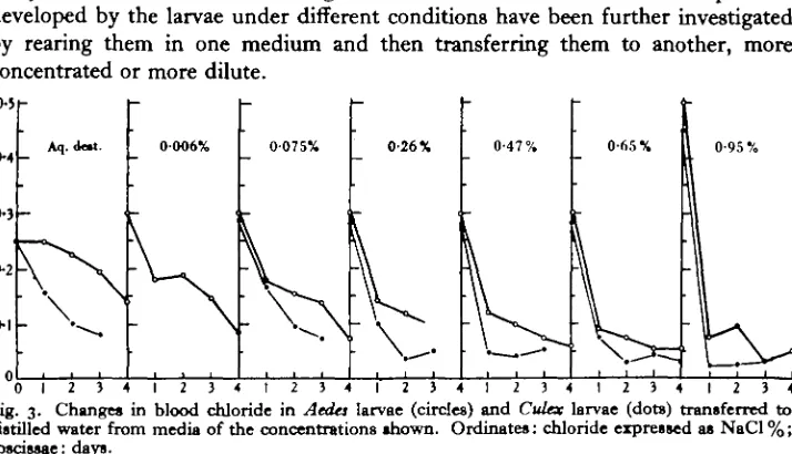
The results given in Figs. 1 and 2 show that the larvae can (i) retain chloride in distilled water, (ii) take up chloride from dilute media, and (iii) prevent the entry of chloride in media with a higher salt content than the blood. The adaptations developed by the larvae under different conditions have been further investigated by rearing them in one medium and then transferring them to another, more concentrated or more dilute.
O-Sr-<M - 0006% 0075% 0-26% 0-47% 0-65% 0-95%
[image:5.451.49.406.254.459.2]" 0 1 2 3 4 1 2 3 4 t 2 3 - 4 1 2 3 4 1 2 3 4 1 2 3 4 1 2 3 4 Fig. 3. Changes in blood chloride in AecUs larvae (circles) and Culex larvae (dote) transferred to distilled water from media of the concentrations shown. Ordinates: chloride expressed as NaCl %; abscissae: days.
240
The loss of chloride from Chironomus larvae in distilled water is believed by Koch (1938) to take place mainly through the general surface of the body. But unlike the cuticle of Chironomus, which is readily permeable to water (Harnisch,
1934; Alexandrov, 1935), the cuticle of Aedes larvae is almost completely im-permeable (Wigglesworth, 19336); it is therefore very improbable that chloride should be lost through the skin. To test this question Aedes larvae reared in 0-9 % salt, the blood chloride being about 0-5, were ligatured with a silk thread around the anal segment and transferred, together with control larvae without a ligature,
05104
03
-0-2
Aq. dclt 0006%
2 3
-0006%
0075%
, _ I 2 3 4 5
0075%
0-34*/ 0 - 9 0 %
2 3 4 5 i _ I 2 3 4
[image:6.451.47.406.184.480.2]O I 2 3 4 5 O 1 2 3 4 5 O I 2 3 4 5 0 1 2 3 4 5 0 1 2 3 4 5
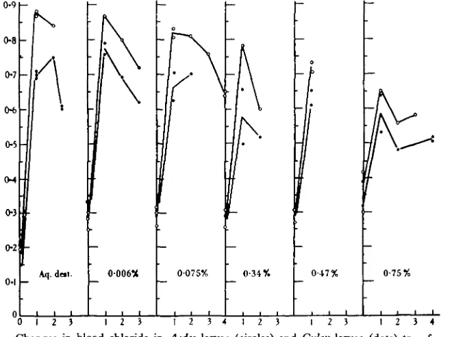
Fig. 4. Changes in blood chloride in Aedes larvae (circles) and Culex larvae (dote) transferred to tap water (0-006 % NaCl) from media of the concentrations shown. Ordinatcs: chloride as NaCl %; abscissae: days.
to distilled water. At the end of 24 hr. the ligatured larvae were dead and their bodies swollen, but the chloride was still from 0*26 to 0*30, whereas in the control larvae it had fallen to 0-07.
Evidently, most if not all the chloride is lost by the anus. Larvae adapted to retain chloride must therefore either produce urine free from chloride or reabsorb chloride from the excreta before they are discharged. In the earlier paper (Wiggles-worth, 1933 c) it was shown that reabsorption of water takes place in the dilated rectum of Aedes larvae, and Koch (1934) has suggested that the rectal epithelium may also be concerned in reabsorbing salts.
e.g. 0-90%, the larvae are at first unable to maintain their chloride even at the normal level, and there is a rapid fall during the first 24 hr. But in the course of a few days they become able to take up chloride and retain it. Aedes is again seen to be more efficient than Culex in this respect; after transfer from 0-90%, the initial loss of chloride is more pronounced in Culex; after transfer from 0-34 and even from 0-075 % there is again a temporary fall in chloride level in Culex but no fall at all in Aedes. The larvae reared in distilled water show once more the tendency to raise the blood chloride even above the normal level, especially during the first 24 hr. (cf. Fig. 1). In these experiments in tap water the factors both of uptake and retention are involved in the maintenance of the chloride level, and it is not possible to say in which of these adaptation has taken place.
The greater efficiency of Aedes larvae in collecting salt from the medium has been shown also by placing eggs of Aedes and Culex in the same vessel of distilled water and adding a very small quantity of powdered dog biscuit. Within a week the Aedes have reached the fourth stage, the Culex show almost no growth and many of them have died, whereas in o-8 % salt solution under the same conditions the Culex larvae grow as rapidly as the Aedes. Roubaud et al. (1935) have pointed out the improvement in the growth of Anopheles maculipenms if the water contains some salt (sea water equivalent to 0*4% NaCl); Pantazis (1935) and Woodhill (1936) have shown that both the optimal concentration of salt for growth and the resistance to high concentrations of salt vary greatly in different species.
(iii) The ability to keep chloride out was tested by transferring the larvae from the same media to 0-95 % salt. Fig. 5 shows that there is much individual variation; many of the larvae reared in distilled water and tap water die in the course of 1 or 2 days; but the results are consistent in showing (a) that the blood chloride rises higher in larvae reared in the more dilute media: these larvae are less efficient in preventing the entry of chloride; (b) that in those larvae which survive transfer, the chloride tends to fall in the course of a few days: the larvae become adapted; and (c) that Culex larvae are rather more efficient than Aedes larvae in keeping chloride out under these conditions (although this difference is not apparent in the experiments given in Fig. 2).
MORPHOLOGICAL CHANGES IN LARVAE REARED IN DIFFERENT MEDIA
It is well known that mosquito larvae occurring naturally in salt water have their anal papillae much reduced. Examples of this reduction in certain African species are given by Gibbins (1932). Martini (1923) showed experimentally that Aedes
meigenanus and A. nemorosus have smaller papillae when reared in 0-45% NaCl
than in tap water and the papillae are larger still in distilled water.1 In Aedes
aegypti, Pagast (1936) found some shortening of the papillae when salt was present,
but the most striking difference was to be seen when larvae from distilled water
1
The anal and ventral papillae of chironomids also vary in size with the water in which the larvae occur, but the conditions which determine these changes are not yet very well denned (Lenz, 1930).
242
[image:8.451.64.394.216.462.2]were compared with larvae from very dilute salt solutions, and as he rightly points out, it was for this reason that the effect was not observed in experiments dealing with more concentrated media (Wigglesworth, 1933 c).
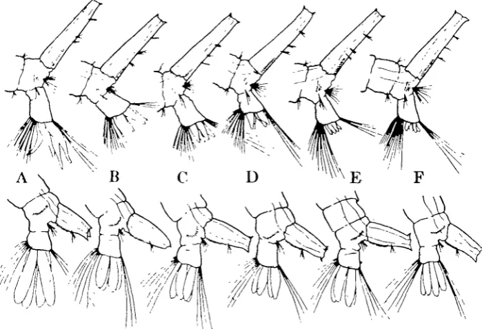
Fig. 6 shows the form of the anal papillae of Culex and Aedes reared in the media used for the present experiments. It is evident that papillae are better developed in Aedes and that the reduction in size with increasing salt concentration is more marked in Culex. As can be seen in the legend to Fig. 6, the individual variation (which is not correlated with the size of the respiratory siphon) is large. But there is little if any constant change in the papillae of Aedes above the concentration of tap water; whereas the reduction in Culex continues at least to 0-34% salt.
2 3 2 3 1 2 3 4
Fig. 5. Changes in blood chloride in Aedes larvae (circles) and Culex larvae (dots) transferred to salt medium with chloride content equivalent to O'gs % NaCl from media of the concentrations shown. Ordinates: chloride as NaCl%; abscissae: days.
These results support the suggestion of Koch (1938) that the enlargement of the papillae in dilute media is a functional hypertrophy connected with the uptake of salt. The smaller degree of reduction in Aedes agrees with the observations (Fig. 4) that they can maintain the chloride level of the blood more successfully than Culex after being reared in saline media. But it is interesting to note that
Culex larvae showing the extreme reduction of the papillae represented in Fig. 6 F
are yet able after a time to absorb chloride from tap water and retain it at the normal level in the blood (Fig. 4, 0-90 %).
in small collections of rain water in cavities of plants, etc. This enlargement has been explained by Martini (1923), Schlieper (1930) and others as a respiratory adaptation. But the papillae have been shown to have almost no respiratory function (Wigglesworth, 19336; Thorpe, 1933). Moreover, the water in rot-holes in trees, the typical breeding place of Aedes aegypti, contains no dissolved oxygen (Beattie, 1929); if, therefore, the papillae were efficient respiratory organs their only effect would be to deprive the larva of the oxygen collected at the water surface.
Fig. 6. Terminal segments of Ctdtx larvae (upper row) and Aedes larvae (lower row) showing typical appearance of anal papillae of larvae reared in different media. Measurements of the papillae in six larvae from each culture gave the following values in mm.; mean values in brackets:
A, distilled water... B, tap water (o-oo6 % NaCl) C,o-o75%NaCl
D, 0-34% E,o-6S% F,o-oo%
Culex 0-66-0-93 (0-83); Aedes 0-65-0-93 (0-89). 0-32-0-52 (0-36);
0-37-0-36 (033); 0-20-0-36 (0-22); 0-15-0-21 (0-20); 0-15-0-21 (0-20);
o-5S-o-77 (o-6o). 0-57-0-75 (o-6o).
0-45-0-58 (0-52). 0-52-0-83 (0-71). 0-38-0-73 (0-50).
THE RELATION BETWEEN OSMOTIC PRESSURE OF THE BLOOD AND THE EXTENT OF AIR IN THE TRACHEOLES
It was shown in an earlier paper (Wigglesworth, 1930) that the terminal parts of the tracheoles in Aedes larvae normally contain fluid, and that when the larvae struggle violently, as they do when held beneath a cover-glass out of contact with the air, the fluid is extracted and air extends into the finest branches. Evidence was brought forward that this effect was due to increased osmotic pressure in the blood as the result of muscular activity; the level of fluid in the tracheoles being
determined by an equilibrium between capillarity and osmotic pressure. With the development of the micro-method for measuring osmotic pressure it has become possible to test this hypothesis directly.
The haemolymph from four resting larvae of Aedes gave values for osmotic
pressure of o, „, „ 0
t
o-86; o-86; 0-89; o-8i.
Four larvae from the same culture, after asphyxiation for ^-1 hr., gave 0-93 (little change in tracheoles);
0-89 (no change in tracheoles); 0-91 (no change in tracheoles);
i-16 (extension of air into fine branches of tracheoles).
Four resting larvae of Culex gave values of o-88; 0-85; 0-89; 0-85.
Four asphyxiated larvae from the same batch, which struggled much more actively than the submerged Aedes larvae, gave
1 -08 (air extends to finest branches of tracheoles); 1 -06 (air extends to finest branches of tracheoles); I*I6 (air extends to finest branches of tracheoles);
0-94 (less struggling; some extension of air, but not into the finest branches).
From these and other similar results it is evident that when the larvae are asphyxiated the osmotic pressure of the blood increases, and that the increase is proportional to the amount of struggling on the one hand and the extension of air into the tracheoles on the other. In the earlier experiments it was found that 1 % of NaCl, or a solution of sodium lactate of the same molecular concentration, introduced into the larva would bring about the extraction of fluid from the tracheoles.
These results therefore support the hypothesis. But there are serious difficulties in the way of accepting this hypothesis in its simplest form. For in the work on adaptation of Aedes larvae to salt water (Wigglesworth, 1933 c) it was observed that larvae accustomed to such concentrations as i-2% of NaCl still had fluid extending to the normal level in the tracheoles of the head and anal papillae. And from this it was inferred that the larvae were homoiosmotic. But we have seen in the present work that at these higher concentrations the osmotic pressure of the blood is, in fact, always a little above that of the external medium. For example, four resting larvae of Aedes in a medium of I-OI gave values
1-05; 1-08; 1-06; 1-05
and in all these the air in the tracheoles extended to about the same level as in normal larvae with an osmotic pressure of 0-85; and four resting Aedes larvae which had been raised in tap water and then transferred for several days to a medium of 0-97 gave values
' ° 1 - 1 2 ; 1 - 1 2 ; i - i i ; 1 1 2
were asphyxiated. The tracheoles in the head and in those parts of the anal papillae which had not been damaged by the salt filled with air. After asphyxiation they gave values
1-27; 1-31.
These results are incompatible with the view that the level of fluid in the tracheoles is determined by a simple relation between capillarity and the osmotic pressure of the blood. A similar difficulty is encountered when other insect larvae are examined. Thus, whereas larvae of all the culicine mosquitoes so far examined
(Aedes aegypti, A. albopictus1 and other spp., Armigeres obturbans1 Culex pipiens, C.fatigans1 and other spp., Lutzia fuscana,1 RacHonotomyia nepenthes1) have fluid
in the terminal parts of the tracheoles during rest, in all the Anophelines examined
(Anopheles maculipenms, hyrcanus,1 barbirostris,1 maculatus,1 sundaicus,1 subpictus1 vagus1) the tracheoles always contain air as far as they can be traced in the head,
and in the anal papillae they contain fluid only in the finest branches. Yet estimations of the osmotic pressure in Anopheles maculipenms larvae gave values of o-8o-o-83 like
Aedes and Culex.
It is not easy to see how these contradictory observations are to be reconciled. But the terminal parts of the tracheoles are always embedded in a layer of cytoplasm of varying thickness, and there are certain experiments which show that fluid may be extracted from the tracheoles by substances which react with this cytoplasmic material without any change in the osmotic pressure of the blood:
(i) If the intact larva of Aedes is exposed to o-i % AgNO3 this enters the cells
of the anal papillae and the cell substance is precipitated. Fluid is rapidly absorbed from the tracheoles and air enters the finest branches. The cells are killed and soon after death the fluid rises in the tracheoles again above the original level.
(ii) As described in connexion with the effect of salts on the anal papillae (Wigglesworth, 1933 a), the same effect, a momentary filling with air followed by a rapid rise of fluid, is produced by iV/50 NaOH.
Perhaps the explanation of the apparent contradictions described above is to be found in some adaptive change in this property of "imbibition" (using the word in the most general sense) of the cytoplasmic walls of the tracheoles. Changes in the composition or osmotic pressure of the blood would be pictured as affecting the fluid in the tracheoles by their action upon this cytoplasmic imbibition, in the same way as hypertonic salt solutions applied to the intact larva cause swelling of the cells of the anal papillae (Wigglesworth, 1933 a).
Thus, we have seen that the haemolymph of a larva in 1-25 % salt solution is practically isotonic with the external medium. Yet if the head of the larva is punctured in such a medium, so that the tissues are exposed to a fluid of the same osmotic pressure as the blood, yet much more rich in salts, the tracheoles im-mediately fill with air (Wigglesworth, 1933 c).
That an adaptation of the kind suggested does occur is shown by introducing the blood of a larva accustomed to 1-25 % salt solution (the tracheoles of such a
1 These tropical species were examined during a visit to the Medical Research Institute, Kuala
larva contain fluid) into a resting larva from tap water. The tracheoles of the tap-water larva at once fill with air. And if this same blood is applied to an intact resting larva from tap water, the cells of its anal papillae assume a vacuolated, swollen appearance (increased cytoplasmic imbibition) and the fluid is instantly extracted from the tracheoles which run through them.
An analogous phenomenon is the expansion and contraction of the wall of the air sacs in Corethra, which appears to be brought about by a controlled imbibition of water (Frankenberg, 1915; Damant, 1924). Immersion of the air sac in the insect's blood plus a trace of NaCl causes it to expand (Christensen, 1928).
That the changes which occur in the tracheoles during asphyxiation are not due to the acidity of the blood has been proved by experiments in which the anal papillae of Aedes larvae were amputated and immersed in dilute Ringer's solution ( 0 7 % NaCl), brought to pH 5-0 with lactic acid. No effect was produced on the tracheoles, whereas these at once fill with air if immersed in Ringer's solution of full strength (0-95 % NaCl) whether neutral or acid.
That the removal of fluid is an active absorption, that is, a secretion by the cytoplasmic layer, induced by oxygen want, such as occurs at the initial filling of the tracheal system (Wigglesworth, 1938), is improbable because (i) the extraction of fluid does not occur in the absence of oxygen unless there is active muscular contraction and a concomitant rise in the osmotic pressure of the blood, and (ii) the blood of asphyxiated larvae whose tracheoles have filled with air will cause instant extraction of fluid from the tracheoles of resting larvae just like hypertonic solutions of NaCl, sodium lactate, glycerol, etc.
A more definite description of the process is impossible at the present time, but it appears as though the extent to which fluid rises in the tracheoles is dependent upon the interaction of several forces: the osmotic pressure of the body fluid, the swelling force of the cytoplasm around the tracheoles and, perhaps, imbibition in the wall of the tracheole itself.
SUMMARY
The osmotic pressure of the haemolymph in well-fed larvae of Aedes aegypti and Culex pipiens is equivalent to o-8o-o-89 % NaCl. During starvation it falls to 070 % NaCl.
The average chloride content of the haemolymph is equivalent to 0-30 % NaCl in Aedes, 0-28 % in Culex.
The non-chloride fraction can be regulated so that the total osmotic pressure remains relatively constant in spite of wide variations in chloride content.
Comparative experiments show that Aedes aegypti is more efficient than Culex
pipiens in absorbing and retaining chloride in dilute media, and Culex is perhaps
a little better at keeping chloride out in more concentrated media. This difference accords with the difference in their natural breeding places.
The anal papillae become greatly enlarged in media almost free from chloride (functional hypertrophy for chloride uptake (Koch, 1938)) and reduced in more concentrated media. The papillae of Culex are more labile in this respect, but the maximum development occurs in Aedes.
The ability of larvae to absorb and retain chloride in dilute media is much reduced if they have been reared in more concentrated media. But after transfer to dilute media they soon recover this ability in spite of the reduction in size of the anal papillae.
When the larva is deprived of oxygen, muscular activity causes a rise in the osmotic pressure of the blood from about 0-83 to i-o or I-I % NaCl, and this change is associated with extraction of fluid from the tracheoles. But reasons are given for doubting that the level of fluid in the tracheoles is determined by a direct relation between capillarity and osmotic pressure of the blood.
I am greatly indebted to Prof. A. Krogh for much helpful discussion in the course of this research, which was carried out during the tenure of a Research Fellowship of the Rockefeller Foundation.
REFERENCES
ALEXANDROV, W. F. (1935). Ada Zool. 16, i. BALDES, E. J. (1934). J. sci. Imtrum. 11, 223. BKATTIE, M. V. F. (1929). Bull. ent. Res. 20, 45.
CHRISTENSEN, P. J. H. (1928). Dansk Naturkist. For. 86, 21. DAMANT, G. C. C. (1924). J. Physiol. 59, 345.
DUVAL, M., PORTIBR, P. & COUHTOIS, A. (1928). C.R. Acad. Set., Paris, 186, 652.
FLORKIN, M. (1937). Mint. Sav. itr. Acad. R. Bdg. 16, 69 pp. FRANKHNBKRG, G. v. (1915). Zool. Jb., Abt. Physiol., 36, 505. FWTZCHE, H. (1917). Int. Rev. Hydrobiol. 8, 22.
GIBBINS, E. G. (1932). Ann. trap. Mtd. Parasit. 26, 551. HARNI8CH, O. (1934). Z. vergl. Physiol. 21, 281.
HOPKINS, G. H. E. (1936). Mosquitoes of the Ethiopian Region, Pt. 1. London. KOCH, H. (1934). Arm. Soc. set. Brux. 54, 346.
(1938). J. exp. Biol. 18, 152.
KROGH, A. (1937). Skand. Arch. Physiol. 76, 60. LKNZ, F. (1930). Arch. Hydrobiol. 21, 447.
MARSHALL, J. F. & STALEY, J. (1937). Proc. roy. ent. Soc. Lond. 12, 17. MARTINI, E. (1923). Verh. int. Ver. theoret. angew. Ltrnnologie, 1, 235. PAOAST, F. (1936). Zool.Jb., Abt. Physiol., 56, 184.
PANTAZIS, G. (1935). Prakt. Acad. Athen. 10, 348.
PORTIER, P. & DUVAL, M. (1927). C.R. Soc. Biol., Paris, 97, 1605.
ROUBAUD, E., COLAS-BELCOUH, J. & TREILLARD, M. (1935). Bull. Soc. Path. exot. 28, 568.
ScHLffiPER, C. (1930). Biol. Rev. 5, 309. THORPE, W. H. (1933). Nature, Lond., 131, 549.
WIOOLESWORTH, V. B. (1930). Proc. roy. Soc. B, 106, 229. (1933a). J. exp. Biol. 10, 1.
(i933ft)- J- «*>• Bid. 10, 16. (i933<0- J- **P- Biol. 10, 27. (i937)- Biochem.J. 31, 1719. (1938). J- exp. Biol. 15, 248.