Original Article
Prognostic value of systemic inflammation response
SIR parameters in unresectable
hepatocellular carcinoma
Sheng-Fei Wang1*, Yan Zhang1*, Meng-Ting Cui1*, Wen-Jie Wang2, Meng-Dan Xu1, Min Jiang1, Lian Lian3, Meng-Yao Wu1, Wei Li1,4, Yuan Gao1, Jie Yu5, Min Tao1
1Department of Oncology, The First Affiliated Hospital of Soochow University, Suzhou 215006, China; 2Department of Radio-Oncology, Nanjing Medical University Affiliated Suzhou Hospital, Suzhou 215001, China; 3Department of Oncology, Suzhou Xiangcheng People’s Hospital, Suzhou 215131, China; 4Comprehensive Cancer Center, Suzhou Xiangcheng People’s Hospital, Suzhou 215131, China; 5Department of Pathology, The First Affiliated Hospital of Soochow University, Suzhou 215006, China. *Equal contributors.
Received December 17, 2018; Accepted January 7, 2019; Epub March 15, 2019; Published March 30, 2019
Abstract: Hepatocellular carcinoma (HCC) is one of the most common malignancies, with increasing incidence and dismal survival. Albumin, globulin, lactate dehydrogenase (LDH), and albumin to globulin ratios (AGR) have been identified as systemic inflammation response (SIR) parameters, correlated with tumor development and metasta-sis. The aim of this study was to investigate the predictive value of systemic inflammation-related parameters in unresectable hepatocellular carcinoma patients. A total of 101 HCC patients were recruited for this study. Patients had histologic or cytologic evidence of locally advanced stage or metastasis. Patients were divided into two groups, according to the median value of ALB, GLB, LDH, and AGR. Post-/pre-chemotherapeutic ratios were defined as the rate of pre-chemotherapeutic SIR parameters values and corresponding ones obtained after chemotherapy (≤ 1 in-dicates ALB, GLB, LDH, and AGR values were not increased after chemotherapy, while > 1 suggests increased ALB, GLB, LDH, and AGR values). Higher GLB levels are correlated with bone metastasis. Patients with lower baseline LDH levels had better overall survival (OS), whereas ALB, GLB, or AGR levels were not correlated with outcomes. Alterations in LDH levels were associated with therapeutic efficacy. Patients with non-increased LDH levels, follow-ing chemotherapy, exhibited improved responses, compared to those with increased LDH levels. Univariate analysis revealed that gender (female), high baseline LDH levels, and non-increased post-/pre-chemotherapy AGR ratios were risk factors affecting OS (overall survival) in patients with HCC. Multivariate analysis indicated that gender (female) and high baseline LDH levels were independent risk factors for HCC prognosis. The area under the curve of baseline LDH was 0.717 (95% CI 0.617-0.818; P < 0.001) and the optimum cutoff point of baseline LDH was 236.500 U/L, with a sensitivity of 73.0% and specificity of 64.6%. The current study shows that baseline LDH levels and changes in AGR levels, after chemotherapy, are correlated with outcomes of patients with HCC.
Keywords: Hepatocellular carcinoma, systemic inflammation response (SIR), prognosis
Introduction
With an estimated 42,220 new cases and 30,200 deaths, hepatocellular carcinoma (HCC) is one of the most common malignant neoplasms. It ranks fourth among causes of cancer mortality in 2018 of United States [1]. Unfortunately, more than half of HCC patients
are first diagnosed in the advanced stage, los -ing opportunities for surgical resection or liver transplantation [2]. To increase the survival rates of patients with unresectable HCC,
alter-native treatments, such as molecular targeted agents, immune therapy, and vaccines, have been widely applied in clinical treatment [3, 4]. However, disease relapse after therapy, as well as drug resistance, are critical issues giving rise to poor prognosis of HCC [5].
Systemic inflammation response (SIR) has
of inflammatory response and malignancies,
as a majority of HCC cases arise from chro-
nic liver inflammation [7]. Albumin (ALB), the major component in serum protein, has be- en used as a predictor of malnutrition and sy-
stemic inflammation [8]. Globulin (GLB) serves as a carrier of hormones, playing an importa-
nt role in inflammation [9]. Albumin to globulin
ratio (AGR) represents both nutrition and
infl-ammatory response [10]. ALB, GLB, and AGR have been used as prognostic indicators for patients with HCCs [11, 12]. Lactic dehydroge-nase (LDH), a key enzyme catalyzing the con-version of pyruvate to lactate, plays a vital role in anaerobic metabolism of malignant cells, even under anaerobic conditions [13]. LDH has been demonstrated to be associated with HCC progression and prognosis [14].
The current study investigated several SIR pa- rameters and evaluated whether these
param-eters could be beneficial for prognosis predic -tion in patients with unresectable HCC. Materials and methods
Subjects and inclusion criteria
This study was conducted as a retrospective investigation of HCC patients that had been
referred to the First Affiliated Hospital of
So-ochow University (Jiangsu, China), between Nov 2000 and Jul 2017. Approval for the study was granted by the Medical Ethics Committee of the
First Affiliated Hospital of Soochow University.
Clinical and pathological records of all patien- ts participating in the study were reviewed periodically.
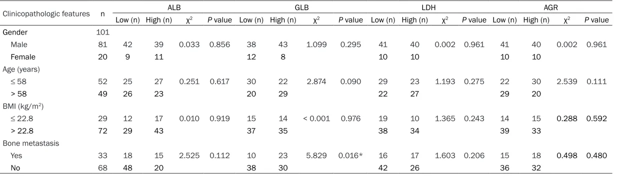
A total of 101 HCC patients were recruited for this study. Patients had histologic or cytologic evidence of locally advanced or metastasis. Patient characteristics are detailed in Table 1. The median age of the 101 patients was 58 years (range, 28-83 years), while 81 patients were male and 20 were female. The median BMI (kg/m2) of the 101 patients was 22.8 kg/ m2 (range, 18.2-31.8 kg/m2) and 33 patients had bone metastasis. Prognostic analyses we- re performed regarding overall survival (OS). Blood samples
Peripheral venous blood (5-7 mL) was collected into sterile ethylenediaminetetraacetic acid
(EDTA) tubes. Blood samples were analyzed using a hematology analyzer (Sysmex XE-2100; Sysmex, Kobe, Japan) or biochemical analyzer (Olympus AU5421+ISE, Olympus, Japan). ALB, GLB, LDH, and AGR levels are recorded in Table 1. The patients were divided into two groups, according to the median values of ALB (low
ALB, < 38.70 or high ALB, ≥ 38.70), GLB (low GLB, < 29.10 or high GLB, ≥ 29.10), LDH (low LDH, < 215.00 or high LDH, ≥ 215.00), and AGR (low AGR, < 1.29 or high AGR, ≥ 1.29). Post/pre-chemotherapeutic ratios are defined
as the rate of pre-chemotherapeutic SIR-related indicator values and corresponding ones obtained after chemotherapy.
Evaluations
Computed tomography (CT) scans were per-formed for assessment of response, every 2 months, and evaluated according to the criteria of Response Evaluation Criteria in Solid Tumors (RECIST) 1.1.
Follow-ups
This study recorded responses to chemothe- rapy, including partial response (PR), stable di- sease (SD), and progressive disease (PD). After
first line chemotherapy, disease progression was defined as a lack of response to chemo -therapy. In contrast, stable disease or partial
response after chemotherapy was defined as a
response to chemotherapy. Survival time was measured from the date of chemotherapy until death or last clinical evaluation. Prognostic analyses were performed regarding overall
sur-vival (OS). OS was defined as the time from
diagnosis date to death from any cause. Statistical analysis
Statistical analyses were performed using SPSS 19.0 software (Chicago, USA). Association between blood parameter status and clinic
pathologic features or chemotherapeutic effi
Table 1. Clinicopathologic features
Clinicopathologic features n ALB GLB LDH AGR
Low (n) High (n) χ2 P value Low (n) High (n) χ2 P value Low (n) High (n) χ2 P value Low (n) High (n) χ2 P value
Gender 101
Male 81 42 39 0.033 0.856 38 43 1.099 0.295 41 40 0.002 0.961 41 40 0.002 0.961
Female 20 9 11 12 8 10 10 10 10
Age (years)
≤ 58 52 25 27 0.251 0.617 30 22 2.874 0.090 29 23 1.193 0.275 22 30 2.539 0.111
> 58 49 26 23 20 29 22 27 29 20
BMI (kg/m2)
≤ 22.8 29 12 17 0.010 0.919 15 14 < 0.001 0.976 19 10 1.365 0.243 14 15 0.288 0.592
> 22.8 72 29 43 37 35 38 34 39 33
Bone metastasis
Yes 33 18 15 2.525 0.112 10 23 5.829 0.016* 16 17 1.603 0.206 15 18 0.498 0.480
No 68 48 20 38 30 42 26 36 32
to identify independent risk factors associated with HCC. Values of P < 0.05 indicate statistical
significance.
Results
Baseline LDH levels correlated with outcomes of HCC patients
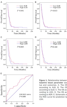
Kaplan-Meier plots were used to determine the effects of baseline ALB, GLB, LDH, and AGR status on OS (Figure 1A-D). The patients were divided into two groups, according to the an values of ALB, GLB, LDH, or AGR. The
medi-off point of baseline LDH was 236.500 U/L,
with a sensitivity of 73.0% and specificity of
64.6%.
Changes in LDH levels, after chemotherapy, associated with chemotherapeutic efficacy in HCC patients
To determine the association between changes in ALB, GLB, LDH, or AGR status and
chemo-therapeutic efficacy, blood samples were
ob-tained and CT evaluations were performed,
simultaneously, after first-line chemotherapy
[image:4.612.90.373.69.521.2]for 2 mouths. Relationships between changes Figure 1. Relationship between
baseline blood parameter sta-tus and outcomes. A. The OS according to ALB. B. The OS according to GLB. C. The OS cording to LDH. D. The OS ac-cording to AGR. E. Schematic of the ROC curve for prediction by LDH.
an OS of the higher ALB group was 14 (95% confidence inter -val [CI] 8.002-19.998) months, while that of the lower ALB group was 10 (95% CI 3.076-16.924) months (P = 0.441). The median OS of the higher GLB group was 15 (95% CI 9.522-20.478) months, while that of the lower GLB group was 10 (95% CI 4.952-15.048) months (P = 0.898). The me- dian OS of the higher LDH group was 7 (95% CI 4.229-9.772) months, while that of the lower LDH group was 18 (95% CI 11.877-24.123) mo- nths (P = 0.003). The median OS of the higher AGR group was 13 (95% CI 7.225-18.775) months, while that of the lo- wer AGR group was 13 (95% CI 8.342-17.658) months (P = 0.268). Therefore, patients wi- th lower baseline LDH levels showed a better prognosis. However, baseline levels of GLB, LDH, or AGR showed no effects on OS.
cut-in blood parameter levels after chemotherapy
and chemotherapeutic efficacy are presented
in Table 2, respectively.
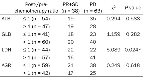
A total of 54 patients had non-increased ALB levels, following chemotherapy, of which 19 patients were PR (partial response) or SD (st- able disease) and 35 patients were PD (pr- ogressive disease). A total of 47 patients had increased ALB levels, following chemotherapy, of which 19 patients were PR or SD and 28 patients were PD (P = 0.588).
A total of 41 patients had non-increased GLB levels, following chemotherapy, of which 18 patients were PR or SD and 23 patients were PD. Moreover, 60 patients had increased GLB levels, following chemotherapy, of which 20 patients were PR or SD and 40 patients were PD (P = 0.282).
A total of 44 patients had non-increased LDH levels, following chemotherapy, of which 22 patients were PR or SD and 22 patients were PD. A total of 57 patients had increased LDH levels, following chemotherapy, of which 16 patients were PR or SD and 41 patients were PD (P = 0.024).
A total of 42 patients had non-increased AGR levels, following chemotherapy, of which 17 patients were PR or SD and 25 patients were PD. A total of 59 patients had increased AGR levels, following chemotherapy, of which 21 patients were PR or SD and 38 patients were PD (P = 0.618).
Thus, changes in LDH levels, after chemother- apy, were associated with chemotherapeutic
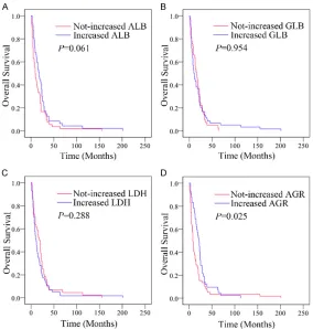
12.601) months (P = 0.061). The median OS of patients with increased GLB levels, following chemotherapy, was 10 (95% CI 5.662-14.338) months, while that of the non-increased group was 16 (95% CI 9.727-22.273) months (P = 0.954). The median OS of patients with incr- eased LDH levels, following chemotherapy, was 11 (95% CI 7.301-14.699) months, while that of the non-increased group was 18 (95% CI 12.312-23.688) months (P = 0.288). The me- dian OS of patients with increased AGR leve- ls, following chemotherapy, was 21 (95% CI 16.463-25.537) months, while that of the non-increased group was 8 (95% CI 6.118-9.882) months (P = 0.025). Thus, patients with incr- eased AGR levels, after chemotherapy, had increased survival rates. However, changes in GLB, LDH, or AGR levels had no effect on OS. Prognostic factors for HCC
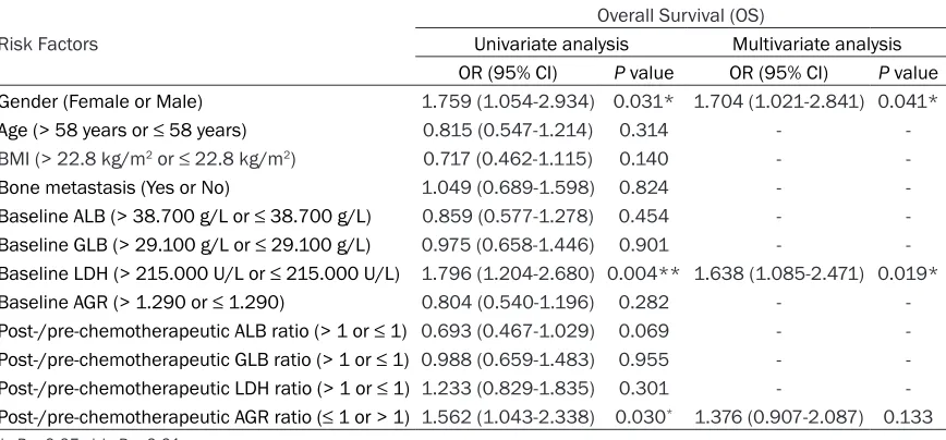
Univariate analyses demonstrated that gen- der (female) (hazard ratio [HR] 1.759; 95% CI 1.054-2.934; P = 0.031), high baseline LDH levels (HR 1.796; 95% CI 1.204-2.680; P = 0.004), and a decreased post-/pre-chemothe- rapeutic AGR ratio (HR 1.562; 95% CI 1.043-2.338; P = 0.030) were significant risk factors
for a poor prognosis (Table 3). According to mul-tivariate analysis, gender (female) (HR 1.704; 95% CI 1.021-2.841; P = 0.041) and high base-line LDH levels ratios (HR 1.638; 95% CI 1.085-2.471; P = 0.019) were found to be indepen-dently associated with poor survival.
Discussion
HCC has been well accepted as the best tum-
[image:5.612.90.331.97.229.2]or type to study the interaction of inflammatory
Table 2. Relationship between changes in the blood parameter status and chemotherapeutic efficacy
Post-/pre-
chemotherapy ratio (n = 38)PR+SD (n = 63)PD χ2 P value
ALB ≤ 1 (n = 54) 19 35 0.294 0.588
> 1 (n = 47) 19 28
GLB ≤ 1 (n = 41) 18 23 1.159 0.282
> 1 (n = 60) 20 40
LDH ≤ 1 (n = 44) 22 22 5.089 0.024*
> 1 (n = 57) 16 41
AGR ≤ 1 (n = 59) 21 38 0.249 0.618
> 1 (n = 42) 17 25
*: P < 0.05.
efficacy of HCC patients. However, ALB, GLB, or AGR had no significant effects on chemotherapeutic efficacy.
Changes in AGR levels, after chemo-therapy, correlated with outcomes of HCC patients
5.399-response and malignancies because of chron-
ic liver inflammation, especially hepatitis B
vi-rus (HBV) or hepatitis C vivi-rus (HCV) infections, leading to a majority of HCC occurrence [7, 15, 16]. Possible mechanisms involved in SIR-induced carcinogenesis are listed as follows. First, as a major etiology for HCC, HBV infe-
ctions may contribute to fibrosis and cirrhosis,
a preneoplastic condition for HCC [17]. More- over, prophylactic HBV vaccinations have alr- eady been proven to prevent the development of hepatomas in children and young adults [18]. Second, SIR may lead to oxidative stress and release of oxygen-free radicals, which pr-
ovokes DNA damage, epigenetic modifications,
and telomerase reactivation [19]. Third,
infla-mmatory cytokines, such as interleukin-6
(IL-6), IL-1β, and tumor necrosis factor-α (TNF-α),
have been proven to be vital lynchpins in in-
flammation-mediated carcinogenesis.
Activa-tion of phosphoinositide 3-kinase-protein kin-
ase B (PI3K-Akt) pathways by inflammatory
cytokines may induce cell proliferation,
malig-ed tumor mortality [24]. Since serum ALB levels
reflect not only nutrition status but also chronic inflammation, recent studies have revealed
that a higher serum ALB level is a favorable prognostic predictor in several types of malig-nant tumors [25]. However, few studies have
shown ALB alone as a significant prognostic
indicator in patients with advanced HCC. In view of the role ALB plays in biological
metabo-lism and chronic inflammation, this study sug -gests ALB as a promising prognosis predictor for advanced HCC. Possible mechanisms are listed as follows. First, as a major protein syn-thesized by hepatocytes, levels of serum ALB
partially reflect the proportion of normal hepa -tocytes and their metabolic activity. Thus, in advanced HCC, a sharpened decrease of serum ALB levels may indicate a decrease in the quan-tity of normal hepatocytes, suggesting an exhausting status and malignant progression. Second, ALB has been demonstrated to play a crucial anticancer role by stabilizing cell growth and DNA replication, as well as maintaining Figure 2. Relationship between changes in blood parameter status after
chemotherapy and outcomes. A. The OS according to changes in ALB. B. The OS according to changes in GLB. C. The OS according to changes in LDH. D. The OS according to changes in AGR.
nant survival, angiogenesis, and metastasis [20]. Consi- dering that SIR is tightly asso-ciated with tumor progression and outcomes, nu-merous studies have obtained an in-depth look at the role systemic
inflammation plays in cancer
progression and outcome pre-diction. Thus, for a more accu-rate and comprehensive prog-nostic analysis of HCC, the current study investigated the predictive value of ALB, GLB, LDH, and AGR in patients with HCC.
ALB, one of the major compon- ents of plasma proteins, is sy- nthesized in the liver and mir-rors individual nutrition stat-
[image:6.612.91.375.72.370.2]Table 3. Univariate and multivariate logistic regression analysis of HCC risk factors
Risk Factors
Overall Survival (OS)
Univariate analysis Multivariate analysis
OR (95% CI) P value OR (95% CI) P value
Gender (Female or Male) 1.759 (1.054-2.934) 0.031* 1.704 (1.021-2.841) 0.041*
Age (> 58 years or ≤ 58 years) 0.815 (0.547-1.214) 0.314 -
-BMI (> 22.8 kg/m2 or ≤ 22.8 kg/m2) 0.717 (0.462-1.115) 0.140 -
-Bone metastasis (Yes or No) 1.049 (0.689-1.598) 0.824 -
-Baseline ALB (> 38.700 g/L or ≤ 38.700 g/L) 0.859 (0.577-1.278) 0.454 -
-Baseline GLB (> 29.100 g/L or ≤ 29.100 g/L) 0.975 (0.658-1.446) 0.901 -
-Baseline LDH (> 215.000 U/L or ≤ 215.000 U/L) 1.796 (1.204-2.680) 0.004** 1.638 (1.085-2.471) 0.019*
Baseline AGR (> 1.290 or ≤ 1.290) 0.804 (0.540-1.196) 0.282 -
-Post-/pre-chemotherapeutic ALB ratio (> 1 or ≤ 1) 0.693 (0.467-1.029) 0.069 -
-Post-/pre-chemotherapeutic GLB ratio (> 1 or ≤ 1) 0.988 (0.659-1.483) 0.955 -
-Post-/pre-chemotherapeutic LDH ratio (> 1 or ≤ 1) 1.233 (0.829-1.835) 0.301 -
-Post-/pre-chemotherapeutic AGR ratio (≤ 1 or > 1) 1.562 (1.043-2.338) 0.030* 1.376 (0.907-2.087) 0.133 *: P < 0.05; **: P < 0.01.
homeostasis of sex hormones to protect against sex hormone-induced cancers [26, 27]. Moreover, in patients with HCC, a lower recur-rence rate and better OS has been proven to be tightly correlated with higher serum ALB levels [28, 29]. Thus, increased ALB levels might lead to relieved tumor loads, improved nutrition sta-tus, and recovery of liver function, suggesting a better prognosis in HCC. However, in the pres-ent study, post-/pre-chemotherapy ALB ratio had no effect on OS. It was speculated that
serum ALB levels could be influenced by multi -ple factors, such as infection, exogenous infu-sion, underlying liver disease, and other accom-panied chronic diseases.
Serum GLB contains acute-phase proteins, such as immunoglobulins, C-reactive protein (CRP), and complements C3 [30]. As a repre-sentative SIR parameter, GLB arises in respo-
nse to various pro-inflammatory cytokines and chemokines, particularly IL-6, IL-1β, and TNF-α
[31, 32]. Therefore, as a parameter which par- tly mirrors the intensity of SIR, high serum GLB levels have been demonstrated to be signi-
ficantly associated with tumor angiogenesis
and recurrence in patients with malignant tu- mors [31, 33]. Actually, previous studies have revealed that an increased serum GLB level si-
gnificantly contributes to poor prognosis in
se-veral types of malignant tumors [9, 30, 34, 35]. Unfortunately, previous studies have focusing on the prognostic value of GLB in patients with
advanced HCC are scarce. Considering serum
GLB levels reflect the inflammatory status, it is
reasonable to classify low GLB levels as a favor-able prognostic indicator in advanced HCC. In the present study, higher GLB levels were cor-related with more possible bone metastasis. However, neither baseline GLB levels nor post-/ pre-chemotherapy GLB ratios were correlated with OS in advanced HCC.
AGR, calculated as the ratio of serum ALB and GLB, has been reported to be an independent
and significant predictor of long-term mortality
in several types of cancer, including HCC [12, 36-38]. The mechanisms by which low serum AGR values impact recurrence and survival have not been fully elucidated. However, favor-able prognosis with a high AGR value can partly explained by the functions of ALB and GLB. Since HCC has been regarded as a typical
malignant tumor derived from chronic
infla-mmation, both increased ALB levels and de- creased serum GLB levels indicate low intensi- ty of SIR, suggesting a higher AGR value and better OS. The present study observed a sig-
nificant improvement of OS in patients with increased AGR values after first-line chemo -therapy. Additionally, non-increased post-/pre-chemotherapy AGR ratios have been proven to be a risk factor affecting OS of unresectable
HCC by univariate analysis. This study defined
AGR as a prognostic indicator in unresectable
the combination of ALB and GLB, this study suggests that it could be less affected by
condi-tions like dehydration and fluid retention, fur -ther enhancing its predictive value.
Unlike normal cells, the phenomenon by which malignant cells preferentially conduct anaero-bic glycolysis to produce most of the energy instead of mitochondrial oxidative phosphoryla-tion, even in the presence of oxygen, has been widely accepted as the Warburg effect [39, 40]. LDH, a key enzyme that plays a crucial role in anaerobic glycolysis, is an indicator for tumor hypoxia, neo-angiogenesis, and worse progno-sis in several types of malignant tumors, includ-ing HCC [41-43]. The mechanisms of high serum LDH levels and poor prognosis involve the following respects. First, malignant cells manifest a high rate of glucose uptake and lac-tate production, presenting a hypermetabolic status and a high serum LDH level [44]. Second, hypoxia, a prominent characteristic of solid tumors, facilitates the process of anaerobic gl- ycolysis and promotes cancer proliferation [45]. Third, hypoxia-mediated hypoxia inducible fa- ctor (HIF) has been demonstrated to upregul- ate LDH activity, as well as further activation of vascular endothelial growth factors (VEGFs) and promotion of tumor angiogenesis [46, 47]. Thus, high serum LDH levels mirror a hyperm- etabolic status, suggesting a high proliferation rate and aggressiveness of malignant tumors. Few studies have obtained an in-depth study of the predictive value of LDH in advanced HCC. For instance, in the retrospective study of Scartozzi M et al., patients with a low pre-trea- tment LDH level (< 450 U/L) had better OS, compared with those with a pre-treatment LDH level above 450 U/L. Patients with a decreased LDH value after transarterial-chemoemboliza-tion (TACE) exhibited better OS, compared with those with an increased LDH level [43].
In the present study, patients with non-in-
creased LDH levels, following first-line
chem-otherapy, exhibited improved responses, comp- ared to those with increased LDH levels. Mult- ivariate analysis revealed that a high baseline LDH level was an independent risk factor aff- ecting OS in patients with advanced HCC. ROC curve analysis showed that pre-treatment LDH values of 236.50 U/L were considered to be the optimal cutoff value for prognosis, with a
sensitivity of 73.0% and specificity of 64.6%. In
summary, since LDH is an inexpensive and co-
nvenient parameter routinely detected in clini-cal laboratories, this study suggests low pre-treatment LDH as a favorable predictive para- meter for HCC prognosis. Additionally, given the correlation between LDH and tumor angioge- nesis, serum LDH levels may be used to screen out patients suitable for anti-VEGF inhibitor treatment.
In conclusion, the current study investigated the predictive value of ALB, GLB, AGR, and LDH, demonstrating that gender (female), pre-treat-ment LDH levels, and post-/pre-chemotherapy AGR ratios are closely correlated with outcomes of unresectable HCC patients. Moreover, this
study defined high pre-treatment LDH levels as
an independent risk factor affecting OS of patients with unresectable HCC. Considering the high rates of HBV infection and HCC mor-bidity in china, these non-invasive, inexpensive,
and convenient parameters may be beneficial
to treatment of HCC. However, the current study
had some limitations, including insufficient
samples, data coming from a single center, and the retrospective design.
Acknowledgements
This work was supported by the National Na- tural Science Foundation of China (grant num-bers 81472199, 81472296, 81602091, 814- 02176, 81402093, 81272542, 81200369), the National Natural Science Foundation of Jiangsu Province (grant number BK20141162), the Six Major Talent Peak Project of Jiangsu Province (grant number 2015-WSN-022), the Project of Invigorating Health Care through Sc- ience, Technology and Education, Jiangsu Prov- incial Medical Youth Talent (grant number QN- RC2016709), the Project of Jiangsu Provincial Commission of Health and Family Planning (gr- ant number H201518), the Science and Educ- ation for Health Foundation of Suzhou for Youth (grant numbers kjxw2015002 and kjxw2015- 003), the Science and Technology Foundation of Suzhou Xiangcheng (grant numbers XJ201- 642 and XJ201702), the Science and Techno- logy Project Foundation of Suzhou (grant nu- mbers SYS201464 and SYS201504), and Bei- jing Xisike Clinical Oncology Research Founda- tion (grant number Y-MX2016-017).
Disclosure of conflict of interest
Address correspondence to: Drs. Min Tao and Yuan Gao, Department of Oncology, The First Affiliated Hospital of Soochow University, Suzhou, China. Fax: 86-051262388798; E-mail: [email protected] (MT); Fax: 86-05126238798; E-mail: shipinlt121@ 163.com (YG); Dr. Jie Yu, Department of Pathology, The First Affiliated Hospital of Soochow University, Suzhou, China. E-mail: [email protected]
References
[1] Siegel RL, Miller KD, Jemal A. Cancer statistics, 2018. CA Cancer J Clin 2018; 68: 7-30. [2] Llovet JM, Burroughs A, Bruix J. Hepatocellular
carcinoma. Lancet 2003; 362: 1907-1917. [3] Greten TF, Wang XW, Korangy F. Current
con-cepts of immune based treatments for pa-tients with HCC: from basic science to novel treatment approaches. Gut 2015; 64: 842-8. [4] Song MJ. Hepatic artery infusion
chemothera-py for advanced hepatocellular carcinoma. World J Gastroenterol 2015; 21: 3843-3849. [5] Tanaka S, Arii S. Molecular targeted therapy for
hepatocellular carcinoma in the current and potential next strategies. J Gastroenterol 2011; 46: 289-96.
[6] McMillan DC. Systemic inflammation, nutri-tional status and survival in patients with can-cer. Curr Opin Clin Nutr Metab Care 2009; 12: 223-226.
[7] Greten TF, Wang XW, Korangy F. Current con-cepts of immune based treatments for pa-tients with HCC: from basic science to novel treatment approaches. Gut 2015, 64: 842-8. [8] Liu X, Meng QH, Ye Y, Hildebrandt MA, Gu J, Wu
X. Prognostic significance of pretreatment se-rum levels of albumin, LDH and total bilirubin in patients with non-metastatic breast cancer. Carcinogenesis 2015; 36: 243-248.
[9] Qu X, Pang Z, Yi W, Wang Y, Wang K, Liu Q, Du J. High percentage of α1-globulin in serum pro-tein is associated with unfavorable prognosis in non-small cell lung cancer. Med Oncol 2014; 31: 238.
[10] Deng Y, Pang Q, Miao RC, Chen W, Zhou YY, Bi JB, Liu SS, Zhang JY, Qu K, Liu C. Prognostic significance of pretreatment albumin/globulin ratio in patients with hepatocellular carcino-ma. Onco Targets Ther 2016; 9: 5317-5328. [11] Zhou T, Zhan J, Hong S, Hu Z, Fang W, Qin T, Ma
Y, Yang Y, He X, Zhao Y, Huang Y, Zhao H, Zhang L. Ratio of C-reactive protein/albumin is an in-flammatory prognostic score for predicting overall survival of patients with small-cell lung cancer. Sci Rep 2015; 5: 10481.
[12] Zhang J, Liu X, Yang Z, Chen Y, Luo R. The pre-treatment albumin to globulin ratio, a validat-ed biomarker, prvalidat-edicts prognosis in hepatocel-lular carcinoma. J BUON 2016; 21: 925-934.
[13] Faloppi L, Bianconi M, Memeo R, Casadei Gardini A, Giampieri R, Bittoni A, Andrikou K, Del Prete M, Cascinu S, Scartozzi M. Lactate dehydrogenase in hepatocellular carcinoma: something old, something new. Biomed Res Int 2016; 2016: 7196280.
[14] Zhang JP, Wang HB, Lin YH, Xu J, Wang J, Wang K, Liu WL. Lactate dehydrogenase is an impor-tant prognostic indicator for hepatocellular car-cinoma after partial hepatectomy. Transl Oncol 2015; 8: 497-503.
[15] Cabillic F, Corlu A. Regulation of transdiffer- entiation and retrodifferentiation by inflamm- atory cytokines in hepatocellular carcinoma. Gastroenterology 2016; 151: 607-615. [16] Shlomai A, de Jong YP, Rice CM. Virus
associ-ated malignancies: the role of viral hepatitis in hepatocellular carcinoma. Semin Cancer Biol 2014; 26: 78-88.
[17] Greten TF, Wang XW, Korangy F. Current con-cepts of immune based treatments for pa-tients with HCC: from basic science to novel treatment approaches. Gut 2015; 64: 842-8. [18] Chang MH, You SL, Chen CJ, Liu CJ, Lee CM,
Lin SM, Chu HC, Wu TC, Yang SS, Kuo HS, Chen DS; Taiwan Hepatoma Study Group. Decreased incidence of hepatocellular carcinoma in hepa-titis B vaccinees: a 20-year follow-up study. J Natl Cancer Inst 2009; 101: 1348-1355. [19] Levrero M, Zucman-Rossi J. Mechanisms of
HBV-induced hepatocellular carcinoma. J Hepatol 2016; 64 Suppl: S84-S101.
[20] Taniguchi K, Karin M. IL-6 and related cyto-kines as the critical lynchpins between inflam-mation and cancer. Semin Immunol 2014; 26: 54-74.
[21] Sonnenschein C, Soto AM, Michaelson CL. Human serum albumin shares the properties of estrocolyone-I, the inhibitor of the prolifera-tion of estrogen-target cells. J Steroid Biochem Mol Biol 1996; 59: 147-54.
[22] McMillan DC, Watson WS, O’Gorman P, Preston T, Scott HR, McArdle CS. Albumin concentra-tions are primarily determined by the body cell mass and the systemic inflammatory response in cancer patients with weight loss. Nutr Cancer 2001; 39: 210.
[23] Liu J, Chen S, Geng Q, Liu X, Kong P, Zhan Y, Xu D. Prognostic value of pretreatment albumin-globulin ratio in predicting long-term mortality in gastric cancer patients who underwent D2 resection. Onco Targets Ther 2017; 10: 2155-2162.
gastric cancer. Medicine (Baltimore) 2016; 95: e3781.
[25] Gupta D, Lis CG. Pretreatment serum albumin as a predictor of cancer survival: a systematic review of the epidemiological literature. Nutr J 2010; 9: 69.
[26] Laursen I, Briand P, Lykkesfeldt AE. Serum al-bumin as a modulator on growth of the human breast cancer cell line, MCF-7. Anticancer Res 1990; 10: 343-51.
[27] Seaton K. Albumin concentration controls can-cer. J Natl Med Assoc 2001; 93: 490-493. [28] Nojiri S, Kusakabe A, Shinkai N, Matsuura K,
Iio E, Miyaki T, Joh T. Factors influencing dis-tant recurrence of hepatocellular carcinoma following combined radiofrequency ablation and transarterial chemoembolization therapy in patients with hepatitis C. Cancer Manag Res 2011; 3: 267-72.
[29] Nojiri S, Joh T. Albumin suppresses human he-patocellular carcinoma proliferation and the cell cycle. Int J Mol Sci 2014; 15: 5163-74. [30] Zhong LT, Wang H, Liang HQ, Su MR, Liu CD,
Wu DH. [An elevated pretreatment serum glob-ulin level predicts a poor prognosis of naso-pharyngeal carcinoma]. Nan Fang Yi Ke Da Xue Xue Bao 2016; 36: 151-6.
[31] Gabay C, Kushner I. Acute-phase proteins and other systemic responses to inflammation. N Engl J Med 1999; 340: 448-54.
[32] Du XJ, Tang LL, Mao YP, Sun Y, Zeng MS, Kang TB, Jia WH, Lin AH, Ma J. The pretreatment al-bumin to globulin ratio has predictive value for long-term mortality in nasopharyngeal carci-noma. PLoS One 2014; 9: e94473.
[33] Alacacioglu A, Tarhan O, Alacacioglu I, Dirican A, Yilmaz U. Depression and anxiety in cancer patients and their relatives. J BUON 2013; 18: 767-774.
[34] Chen J, Zhou Y, Xu Y, Zhu HY, Shi YQ. Low pre-treatment serum globulin may predict favor-able prognosis for gastric cancer patients. Tumour Biol 2016; 37: 3905-3911.
[35] Li Q, Meng X, Liang L, Xu Y, Cai G, Cai S. High preoperative serum globulin in rectal cancer treated with neoadjunctive chemoradiation therapy is a risk factor for poor outcome. Am J Cancer Res 2015; 5: 2856-64.
[36] Li Y, Jia H, Yu W, Xu Y, Li X, Li Q, Cai S. Nomograms for predicting prognostic value of inflammatory biomarkers in colorectal cancer patients after radical resection. Int J Cancer 2016; 139: 220-31.
[37] Park S, Park S, Lee SH, Suh B, Ock CY, Keam B, Kim TM, Kim DW, Kim YW, Heo DS. Pretrea- tment albumin-to-globulin ratio as a predictive marker for tyrosine kinase inhibitor in non-small cell lung cancer. Cancer Biomark 2016; 16: 425-33.
[38] Azab BN, Bhatt VR, Vonfrolio S, Bachir R, Rubinshteyn V, Alkaied H, Habeshy A, Patel J, Picon AI, Bloom SW. Value of the pretreatment albumin to globulin ratio in predicting long-term mortality in breast cancer patients. Am J Surg 2013; 206: 764-70.
[39] Warburg O. On the origin of cancer cells. Science 1956; 123: 309-314.
[40] Weinhouse S. On respiratory impairment in cancer cells. Science 1956; 124: 267-269. [41] Sougioultzis S, Syrios J, Xynos ID, Bovaretos N,
Kosmas C, Sarantonis J, Dokou A, Tzivras D, Zografos G, Felekouras E, Papalambros E, Tsavaris N. Palliative gastrectomy and other factors affecting overall survival in stage IV gastric adenocarcinoma patients receiving chemotherapy: a retrospective analysis. Eur J Surg Oncol 2011; 37: 312-318.
[42] Zhao Z, Han F, Yang S, Hua L, Wu J, Zhan W. The clinicopathologic importance of serum lac-tic dehydrogenase in patients with gastric can-cer. Dis Markers 2014; 2014: 140913. [43] Faloppi L, Scartozzi M, Bianconi M, Svegliati
Baroni G, Toniutto P, Giampieri R, Del Prete M, De Minicis S, Bitetto D, Loretelli C, D’Anzeo M, Benedetti A, Cascinu S. The role of LDH serum levels in predicting global outcome in HCC pa-tients treated with sorafenib: implications for clinical management. BMC Cancer 2014; 14: 110.
[44] Sun X, Sun Z, Zhu Z, Guan H, Zhang J, Zhang Y, Xu H, Sun M. Clinicopathological significance and prognostic value of lactate dehydrogenase a expression in gastric cancer patients. PLoS One 2014; 9: e91068.
[45] Vander Heiden MG, Cantley LC, Thompson CB. Understanding the warburg effect: the meta-bolic requirements of cell proliferation. Science 2009; 324: 1029-33.
[46] Lu H, Forbes RA, Verma A. Hypoxia-inducible factor 1 activation by aerobic glycolysis impli-cates the Warburg effect in carcinogenesis. J Biol Chem 2002; 277: 23111-5.