www.ijcem.com /ISSN:1940-5901/IJCEM0092087
Original Article
Role of Toll-like receptor 2/NF-κB signaling
pathway in the pathogenesis of allergic rhinitis
Min Zhang, Dan Wu, Li-Xing Liu, Sai-Rong Hou, Li-Xi Cai
Department of Human Physiology, Basic Medical College of Putian University, Putian 351100, Fujian Province, China
Received January 30, 2019; Accepted May 9, 2019; Epub July 15, 2019; Published July 30, 2019
Abstract: We investigated the expression and role of the toll-like receptor 2 (TLR2)/nuclear factor (NF)-κB signaling pathway in the pathogenesis of allergic rhinitis (AR). One hundred rats were randomly divided into five groups: con -trol (Group A), AR (Group B), AR + peptidoglycan (Group C), AR + peptidoglycan + pyrrolidine dithiocarbamate (PDTC; Group D), and AR + peptidoglycan + beclomethasone (Group E). Changes in nasal mucosa morphology, inflamma -tory cell infiltration, interleukin (IL)-12, IL-13, and immunoglobulin E (IgE) levels, TLR2 mRNA expression levels, and NF-κB protein expression levels were detected. Allergic injury was clearly observed in AR rat models. Rats stimu -lated with peptidoglycan demonstrated an increase in T helper 1 (Th1) cytokine IL-12 (interleukin-12) expression and a significant decrease in T helper 2 (Th2) cytokine IL-13 (interleukin-13) and IgE expression. Additionally, the quantity of neutrophils detected in Group C significantly increased in comparison to Group B; this was associated with enhanced TLR2 and NF-κB expression, indicating that the Th1/Th2 ratio changed after allergen stimulation, provoking cellular immunity. When the rats were injected with PDTC or beclomethasone, two inhibitors of NF-κB, the expression of NF-κB in Groups D and E was decreased compared to Group B. Increased expression of IL-12 and reduced expression of IL-13 and IgE were also detected; however, the mean neutrophil counts in groups D and E were significantly decreased in comparison to Group B, suggesting that lower expression of NF-κB inhibited exces -sive cellular immunity in nasal mucosa, and that allergic damage was alleviated accordingly. Our findings suggest that inhibition of the TLR2/NF-κB signaling pathway could correct the imbalance of Th1/Th2 cytokines in AR rats, which would provide a novel means to prevent and treat allergic diseases.
Keywords: NF-κB, Toll-like receptor, peptidoglycan, allergic rhinitis, signaling pathway
Introduction
In recent years, the prevalence of allergic
rhini-tis (AR) has increased significantly. Nearly
one-third of AR patients develop concurrent asth-ma, which seriously affects quality of life and
work efficiency [1, 2]. As an inflammatory disor
-der, AR causes inflammation in the nose and
impacts the peripheral blood, bone marrow,
and lungs. AR is reported to influence 10-25% of the world population [3]. The pathogenesis of AR is mainly a type I allergic reaction influ -enced by an imbalance in T helper 1 (Th1)/T helper 2 (Th2) immune response, cytokine re-
lease, and allergen-specific immunoglobulin production [4-6]. Rebalancing Th1/Th2 is, thus,
useful for preventing and treating AR. For exam-ple, by decreasing the expression of Th2 cyto-kines and increasing the expression of Th1, the Th1/Th2 ratio could be rebalanced, reducing
the pathogenesis of AR. The low morbidity of AR was reported to cause effective immune reactions caused by bacteria or virus infections
during childhood development [7, 8].
The pathogenesis of AR is also impacted by toll-like receptors (TLRs), which can bind the patho-gen-associated molecules to initiate intracellu-lar signaling pathways and induce the activation
of nuclear factor (NF)-κB [9, 10]. NF-κB is a
ubiquitous transcription factor that regulates
the expression of various proinflammatory
ge-nes and mediates responses to stimuli in the
inflammatory process [11-14].
The role and mechanism of NF-κB in several
diseases, such as esophageal carcinoma and
viral encephalitis, have been explored [14, 15].
However, to the best of our knowledge, the
8703 Int J Clin Exp Med 2019;12(7):8702-8709 AR has not been reported. Thus, the role of the
TLR/NF-κB pathway in the pathogenesis of AR
is not fully understood. More than 10 known TLR family members exist, among which TLR2 is an important component that can recognize gram-positive bacteria, protozoan parasites,
and microbial lipoproteins [16]. The ability of
TLR to recognize and bind to foreign antigens
are prerequisites for a body-specific immune
response, and studies suggest that TLR may be a target for AR prevention and treatment;
how-ever, the detailed mechanism is unclear [17]. In order to explore the impact of NF-κB expres -sion levels on the development of AR, one
hun-dred rats were divided into five groups for rat
nasal control experiments with different rea- gents, and the results of the different groups were compared and discussed in terms of the
variations in nasal mucosa morphology, inflam
-matory cell infiltration, interleukin (IL)-12, IL-13,
and immunoglobulin E (IgE) levels, TLR2 mRNA
expression levels, and NF-κB protein expres
-sion levels. The findings of this study can be
used to enhance the understanding of the role
of the TLR2/NF-κB signaling pathway in treat -ing allergic diseases.
Materials and methods
Methods
First, peptidoglycan from gram-positive bacte-rial cell walls was applied to rat nostrils to sti- mulate the nasal mucosa. Then, the stimulated mucosal epithelial cells and TLR2 on the
sur-face of local inflammatory cells activated NF-κB and induced the release of related Th cyto -kines. Two chemical compounds, pyrrolidine dithiocarbamate (PDTC) and beclomethasone,
were used to inhibit the activation of NF-κB. One hundred rats divided into five groups were
tested with different reagents (peptidoglycan, PDTC, and beclomethasone) to promote or
inhibit the expression of NF-κB. Changes in the
morphology of the nasal mucosa, the quantity
of inflammatory cell infiltration, the levels of
interleukin (IL)-12, IL-13, and immunoglobulin E (IgE), as well as the expression of TLR2 and
NF-κB between the different groups were com
-pared to explore the impact of NF-κB expres -sion levels on the development of AR.
Experimental animals
A total of 100 healthy Wistar rats (250-300 g) of both sexes provided by the Laboratory Ani-
mal Center of Fujian Medical University were used. All experiments were approved by the Ethics Committee of Fujian Medical University.
Rat model establishment and grouping
Rats were randomly divided into five groups
with 20 rats in each group and were placed on ad libitum feeding for one week. Except for Group A, the rats were sensitized every other day seven times by intraperitoneal injection of 1 mL of normal saline containing 0.3 mg of ovalbumin (Sigma-Aldrich) and 330 mg of Al(OH)3. Afterwards, enhanced sensitization of the nasal cavity was induced for seven
consec-utive days by instilling 50 μL of ovalbumin (10 μg/100 μL) into each nostril to produce the AR
models.
Rats were grouped as follows: Group A (normal control group): Intraperitoneal injection was performed using 1 mL of normal saline and 50
μL nasal drops; Group B (AR group): Rats were
sensitized with ovalbumin (0.3 mg/1 mL) to establish an AR model. After the model was
established, 50 μL nasal drops of ovalbumin
(10 mg/100 uL) were administered every other day to maintain the allergic state; Group C
(AR + peptidoglycan group): AR models were created as in Group B. While the allergic state was maintained, 50 μl nasal drops of peptido
-glycan (10 μg/100 μL) were applied daily for seven consecutive days; Group D (AR + pepti
-doglycan + PDTC group): Rats were given the
same treatment as Group C. While peptidogly-can was applied, 1 mL PDTC (1 mg/kg) was intramuscularly injected into the hind limb ev- ery day for seven consecutive days; Group E
(AR + peptidoglycan + beclomethasone): Rats
were given the same treatment as Group C.
While peptidoglycan was applied, 50 μL beclo -methasone (0.8 mg/2 mL) nasal drops were applied daily for seven consecutive days.
The behavioral score of the rats
Ten minutes after the first nasal irrigation with
Isolation and histology observation of nasal mucosa
After the last nasal drop treatment, the rats were intraperitoneally injected with 1 mL chlo-ral hydrate (10 g/L) for anesthetization. The chest was cut open to expose the heart and then the left ventricle was perfused with nor-mal saline. The nasal cavities were dissected from the middle nasal concha (middle turbinal), and nasal mucosa was removed. Nasal
muco-sa from one namuco-sal cavity was fixed in 10 g/L
neutral paraformaldehyde at 4°C for 8 hours and the specimens were serially sliced. One slice was chosen from every four sections, and four slices in total were acquired from each specimen for hematoxylin and eosin staining and immunohistochemical staining. The struc-ture change of the nasal mucosa was observ- ed with an electron microscope (Leica XS- T10). Eosinophils and neutrophils were count- ed with a 40× objective lens.
Determination of IL-12, IL-13 and IgE expres-sion in nasal mucosa
Nasal mucosa from the other nasal cavity was submerged in 0.01 M citrate antigen repair
solution (pH 6.0) for 10 minutes, then rinsed with phosphate buffered saline (PBS) three times for 3 minutes, and then incubated in 3%
H2O2 at room temperature for 5-10 mins to eliminate endogenous peroxidase activity,
fol-37°C for 30 minutes. 100 uL of DAB solution
was added for coloration, and 2 minutes after, the slices were counterstained with hematoxy-lin for 1 minute. The positive reaction of the substance under the light microscope was brownish yellow, and the negative control was not colored. The average optical density values
were determined using Image-Pro Plus 6.0
image analysis software.
Detection of TLR2 mRNA expression levels in nasal mucosa
Total RNA was extracted using the Trizol single-step method. The primer sequences used for
TLR2 were: F5-GGA AGC AGG TGA CAA CCA TT-3 and R5-AAT CCT GCT CGC TGT AGG AA-3. Polymerase chain reaction (PCR) was perfor-
med on a fluorescence quantitative PCR sys -tem (StepOne™ Real-Time PCR Sys-tem, Appli-
ed Biosystems). A two-step PCR amplification
standard procedure was used, and real-time quantitative PCR reaction was performed using
SYBR Green I fluorescent dye technology to obtain amplification curves of each group of samples. We used the 2^-ΔΔCt method to cal -culate the relative mRNA expression levels of the target gene.
Detection of NF-κB protein expression levels
Following the steps described in the Protein
Extraction Kit, the NF-κB protein was
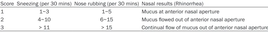
denatur-Table 1. Assessment form of rats with nasal symptoms
Score Sneezing (per 30 mins) Nose rubbing (per 30 mins) Nasal results (Rhinorrhea)
1 1~3 1~5 Mucus at anterior nasal aperture
2 4~10 6~15 Mucus flowed out of anterior nasal aperture
3 > 11 > 15 Continual flow of mucus out of anterior nasal aperture
Table 2. Nasal symptom assessment scores according to the number of days rats were subjected to nasal drop treatment using normal saline, ovalbumin, peptidoglycan, pyrrolidine dithiocarba-mate, or beclomethasone
Day Group A Group B Group C Group D Group E Day 1 0.84 ± 0.42 5.56 ± 1.24 3.33 ± 1.01 3.45 ± 1.76 4.86 ± 1.04 Day 2 0.73 ± 0.48 6.17 ± 0.84 3.26 ± 1.57 3.32 ± 1.00 3.94 ± 1.21 Day 3 1.69 ± 0.93 8.07 ± 1.12 3.56 ± 1.43 3.90 ± 1.20 4.92 ± 1.02 Day 4 1.58 ± 0.72 9.67 ± 0.48 4.28 ± 1.83 4.91 ± 1.25 3.75 ± 1.28 Day 5 0.96 ± 0.26 8.46 ± 1.35 5.42 ± 0.64 3.61 ± 0.43 5.55 ± 0.68 Day 6 0.75 ± 0.89 9.38 ± 1.20 4.83 ± 1.48 5.31 ± 1.24 4.74 ± 1.12 Day 7 0.66 ± 0.34 9.67 ± 1.23 4.65 ± 1.01 4.76 ± 1.12 4.42 ± 0.73
lowed by another three rinses
with PBS for 3 minutes. IL-12
[image:3.612.89.520.86.139.2]8705 Int J Clin Exp Med 2019;12(7):8702-8709
ed and quantified. The protein samples and protein markers were applied on top of a 4% stacking gel and 10% separating gel. The elec -trophoresis apparatus was operated at 80 V for 30 minutes. After the dye front transferred to the stacking gel, the voltage was increased to 120 V. The marker strip bands showed up clearly. The proteins were then transferred to a
polyvinylidene fluoride (PVDF) membrane using a semi-dry transfer unit at 160 mV for 70 min
-and the relative value of NF-κB protein was
obtained by dividing the two average gray values.
Statistical analysis
The experimental data were expressed as the mean ± standard deviation (SD). Statistical analysis was conducted using the SPSS13.0
statistical software package. The significance
test for the differences between the experi-mental groups was performed using an analy-sis of variance (ANOVA). When the variance was
homogeneous, the Fisher’s least significant dif -ference (LSD) procedure was used to perform pairwise comparisons. When the variance was not homogeneous, the Tamhane’s T2 method was used to perform pairwise comparisons. P <
0.05 was considered statistically significant.
Results
Symptom assessment scores
The symptom assessment scores after differ-ent days of nasal drop treatmdiffer-ent are shown in Table 2 (mean ± SD). After 14 days of sensiti-
zation, as expected, Group B showed obvious symptoms of AR on the first day of nasal drip
[image:4.612.87.377.71.370.2]Figure 1. Hematoxylin and eosin (H&E) staining of rat nasal mucosa (200× magnification). (A) Group A: normal control; (B) Group B: allergic rhinitis (AR) group; (C) Group C: AR + peptidoglycan; (D) Group D: AR + peptidoglycan + pyrrolidine dithio -carbamate (PDTC); (E) Group E: AR + peptidoglycan + beclomethasone. Neu, neutrophil; Eos, eosinophil.
Table 3. Results of nasal mucosal eosinophil count and neutrophil count (5 high power
field)
Group Average Eosinophil Count Average Neutrophil Count A 0.81 ± 0.33# 1.33 ± 0.41#
B 13.41 ± 2.37* 5.32 ± 2.25*
C 7.42 ± 2.35*,# 12.37 ± 2.58*,#
D 9.24 ± 1.39*,# 4.38 ± 2.34*,#
E 8.54 ± 1.47*,# 4.11 ± 2.36*,#
Values are mean ± standard deviation (SD), n = 20.
Group A: normal control; Group B: allergic rhinitis (AR); Group C: AR + peptidoglycan; Group D: AR + peptidogly
-can + pyrrolidine dithiocarbamate (PDTC); Group E: AR + peptidoglycan + beclomethasone. “*” indicates signifi
-cant differences compared to Group A, and “#” indicates significant differences compared to Group B (P < 0.05).
utes. After the transfer, one membrane was incubated wi-
th the NF-κB primary
antibo-dy (1:200) and another me- mbrane was incubated with the internal reference protein, GAPDH, primary antibody (1: 4000) at 4°C overnight. The membranes were then incu-bated with the secondary anti-body (1:10000) at 4°C over-night. Reagents were then ap- plied on the incubated
mem-branes, and light green fluo -rescent strips showed up af-
ter five minutes. The mem
-branes were dried using filter paper and covered by film.
Then, these membranes were exposed to light and rinsed with water. The markers were calibrated for analysis and scanning. Quantity One image analysis software was used to determine the average gray
value of NF-κB protein and
[image:4.612.90.289.428.518.2]stimulation, i.e., the rats showed clear symp-toms of nose rubbing, sneezing, and rhinor-rhea. Two days after nasal drip, the behavioral
scores of Group B were also significantly
hig-her than those of the othig-her groups. Groups C, D, and E also showed nasal symptoms of AR during the stimulation phase, but the nasal
symptom score was significantly lower than th-at of Group B. These results show thth-at peptido -glycan, PDTD, and beclomethasone reduced the degree of AR symptoms.
Values are mean ± standard deviation (SD), n =
20. Group A: normal control; Group B: allergic rhinitis (AR); Group C: AR + peptidoglycan; Gr-oup D: AR + peptidoglycan + pyrrolidine dithio
-carbamate (PDTC); Group E: AR + peptidoglycan + beclomethasone.
Changes in histomorphology
The nasal mucosa of Group A was normal
pseu-dostratified ciliated columnar epithelium. The dense fibrous connective tissue of the submu
-Eosinophil and neutrophil counts
The mean eosinophil and neutrophil counts in
Group B were significantly higher (16.6 times)
than in Group A (P < 0.05) (Table 3). The qu-
antities of eosinophils were significantly
reduc-ed after treating rats with peptidoglycan and PDTC or beclomethasone in comparison to
Group B. In contrast, in comparison to Group B, the neutrophil count was significantly
in-creased after allergen stimulation in Group C, while it was decreased in Groups D and E (P > 0.05).
Distribution and level of IL-12, IL-13, and IgE in nasal mucosa
The results showed that IL-12, IL-13, and IgE
were mainly distributed in the
pseudostratifi-ed ciliatpseudostratifi-ed columnar epithelium and glandular cells of the lamina propria of the nasal mucosa (Figure 2). The expression (200×) of IL-12, IL-13,
and IgE in Group B was significantly higher
th-an in Group A (P < 0.05). The expression of IL-12 in Group C was further elevated than that
cosa was thin. The mucosal epithelial structure was intact with neat arrangement, and the cilia thickness was
consis-tent without observed inflam
-matory cell infiltration (Figure 1). The epithelium of the nasal
mucosa in Group B was
she-dding and incomplete. Many lymphocytes and eosinophils
infiltrated the lamina propria,
indicating an obvious allergic injury. The nasal mucosa of Groups C, D, and E were thick-ened. The mucosal epithelium was exfoliated and incomp- lete after treating the AR rats with peptidoglycan and PDTC or beclomethasone; however, the damage to the mucosal epithelium was less than th-
at of Group B. Moreover, the
lamina propria of the mucous membrane showed a large
amount of inflammatory cell infiltration, with an obvious
[image:5.612.88.376.70.407.2]expanded gland in Groups C, D, and E. The allergic dama- ge was then alleviated. Figure 2. The expression and
8707 Int J Clin Exp Med 2019;12(7):8702-8709
TLR2 in nasal mucosa
The expression of TLR2 in Group B was 2.2
times higher than in Group A (P < 0.05). The expression of TLR2 was further elevated in
Groups C, D, and E in comparison to Group B (P
< 0.05) (Figure 3).
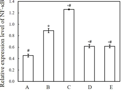
Expression level of NF-κB in nasal mucosa
The NF-κB protein expression level in the
na-sal mucosa of AR rats was higher than in the control group (P < 0.05). The expression level
of NF-κB was further increased after peptido -glycan stimulation but decreased after inject-ing rats with PDTC or beclomethasone (P < 0.05; Figure 4).
Discussion
The behavioral scores of AR rats were higher than those of the rats in the normal control group. The mucosal structure of rats in Group
B was also seriously damaged. These results clearly confirm that the allergic reaction
ap-peared in the AR rats with increased quantiti- es of eosinophils and neutrophils, as well as
increased expression of inflammatory factors [18].
The allergen, peptidoglycan, was used to sti- mulate the nasal mucosa in our study. The study carried out by Fu showed that TLR and
NF-κB can be potential targets for the treat
[image:6.612.92.408.96.177.2]-ment of allergic diseases [19]. As a pattern rec -ognition receptor, TLR is an important factor in the body’s activation of innate immunity and induction of adaptive immunity against patho-genic microorganisms through stimulation of NF-κB expression [20]. Our results show that
Table 4. Average optical density values of IL-12, IL-13, and IgE in nasal mucosa of groups A-E
Group IL-12 IL-13 IgE
A 0.09 ± 0.009015# 0.10171 ± 0.006957# 0.06217 ± 0.00401#
B 0.12378 ± 0.007633* 0.44242 ± 0.009588* 0.387 ± 0.005948*
C 0.19186 ± 0.007904*,# 0.14556 ± 0.005906*,# 0.217841 ± 0.006673*,#
D 0.14567 ± 0.006995*,# 0.2441 ± 0.006134*,# 0.24317 ± 0.00739*,#
E 0.15722 ± 0.008546*,# 0.26056 ± 0.009719*,# 0.2858 ± 0.00579*,#
Values are mean ± standard deviation (SD), n = 20. Group A: normal control; Group B: allergic rhinitis (AR); Group C: AR + peptidoglycan; Group D: AR + peptidoglycan + pyrrolidine dithio
[image:6.612.91.289.251.389.2]-carbamate (PDTC); Group E: AR + peptidoglycan + beclomethasone. “*” indicates significant differences compared to Group B, and “#” indicates significant differences compared to Group B(P < 0.05).
Figure 3. Relative mRNA expression levels of TLR2
in rat nasal mucosa. Group A: normal control; Group B: allergic rhinitis (AR); Group C: AR + peptidoglycan; Group D: AR + peptidoglycan + pyrrolidine dithiocar -bamate (PDTC); Group E: AR + peptidoglycan + beclo -methasone. Values are mean ± standard deviation (SD), n = 20.
Figure 4. Relative protein expression level of NF-κB in rat nasal mucosa. Group A: normal control; Group B: allergic rhinitis (AR); Group C: AR + peptidoglycan; Group D: AR + peptidoglycan + pyrrolidine dithiocar -bamate (PDTC); Group E: AR + peptidoglycan + beclo -methasone. Values are mean ± standard deviation (SD), n = 20.
in Group B (P < 0.05), while the expression of IL-13 and IgE in Gr- oup C was lower than
that in Group B (P < 0.05). For Groups D and E, the expression of IL-12 was increas- ed, while the expres-sion of IL-13 and IgE was reduced in
com-parison to Group B
(Table 4).
[image:6.612.92.289.494.640.2]expression of TLR2 and NF-κB increased sig
-nificantly in the peptidoglycan group (Group C) compared to Group B, without peptidoglycan treatment, suggesting that peptidoglycan in- duces the expression of TLR2 and NF-κB and affects T cell differentiation and cytokine pro-duction by activating the TLR2/NF-κB signaling pathway.
The balance of Th1/Th2 cytokines plays a vital
role in the pathogenesis of AR [21]. Th1 cyto -kine, IL-12, has been reported to inhibit IgE
syn-thesis and mediate cellular immunity [22, 23].
This agrees with our results as IL-12 had a
high-er expression level in Group C than in Group B,
but the level of IgE was lower in Group C. Th2 cytokine, IL-13, plays an important role in
pro-moting the synthesis of IgE in B lymphocytes [24]. Increased secretion of IgE by B lympho -cytes is associated with humoral immunity. These results indicated that humoral immunity was converted to cellular immunity after pepti-doglycan stimulation, producing many neutro-phils and reducing nasal mucosal damage in comparison to AR rats.
Lower expression levels of TLR2 and NF-κB
were observed in rats treated with PDTC (Group D) and beclomethasone (Group E) than in Gr- oup C, indicating that PDTC and beclometha-sone inhibit over-expression of TLR2 and NFκB,
which resulted in reduced numbers of neutro-phils, thus, preventing excessive increased cel-lular immunity in the nasal mucosa of AR rats. Our experiments have shown that peptidogly-can acts on TLR receptors on the surface of
effector cells and activates NF-κB through the TLR2/NF-κB signaling pathway, which further activates NF-κB transcription-related genes
and stimulates the release of many Th1-relat- ed cytokines, such as IL-12, and inhibits the synthesis of Th2-related cytokines, such as IL- 13. Increasing the Th1/Th2 ratio can correct the Th1/Th2 imbalance in the pathogenesis of AR and can convert humoral immunity into
cel-lular immunity. NF-κB inhibitors and glucocorti -coids can effectively reduce the expression of
NF-κB and inhibit the excessive enhanced cel
-lular immunity. Thus, the expression of NF-κB
plays an important regulatory role in the pa- thogenesis of allergic diseases and in the tr- ansformation from humoral immunity to cellu-lar immunity in the development of allergic
dis-eases. The application of NF-κB inhibitors
co-uld provide a new way to prevent and treat aller-gic diseases.
Acknowledgements
We would like to acknowledge the funding from Yumiao Funding of Putian University (Project No. 2014058).
Disclosure of conflict of interest
None.
Address correspondence to: Dr. Min Zhang, De- partment of Human Physiology, Basic Medical Col-lege of Putian University, Putian 351100, Fujian Province, China. Tel: +(86) 137 0606-5995; E-mail: zm06065995@163.com
References
[1] Di R, Lou X, Ye L, Miao J and Zhao Y. Preva-lence of allergic rhinitis and its effect on the quality of life of middle school students. Inter-national Journal of Clinical and Experimental Medicine 2016; 9: 15772-15779.
[2] Incorvaia C, Masieri S, Cavaliere C, Makri E, Sposato B and Frati F. Asthma associated to rhinitis. J Biol Regul Homeost Agents 2018; 32 Suppl 1: 67-71.
[3] Aberg N, Sundell J, Eriksson B, Hesselmaf B and Aberg B. Prevalence of allergic diseases in schoolchildren in relation to family history, up-per respiratory infections, and residential char-acteristics. Allergy 1996; 51: 232-237. [4] Luo Y, Deng Y, Tao Z, Chen S, Xiao B, Ren J,
Chen Z, Han J, Kong Y, Xu Y, Deng M. Regula-tory effect of microRNA-135a on the Th1/Th2 imbalance in a murine model of allergic rhini-tis. Exp Ther Med 2014; 8: 1105-1110. [5] Strachan DP. Family size, infection and atopy:
the first decade of the ‘hygiene hypothesis’. Thorax 2000; 55: S2-S10.
[6] Zhu D, Hu Y, Sun R, Sha J, Meng C, Cui N, Xiu Q and Li L. JNJ7777120, the histamine receptor 4 antagonist, decreases the allergic remodel-ing and Th2 inflammation in a rat model of al -lergic rhinitis. International Journal of Clinical & Experimental Medicine 2017; 10: 489-497. [7] Shpakou A, Brożek G, Stryzhak A, Neviartovich
T, Zejda J. Allergic diseases and respiratory symptoms in urban and rural children in Grod-no Region (Belarus). Pediatr Allergy ImmuGrod-nol 2012; 23: 339-346.
8709 Int J Clin Exp Med 2019;12(7):8702-8709 [9] Akira S, Takeda K and Kaisho T. Toll-like
recep-tors: critical proteins linking innate and ac-quired immunity. Nat Immunol 2001; 2: 657-680.
[10] Li L, Jin G, Jiang J, Zheng M, Jin Y, Lin Z, Li G, Choi Y and Yan G. Cornuside inhibits mast cell-mediated allergic response by down-regulating MAPK and NF-κB signaling pathways. Biochem Biophys Res Commun 2016; 473: 408-414. [11] Azzolina A, Bongiovanni A and Lampiasi N.
Substance P induces TNF-α and IL-6 produc -tion through NFκB in peritoneal mast cells. Bio -chim Biophys Acta 2003; 1643: 75-83. [12] Kim SH, Jun CD, Suk K, Choi BJ, Lim H, Park S,
Lee S, Shin HY, Kim DK and Shin TY. Gallic acid inhibits histamine release and pro-inflammato -ry cytokine production in mast cells. Toxicol Sci 2006; 91: 123-131.
[13] Park HH, Lee S, Oh JM, Lee MS, Yoon KH, Park BH, Kim JW, Song H and Kim SH. Anti-inflam -matory activity of fisetin in human mast cells (HMC-1). Pharmacol Res 2007; 55: 31-37. [14] Yang C, Li F, Wu Z, Shen W, Shen X and Huang
J. Molecular mechanisms of NF-κB signaling pathway in the development and progression of esophageal carcinoma. International Jour-nal of Clinical and Experimental Medicine 2018; 11: 488-499.
[15] Zhang D, Zheng N, Liu X. The role and mecha-nism of NF-κB in viral encephalitis of children. Exp Ther Med 2017; 13: 3489-3493.
[16] Aliprantis AO, Yang RB, Mark MR, Suggett S, Devaux B, Radolf JD, Klimpel GR, Godowski P and Zychlinsky A. Cell activation and apoptosis by bacterial lipoproteins through toll-like recep-tor-2. Science 1999; 285: 736-739.
[17] Aryan Z, Holgate ST, Radzioch D and Rezaei N. A new era of targeting the ancient gatekeepers of the immune system: toll-like agonists in the treatment of allergic rhinitis and asthma. Int Arch Allergy Immunol 2014; 164: 46-63.
[18] Xi L, Fan E, Zhao Y, Li Y, Zhang Y and Zhang L. Role of aluminum adjuvant in producing an al-lergic rhinitis animal model. Genet Mol Res 2014; 13: 5173-5181.
[19] Fu W, Zhao J, Liu X, Gao Y and Zheng C. The roles of the TLR/NF-κB signaling pathway in the mutual interactions between the lung and the large intestine. Mol Med Rep 2018; 18: 1387-1394.
[20] Zhang M, Lin JM, Li XS and Li J. Quercetin ame-liorates LPS-induced inflammation in human peripheral blood mononuclear cells by inhibi-tion of the TLR2-NF-κB pathway. Genet Mol Res 2016; 15.
[21] Shao YY, Zhou YM, Hu M, Li JZ, Chen CJ, Wang YJ, Shi XY, Wang WJ, Zhang TT. The anti-allergic rhinitis effect of traditional Chinese medicine of Shenqi by regulating mast cell degranula-tion and th1/th2 cytokine balance. Molecules 2017; 22: 504.
[22] Gour N and Wills-Karp M. IL-4 and IL-13 signal-ing in allergic airway disease. Cytokine 2015; 75: 68-78.
[23] Sogut A, Yilmaz O, Kirmaz C, Ozbilgin K, Onur E, Celik O, Pinar E, Vatansever S, Ding G and Yuksel H. Regulatory-T, T-helper 1, and T-helper 2 cell differentiation in nasal mucosa of aller-gic rhinitis with olive pollen sensitivity. Int Arch Allergy Immunol 2012; 157: 349-353.