Original Article
D-4F alleviates renal lipid abnormalities and increases
the protein levels of renal LXRβ in LDL receptor-null
mice fed a western diet
Jie Han1, Song Zhang2, Xiao-Han Ding3, Ping Ye3
1Department of Cardiology, The Second Affiliated Hospital of Xi’an Jiaotong University, Xi’an, China; 2Department of Geriatrics, Tianjin Medical University General Hospital, Tianjin, China; 3Department of Geriatric Cardiology, Chinese PLA General Hospital, Beijing, China
Received May 22, 2018; Accepted December 10, 2018; Epub May 15, 2019; Published May 30, 2019
Abstract: Objectives: Lipid abnormalities appear to contribute to both the initiation and progression of renal injury, regardless of etiology. D-4F contains a class A amphipathic helix with a polar face and a nonpolar face that allows it to bind lipids similar to apoA-I. We designed the present study to test whether D-4F treatment could alleviate lipid abnormalities in the kidneys and determine the role of liver X receptors (LXRs) in D-4F treatment. Methods: Female LDL receptor-null (LDLR-/-) mice 3-4 weeks old were administered a Western diet (21% fat and 0.3% cholesterol) for
18 weeks. Then, all the mice were randomly assigned to 3 groups for treatment with D-4F given in drinking water-a high-dose group (0.5 mg/ml), a low-dose group (0.3 mg/ml), and a control group (just drinking water)-for 6 weeks before examination. Hematoxylin-eosin (HE), periodic acid-Schiff (PAS), and Masson trichrome staining were used to evaluate renal histology. Lipid levels were determined by automatic biochemical analyzer and an enzyme-linked immunosorbent assay. Western blotting analysis was used to determine the expression of the LXR protein levels.
Results: D-4F treatment displayed, after HE staining, a significant decrease in the glomerular tuft-to-Bowman’s capsule diameter ratio and the number of cells per glomerulus. The cells also showed decreased PAS staining and Masson trichrome staining. The plasma levels of low-density lipoprotein cholesterol (LDL-C), high-density lipoprotein cholesterol (HDL-C), total cholesterol (TC), and triglyceride (TG) in the mice treated with D-4F did not show any sig-nificant changes. However, the addition of D-4F to the drinking water decreased the levels of LDL-C, TC and TG and increased the level of HDL-C in the kidney. The protein levels of LXRβ were significantly increased in the high-dose group, and the protein levels of LXRα did not change significantly after D-4F treatment. Conclusions: Our study sug-gests that D-4F could alleviate renal lipid accumulation, improve lipid metabolism and prevent renal histological de-terioration. These protective effects might be associated with LXRβ in the kidney instead of the plasma lipid levels. Keywords: Lipid abnormalities, renal disease, apoA-I mimetic peptide, D-4F, liver X receptors
Introduction
There has been a longstanding recognition of the potential relationship between lipoproteins and renal disease [1-4]. Numerous animal stud-ies have examined the role of lipid abnormali-ties in renal disease. These studies demon-strated that lipid abnormalities appear to con-tribute to both the initiation and progression of renal injury, regardless of etiology [4]. LDL receptor-null (LDLR-/-) C57BL/6J mice fed a
high-fat/high-cholesterol diet develop severe hyperlipidemia and serve as models for lipid abnormalities as the initiators of renal disease [5].
D-4F, an 18-amino acid mimetic peptide of apoA-I, contains a class A amphipathic helix with a polar and a nonpolar face that allows it to bind lipids similar to apoA-I [6-8]. We previously reported that when administered in drinking water for 6 weeks, D-4F increased cardiac high-density lipoprotein cholesterol (HDL-C) levels and decreased cardiac low-density lipoprotein cholesterol (LDL-C), total cholesterol (TC), and triglyceride (TG) levels in LDLR-/- C57BL/6J mice
consideration that liver X receptors (LXRs) are activated when cells accumulate excess cho-lesterol and that the expression of LXRα at pro-tein levels is significantly increased after D-4F treatment [8-10], we designed the present study to test whether D-4F treatment could alleviate lipid abnormalities in the kidneys and determine the role of LXRs in D-4F treatment.
Materials and methods
Materials
D-4F with the primary sequence Ac-DWFK- AFYDKVAEKFKEAF-NH2 was synthesized by the Shanghai Ketai Bio-Technique Co Ltd. (Shanghai, China). The purity of synthetic pep-tides (typically > 98%) was established by analytical high-performance liquid chromatog-raphy and confirmed by mass spectral analysis. Sodium pentobarbitone was purchased from Merck KGaA (Darmstadt, Germany). Enzyme-linked immunosorbent assay (ELISA) kits were obtained from Abcam (Cambridge, United King- dom). A bicinchoninic acid (BCA) protein assay kit was purchased from Pierce Biotechnolo- gy (Rockford, IL, USA). Protease inhibitor cock-tails were purchased from Roche Diagnostics GmbH (Mannheim, Germany). Antibodies were all purchased from Abcam (Cambridge, United Kingdom).
Animals and experimental protocol
Female LDLR-/- C57BL/6J mice were provided
by the Model Animal Research Center of Nanjing University and all the mice were car- ed for in accordance with the standards for laboratory animals established by the People’s Republic of China (GB14925-2001). The WD (21% fat and 0.3% cholesterol, Beijing Keao- xieli Co., Ltd. (Beijing, China)) was administered to the mice when they were 3-4 weeks old, and the treatment continued for 18 weeks. Then, all the mice were randomly assigned to 3 groups for treatment with D-4F given in drink-ing water-the high-dose group (0.5 mg/ml), the low-dose group (0.3 mg/ml), and the control group (just drinking water)-for 6 weeks before examination. Each group contained 10 mice. To ensure the efficacy of D-4F, the drinking wa- ter was changed every day. Each mouse con-sumed approximately 2.5 ml of water per day, and there was no significant difference in water or food consumption among the groups. All the mice were continued on the WD. After the
experimental period, the mice were anesthe-tized by an intraperitoneal injection of sodium pentobarbitone (50 mg/kg). Blood samples were collected for measurements of plasma biochemical indexes. All the kidneys were rap-idly excised, immersed in an ice-cold saline solution, and blotted dry. After removing any excess connective tissue, the kidneys were fixed in 10% neutral-buffered formalin and embedded in paraffin. The remaining kidneys were stored at -80°C after liquid nitrogen freezing.
Biochemical analysis
The plasma levels of LDL-C, HDL-C, TC and TG were determined by an automatic biochemi- cal analyzer (Roche 6000; Roche Diagnostics, Indianapolis, IN). The renal levels of LDL-C, HDLC, TC and TG were determined by ELISA using a commercial kit.
Histological studies of kidney
Transverse sections of the kidneys were fixed in 10% buffered formalin and embedded in pa- raffin. Two-micron-thick paraffin sections were prepared for histological examination with he- matoxylin-eosin (HE), periodic acid-Schiff (PAS) and Masson trichrome staining. Semiquanti- tative evaluation was performed at ten non-overlapping vision fields that were randomly selected under an Olympus microscope (Oly- mpus BX51T-PHD-J11, Japan) and an image analysis program (Image-Pro Plus 3.0, Media Cybernetics, Rockville, MD, USA).
Western analysis
with anti-LXRα (1:500) and anti-LXRβ (1:1000). Then, the membranes were incubated for 1 hour with horseradish peroxidase-labeled
anti-oup, the renal paraffin sections of the mice with D-4F treatment displayed, after HE staining, a significant decrease in the glomerular
tuft-to-Table 1. Effect of D-4F on WD-induced Weight Gain in LDLR-/- mice
(n=10/group)
0.5 mg/ml D-4F 0.3 mg/ml D-4F Control Body weight, g 21.51±0.60a,b 23.00±0.46a 24.79±0.66
Results are expressed as the mean ± SEM. aP<0.05, compared with the control
group. bP<0.05, compared with the 0.3 mg/ml D-4F group. Each group contained
[image:3.612.89.377.175.610.2]10 mice.
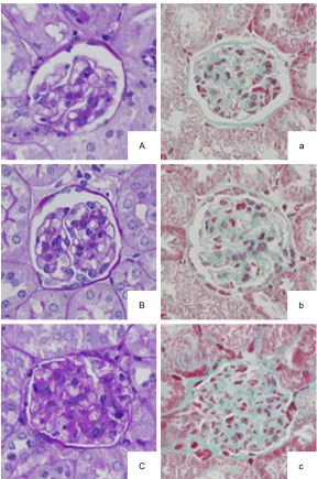
Figure 1. The sections were stained with periodic acid-Schiff (PAS) staining
(A-C) or Masson trichrome staining (a-c) and visualized at 400× magnifica-tions. (A and a) are from the high-dose group (0.5 mg/ml D-4F), (B and b) are from the low-dose group (0.3 mg/ml D-4F), and (C and c) are from the control group.
IgG (1:1000) at 37°C. The an- tigen-antibody complexes we- re revealed by an electroche-miluminescence detection sy- stem (Santa Cruz Biotechno- logy, Santa Cruz, CA). Glycer- aldehyde-3-phosphate dehyd- rogenase (GAPDH) was used as a loading control to normal-ize the data. The results were expressed as fold increases compared with the control. Statistical analysis
Continuous variables were ex- pressed as the mean values ± SEM. The differences be- tween the groups were com-pared by Student’s t test for phenotypes with equal vari-ance and by adjusted Stu- dent’s t test for those with unequal variance. All analy-ses were conducted using the SPSS software for Windows, version 13.0 (SPSS, Chica- go, IL). Statistical significance was set at P<0.05.
Results
The effect of D-4F on
WD-induced weight gain in LDLR-/- Mice
Compared with the control group, body weight was signif-icantly decreased, with a tr- end toward dose-responsivity, in the WD-fed mice treated with D-4F (Table 1).
The effect of D-4F on renal
histology in LDLR-/- mice
Bowman’s capsule diameter ratio and the num-ber of cells per glomerulus. They also showed decreased PAS staining, which is a hallmark of mesangial expansion with decreased extracel-lular matrix protein content (Figure 1). Further- more, Masson trichrome staining showed that collagen deposition and fibrotic areas were significantly decreased in the D-4F treatment groups (Figure 1). All these changes showed a trend toward dose-responsivity (Table 2). The effect of D-4F on plasma and renal lipids in LDLR-/- mice
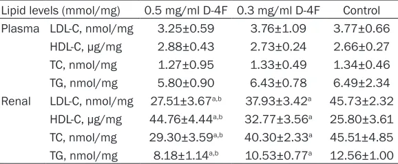
To investigate a possible relationship between renal histological changes and lipid levels, we measured the lipid levels in both the plasmas and kidneys. Compared with the levels in the control group, the plasma levels of LDL-C, HDL-C, TC and TG in mice treated with D-4F did not show any significant changes. However, the addition of the D-4F to the drinking water decreased the levels of LDL-C, TC and TG, and increased the level of HDL-C in the kidneys, and these changes showed a trend toward dose-responsivity (Table 3).
The purpose of our current study was to investi-gate the role of D-4F in renal histology and lipid metabolism changes in LDLR-/- mice and then
further explore the possible underlying mecha-nism. We examined 1) whether there were altered renal histology and lipid metabolisms in LDLR-/- mice treated with D-4F and 2) whether
LXRs were involved in D-4F induced changes of renal histology and lipid metabolism.
The C57BL/6J mice that were fed a high-fat diet (HFD) for 12 weeks developed metabolic syndrome, including obesity and hyperglyce-mia, and had an increase in renal lipid accu- mulation, which correlated well with the in- creased renal triglyceride and cholesterol con-tent [11-14]. Previous studies have also demon-strated that the kidneys of metabolic syndro- me patients have lipid accumulation that plays an important role in the pathogenesis of renal disease [15-17]. Buga and colleagues subse-quently found that these changes could be pre-vented partly by adding D-4F to drinking water (0.3 mg/ml D-4F) for 7 weeks. Compared with the LDLR-/- mice on a HFD without D-4F
treat-ment, the renal total triglyceride levels were
sig-Table 2. Effect of D-4F in LDLR-/- mice (n=10/group)
0.5 mg/ml D-4F 0.3 mg/ml D-4F Control Glomerular tuft/Bowman’s capsule diameter ratio, % 0.58±0.03a,b 0.69±0.03a 0.77±0.02
Number of cells per glomerulus 28.60±1.03a,b 37.70±1.16a 57.80±2.30
ECM protein content, % 14.48±0.76a,b 17.85±0.79a 20.20±0.51
Collagen deposition and fibrosis areas, % 23.83±4.08a,b 27.03±5.04a 32.28±4.17
Results are expressed as the mean ± SEM. aP<0.05, compared with the control group. bP<0.05, compared with the 0.3 mg/ml
[image:4.612.92.526.85.152.2]D-4F group. Each group contained 10 mice. A semiquantitative evaluation was performed at ten nonoverlapping vision fields that were randomly selected per mouse. Abbreviations: extracellular matrix (ECM).
Table 3. Effect of D-4F on plasma and renal lipid levels in LDLR
-/-mice (n=10/group)
Lipid levels (mmol/mg) 0.5 mg/ml D-4F 0.3 mg/ml D-4F Control Plasma LDL-C, nmol/mg 3.25±0.59 3.76±1.09 3.77±0.66
HDL-C, μg/mg 2.88±0.43 2.73±0.24 2.66±0.27 TC, nmol/mg 1.27±0.95 1.33±0.49 1.34±0.46 TG, nmol/mg 5.80±0.90 6.43±0.78 6.49±2.34 Renal LDL-C, nmol/mg 27.51±3.67a,b 37.93±3.42a 45.73±2.32
HDL-C, μg/mg 44.76±4.44a,b 32.77±3.56a 25.80±3.61
TC, nmol/mg 29.30±3.59a,b 40.30±2.33a 45.51±4.85
TG, nmol/mg 8.18±1.14a,b 10.53±0.77a 12.56±1.00
Results are expressed as the mean ± SEM. aP<0.05, compared with the control
group. bP<0.05, compared with the 0.3 mg/ml D-4F group. Every group contained 10
mice. Abbreviations: low-density lipoprotein cholesterol (LDL-C), high-density lipopro-tein cholesterol (HDL-C), total cholesterol (TC), and triglyceride (TG).
The effect of D-4F on the
expression of LXRs in LDLR-/- mice
Western blotting analysis was performed to investi-gate the change of LXRs after D-4F treatment. Com- pared with the control group, the protein levels of LXRβ increased in the high-dose group and the protein levels of LXRα did not change sig-nificantly after D-4F treat-ment (Figure 2).
[image:4.612.92.381.231.350.2]nificantly decreased in the LDLR-/- mice on a
HFD treated with D-4F. However, they did not see a change in renal total cholesterol levels [18]. Similar to their findings, we confirmed that the addition of D-4F to the drinking water decreased the renal total triglyceride levels. However, the difference was that, in our study, the renal total cholesterol levels also decreased after D-4F treatment. This effect was more obvious in the high-dose group (0.5 mg/ml D-4F). This might be partly because the mice in Buga’s study were slightly older at the st- art of the study (7 versus 3 weeks) and were maintained on the high-fat diet for less than half as long as in our study (7 versus 18 we- eks). Additionally, we also observed decreas- ed LDL-C levels, as well as increased HDL-C lev-els. These altered lipid levels induced by the D-4F treatment were the same as with our pre-vious work in cardiac tissue and are associated with alleviative lipid accumulation and improved cardiac function [8].
The effects of D-4F treatment on the plasma lipid levels in our study were the same as some other earlier studies [19]. Although D-4F treat-ment affected lipid accumulation in renal tis-sue, the plasma levels of LDL-C, HDL-C, TC and TG in the D-4F-treated mice were not influ-enced. The underlying mechanisms need to be further explored. Some studies assumed that the ability of D-4F to exert its positive effect might be independent of changes in circulating lipoprotein cholesterol levels [18, 19]. Aside from this, we speculated that the doses of 0.3 mg/ml and 0.5 mg/ml of D-4F in drinking water
rosis and proteinuria were also observed by Jiang and colleagues [14]. In the study by Buga and colleagues, the addition of D-4F in drinking water prevented the increase in the glomerular tuft-to-Bowman’s capsule diameter ratio, the number of cells per glomerulus, and mesangial cells that is observed in LDLR-/- mice on a WD
without D-4F treatment [18]. We confirmed these findings in our study. Furthermore, with the use of Masson trichrome staining, we also found that collagen deposition and fibrotic areas were significantly decreased in the D-4F treatment groups. Since we added two differ-ent doses of D-4F in the drinking water, all these changes showed a trend toward dose-responsivity and the higher dose of D-4F in drinking water seemed to exert a more obvious effect. Therefore, we suggested that through alleviating lipid accumulation and structure, D-4F treatment could delay or even reverse the process of renal disease induced by a HFD to a certain extent in the LDLR-/- mice.
[image:5.612.90.374.70.213.2]LXRs, which belong to the nuclear receptor superfamily, are activated when cells accumu-late excess cholesterol. After ligand binding, LXRs form heterodimers with retinoid X recep-tors and then regulate the transcription of genes involved in lipid/glucose metabolism and inflammation. LXRα is highly expressed in the liver, intestine, kidney, adipose tissue and mac-rophages, while LXRβ is ubiquitously expressed [20-23]. LXRs exert distinct effects on choles-terol and lipid metabolism in discrete cell types. The key biological effects of LXRs include the promotion of cellular cholesterol efflux from
Figure 2. Western blotting analysis of LXRα (A) and LXRβ (B) proteins in the
kidneys of the D-4F treatment and control groups. Every group contained 6 mice. GAPDH was included as a loading control. The quantitative data rep-resent the band intensity and are given as the means ± SEM. *P<0.05,
com-pared with the control group.
glomeruloscle-macrophages and foam cells, an improvement in cholesterol flux from the periphery to the liver, the inhibition of inflammation, and the pre-vention of cell death [24]. We previously sug-gested that the level of cardiac tissue HDL-C was increased after D-4F treatment and that the protein expressions of LXRα but not LXRβ were consistently elevated as well. In the pres-ent study, the level of renal tissue HDL-C was also increased, but the protein expressions of LXRα did not rise. In contrast, the protein expressions of LXRβ increased in the higher D-4F dose treatment group. These results imply that D-4F might exert protective effects in dif-ferent ways between difdif-ferent organs.
Conclusion
In conclusion, our study suggests that D-4F could alleviate renal lipid accumulation, im- prove lipid metabolism, and prevent renal histo-logical deterioration. These protective effects might be associated with LXRβ in the kidneys instead of in the plasma lipid levels.
Acknowledgements
We would like to thank colleagues at the De- partment of Laboratory Medicine, the PLA General Hospital, for their help with the bio-chemical measurements. We are also grateful to all study participants for their participation in the study. This research was supported by a grant from the National Natural Science Foundation of China (81801382) to Xiao-Han Ding.
Disclosure of conflict of interest
None.
Address correspondence to: Ping Ye, Department of Geriatric Cardiology, Chinese PLA General Hospital, Beijing, China. Tel: +86 10 66876369; Fax: +86 10 66876349; E-mail: [email protected]
References
[1] Moorhead JF, Chan MK, El-Nahas M and Var-ghese Z. Lipid nephrotoxicity in chronic pro-gressive glomerular and tubulo-interstitial dis-ease. Lancet 1982; 2: 1309-1311.
[2] Keane WF, Mulcahy WS, Kasiske BL, Kim Y and O’Donnell MP. Hyperlipidemia and progressive renal disease. Kidney Int Suppl 1991; 31: S41-48.
[3] Kamanna VS, Roh DD and Kirschenbaum MA. Hyperlipidemia and kidney disease: concepts derived from histopathology and cell biology of the glomerulus. Histol Histopathol 1998; 13: 169-179.
[4] Dalrymple LS and Kaysen GA. The effect of li-poproteins on the development and progres-sion of renal disease. Am J Nephrol 2008; 28: 723-731.
[5] Kennedy AJ, Ellacott KL, King VL and Hasty AH. Mouse models of the metabolic syndrome. Dis Model Mech 2010; 3: 156-166.
[6] Navab M, Anantharamaiah GM, Reddy ST, Hama S, Hough G, Grijalva VR, Yu N, Ansell BJ, Datta G, Garber DW and Fogelman AM. Apoli-poprotein A-I mimetic peptides. Arterioscler Thromb Vasc Biol 2005; 25: 1325-1331. [7] Navab M, Anantharamaiah GM, Reddy ST, Van
Lenten BJ, Buga GM and Fogelman AM. Pep-tide mimetics of apolipoproteins improve HDL function. J Clin Lipidol 2007; 1: 142-147. [8] Han J, Zhang S, Ye P, Liu YX, Qin YW and Miao
DM. Apolipoprotein A-I mimetic peptide D-4F reduces cardiac hypertrophy and improves apolipoprotein A-I-mediated reverse cholester-ol transport from cardiac tissue in LDL recep-tor-null mice fed a western diet. J Cardiovasc Pharmacol 2016; 67: 412-417.
[9] Repa JJ and Mangelsdorf DJ. The role of or-phan nuclear receptors in the regulation of cholesterol homeostasis. Annu Rev Cell Dev Biol 2000; 16: 459-481.
[10] Janowski BA, Willy PJ, Devi TR, Falck JR and Mangelsdorf DJ. An oxysterol signalling path-way mediated by the nuclear receptor LXR al-pha. Nature 1996; 383: 728-731.
[11] Parekh PI, Petro AE, Tiller JM, Feinglos MN and Surwit RS. Reversal of diet-induced obesity and diabetes in C57BL/6J mice. Metabolism 1998; 47: 1089-1096.
[12] Fried LF, Orchard TJ and Kasiske BL. Effect of lipid reduction on the progression of renal dis-ease: a meta-analysis. Kidney Int 2001; 59: 260-269.
[13] Molnar J, Yu S, Mzhavia N, Pau C, Chereshnev I and Dansky HM. Diabetes induces endotheli-al dysfunction but does not increase neointi-mal formation in high-fat diet fed C57BL/6J mice. Circ Res 2005; 96: 1178-1184.
[14] Jiang T, Wang Z, Proctor G, Moskowitz S, Lieb-man SE, Rogers T, Lucia MS, Li J and Levi M. Diet-induced obesity in C57BL/6J mice causes increased renal lipid accumulation and glo-merulosclerosis via a sterol regulatory ele-ment-binding protein-1c-dependent pathway. J Biol Chem 2005; 280: 32317-32325.
in the kidney in the early stages of obesity. J Am Soc Nephrol 2001; 12: 1211-1217. [16] Praga M. Obesity--a neglected culprit in renal
disease. Nephrol Dial Transplant 2002; 17: 1157-1159.
[17] Bagby SP. Obesity-initiated metabolic syn-drome and the kidney: a recipe for chronic kid-ney disease? J Am Soc Nephrol 2004; 15: 2775-2791.
[18] Buga GM, Frank JS, Mottino GA, Hakhamian A, Narasimha A, Watson AD, Yekta B, Navab M, Reddy ST, Anantharamaiah GM and Fogelman AM. D-4F reduces EO6 immunoreactivity, SREBP-1c mRNA levels, and renal inflamma-tion in LDL receptor-null mice fed a western diet. J Lipid Res 2008; 49: 192-205.
[19] Navab M, Anantharamaiah GM, Hama S, Gar-ber DW, Chaddha M, Hough G, Lallone R and Fogelman AM. Oral administration of an Apo A-I mimetic peptide synthesized from D-amino acids dramatically reduces atherosclerosis in mice independent of plasma cholesterol. Cir-culation 2002; 105: 290-292.
[20] Willy PJ, Umesono K, Ong ES, Evans RM, Hey-man RA and Mangelsdorf DJ. LXR, a nuclear receptor that defines a distinct retinoid re-sponse pathway. Genes Dev 1995; 9: 1033-1045.
[21] Goldstein JL, DeBose-Boyd RA and Brown MS. Protein sensors for membrane sterols. Cell 2006; 124: 35-46.
[22] Apfel R, Benbrook D, Lernhardt E, Ortiz MA, Salbert G and Pfahl M. A novel orphan receptor specific for a subset of thyroid hormone-re-sponsive elements and its interaction with the retinoid/thyroid hormone receptor subfamily. Mol Cell Biol 1994; 14: 7025-7035.
[23] Auboeuf D, Rieusset J, Fajas L, Vallier P, Frering V, Riou JP, Staels B, Auwerx J, Laville M and Vi-dal H. Tissue distribution and quantification of the expression of mRNAs of peroxisome prolif-erator-activated receptors and liver X receptor-alpha in humans: no alteration in adipose tis-sue of obese and NIDDM patients. Diabetes 1997; 46: 1319-1327.