R E S E A R C H
Open Access
Galectin-9 prolongs the survival of septic mice by
expanding tim-3-expressing natural killer T cells
and PDCA-1
+
CD11c
+
macrophages
Takashi Kadowaki
1†, Asahiro Morishita
2†, Toshiro Niki
1, Junko Hara
3, Miwa Sato
3, Joji Tani
2, Hisaaki Miyoshi
2,
Hirohito Yoneyama
2, Tsutomu Masaki
2, Toshio Hattori
4, Akihiro Matsukawa
3and Mitsuomi Hirashima
2,5*Abstract
Introduction:Galectin-9 ameliorates various inflammatory conditions including autoimmune diseases by regulating T cell and macrophage/dendritic cell (DC) functions. However, the effect of galectin-9 on polymicrobial sepsis has not been assessed.
Methods:We induced polymicrobial sepsis by cecal ligation and puncture (CLP) in mice. The survival rate was compared between galectin-9- and PBS-treated CLP mice. An ELISA was used to compare the levels of various cytokines in the plasma and culture supernatants. Fluorescence-activated cell sorting analysis was further performed to compare the frequencies of subpopulations of spleen cells.
Results:Galectin-9 exhibited a protective effect in polymicrobial sepsis as demonstrated in galetin-9 transgenic mice and therapeutic galectin-9 administration. In contrast, such effect was not observed in nude mice, indicating the involvement of T cells in galectin-9-mediated survival prolongation. Galectin-9 decreased TNFα, IL-6, IL-10 and, high mobility group box 1 (HMGB1) and increased IL-15 and IL-17 plasma and spleen levels. Galectin-9 increased the frequencies of natural killer T (NKT) cells and PDCA-1+CD11c+macrophages (pDC-like macrophages) but did not change the frequency of CD4 or CD8 T cells,γδT cells or conventional DC. As expected, galectin-9 decreased the frequency of Tim-3+CD4 T cells, most likely Th1 and Th17 cells. Intriguingly, many spleen NK1.1+NKT cells and pDC-like macrophages expressed Tim-3. Galectin-9 increased the frequency of Tim-3-expressing NK1.1+NKT cells and pDC-like macrophages. Galectin-9 further increased IL-17+NK1.1+NKT cells.
Conclusion:These data suggest that galectin-9 exerts therapeutic effects on polymicrobial sepsis, possibly by expanding NKT cells and pDC-like macrophages and by modulating the production of early and late proinflammatory cytokines.
Introduction
Sepsis is the leading cause of death in critically ill patients, and the incidence of sepsis is increasing. The mortality rate of severe sepsis is very high, up to 70%. Two types of animal sepsis model have been established: the lipo-polysaccharide(LPS)-induced inflammation, and the cecal ligation and puncture (CLP) model of microbial sepsis.
LPS stimulates macrophages to release large amounts of TNFα and IL-1β that can precipitate tissue injury and lethal shock. Antagonists of TNFαand IL-1βhave shown limited efficacy in clinical trials, most likely because these cytokines are early mediators in sepsis pathogenesis [1,2].
On the other hand, high mobility group box 1 (HMGB1) is thought to be a late mediator of endotoxin lethality in mice, and HMGB1 is first detectable in the circulation 8 hours after the onset of sepsis disease, subsequently increasing to plateau levels from 16 to 32 hours [3]. Ad-ministration of HMGB1-specific neutralizing antibodies beginning 24 hours after the onset of sepsis induced by CLP was shown to lead to a dose-dependent rescue of mice from lethal sepsis [4-6].
* Correspondence:mitsuomi@kms.a
†Equal contributors 2
Department of Gastroenterology and Neurology, Kagawa University Faculty of Medicine, Kagawa, Japan
5
Laboratory of Biodefense Research, Faculty of Pharmaceutical Sciences at Kagawa Campus, Tokushima Bunri University, Kagawa, Japan
Full list of author information is available at the end of the article
Recent studies have also shown that programmed death-1 (PD-1) expression on macrophages is critically associated with altering microbial clearance and the innate inflammatory response to sepsis in CLP mice [7]. Upregulation of PD-1 on T cells and the PD-ligand (L) 1 on monocytes in patients with septic shock has also been observed [8], and it has been shown that PD-1 levels correlate with increased mortality, nosocomial in-fections, and immune dysfunction in patients with septic shock [9]. Moreover, blockade of the PD-1/PD-L1 path-way improves survival in CLP mice by reversing immune dysfunction [10-12].
Galectin-9 (Gal-9) is a member of the galectin family that selectively binds to β-galactoside [13]. Gal-9 was first identified as an apoptosis-inducing factor for thymocytes [14] and an eosinophil-activating factor [15]. However, recent experiments have revealed that Gal-9 is a ligand of Tim-3 that is expressed on Th1 and Th17 cells, and that Gal-9 signaling induces death of these cells, resulting in the suppression of Th1- and Th17-related cytokine production in vivo andin vitro[16,17]. More recently, it has been shown that Gal-9 derived from surface Gal-9-expressing Th cells (ThGal-9) suppresses the development of Th17 cells and enhances the development of FoxP3+ regulatory T cells (Tregs) in a Tim-3-independent manner [18]. In addition to the above mechanisms, Gal-9 modulates immune responses by expanding myeloid suppressor cells [19] and plasma-cytoid dendritic cell (pDC)-like macrophages [20-23]. We have also shown that Gal-9 ameliorates LPS-induced inflammation and protects mice from an LPS-induced Schwartzman reaction by suppressing the production of proinflammatory cytokines from LPS-stimulated macro-phages and activating PGE2-producing neutrophils, respectively [24]. Therefore, it is of interest to examine the role of Gal-9 in a murine model of polymicrobial sepsis induced by CLP. The purpose of the present experiment is to show a beneficial effect of Gal-9 in a murine sepsis model induced by CLP and to clarify possible functions of Gal-9 in this model.
Materials and methods Mice
Female wild-type (WT) Balb/c mice and Gal-9 trans-genic (TG) mice were used in the present study [24].
These mice were maintained on a 12:12-hour light–
dark cycle under specific pathogen-free conditions at Kagawa University. The animals were fed a standard
laboratory diet and were provided water ad libitum.
All experimental procedures were approved by the Animal Care and Use Committee of Kagawa University. The animals used in this research received humane care in accordance with international guidelines and national law.
Induction of sepsis
CLP appears to be a reliable and clinically relevant animal model of the human septic condition because it produces an endogenous polymicrobial infection similar to clinical peritonitis and sepsis. Thus, sepsis was in-duced by CLP in the present experiments as described elsewhere [25]. Briefly, mice were anesthetized, and the cecum was exposed, ligated with a 3–0 silk suture and punctured once with an 18-gauge needle. The cecum was then replaced in the peritoneal cavity, and the inci-sion was closed with surgical staples. All mice received a subcutaneous injection of 1 ml of sterile saline to avoid dehydration and were placed on a heating pad until they recovered from anesthesia. All mice were allowed free access to food and water throughout the experiments. We used stable human Gal-9 [26] to assess the effects of Gal-9 on the survival of CLP mice. The purity of Gal-9 was >95%. The endotoxin level was determined using a Limulus assay (BioWhittaker, Tokyo, Japan) and was found to be <0.008 endotoxin units. CLP mice were treated with Gal-9 intravenously or subcutaneously im-mediately after CLP unless otherwise specified. To deter-mine the survival of the mice, CLP mice were monitored for 7 days after CLP. In a different set of experiments, CLP mice were anesthetized, bled and euthanized at indicated time points after CLP.
Bacterial colony-forming unit determination
The peritoneal cavities were washed with 2 ml of sterile saline, and the lavage fluids were harvested. A 10 μl aliquot of lavage fluids was used to assess bacterial colony-forming units (CFU). Peritoneal fluids (PF, 10μl) from each mouse were serially diluted with sterile saline. Each PF dilution was plated on tryptic soy blood agar plates and incubated overnight at 37°C, after which the number of aerobic colonies was counted. Data were expressed as CFU per 10μl PF.
Cell preparation
Spleens were obtained at 24 hours after CLP from mice treated with PBS or Gal-9 immediately after CLP and dispersed into single-cell suspensions. Red blood cells (RBC) were lysed with 0.1 N NH4Cl, and the spleen cells
were washed three times with Roswell Park Memorial Institute medium (RPMI) 1640 and cultured in RPMI 1640 solution supplemented with 5% FCS, glutamine and antibiotics in a 5% CO2incubator for 48 hours.
Measurement of cytokines
study did not cross-react with other available murine cytokines.
Flow cytometry analysis
Spleen cells were harvested from WT and Gal-9 TG CLP mice at 24 hours after CLP, and the cells (1 × 10⁶ cells/ml) were suspended in PBS supplemented with 2% FCS and 0.1% sodium azide. Cells were stained with mAbs specific for mouse CD3, CD4, CD8, GL-3, NK1.1 and Tim-3 (BioLegend, Tokyo, Japan). Stained cells were analyzed using a FACSCalibur (BD Biosciences, Tokyo, Japan). To assess intracellular IL-17 production, spleen cells were cultured for 48 hours in the presence of 1μg/ml brefeldin A (Sigma, Tokyo, Japan) and were permeabilized overnight with permeabilization buffer (Cytofix/Cytoperm; eBioscience, San Diego, CA, USA).
Statistics
Statistical significance was determined using the Mann– WhitneyU-test. In the case of survival curves, the data were analyzed using the log-rank test. A P-value <0.05 was regarded as statistically significant. All data were expressed as mean ± standard error of the mean (SEM).
Results and discussion
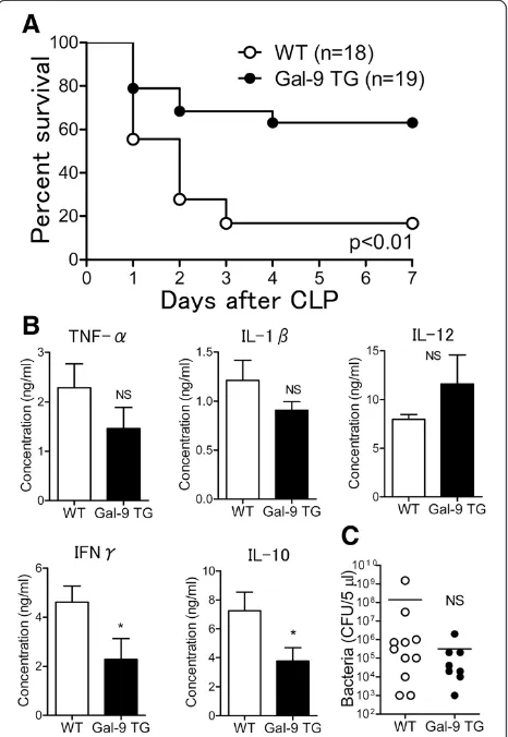
Improved survival in CLP-induced sepsis in Gal-9 TG mice We first compared the survival rate after CLP between (WT and Gal-9 TG mice. The survival rate of Gal-9 TG mice was significantly higher than that of WT mice. Of the 19 Gal-9 TG mice, 12 survived to day 7 after CLP (63.2%), whereas only 3 of 18 WT mice survived to day 7 (16.7%) (P<0.01). Thus, Gal-9 TG mice were resistant to the lethality induced by CLP, thereby suggesting a beneficial effect of Gal-9 administration in mice under-going CLP (Figure 1A).
To uncover the mechanism by which Gal-9 prolongs the survival of CLP mice, we assessed the levels of pro-inflammatory cytokines such as TNF-αand IL-1βin the PF of WT and Gal-9 TG mice at 24 hours after CLP.
Figure 1B shows that the levels of TNF-α and IL-1β
were relatively decreased at this time point and that the level of IL-12 was relatively increased in Gal-9 TG mice compared to WT mice. However, we previously showed that the levels of TNF-α and IL-12 in PF were signifi-cantly suppressed in Gal-9 TG mice during early periods (1 to 6 hours) of LPS-induced peritoneal inflammation [24]. In contrast, the levels of IFNγ and IL-10 were significantly decreased in Gal-9 TG mice.
We further tested whether Gal-9 could decrease the bacterial load in PF at 24 hours after CLP. The bacterial load in Gal-9 TG mice tended to be lower than the bacterial load in WT mice but the difference was not statistically significant (Figure 1C). No bacterial CFU or few bacterial CFU were found at 7 days after CLP in
the PF of Gal-9-treated surviving mice, as expected (data not shown).
[image:3.595.306.539.90.428.2]Delayed Gal-9 treatment prolongs the survival of CLP mice To determine whether Gal-9 exhibits protective effects in CLP mice, we treated CLP mice with a single Gal-9 administration (intravenous (i.v.), 30μg/mouse) immedi-ately after CLP. As shown in Figure 2A, 7 of 16 Gal-9-treated CLP mice survived at day 7 after CLP (43.8%), whereas only 2 of 16 PBS-treated mice survived at this time point (12.5%) (P<0.05). We further failed to detect any significant difference in the bacterial loads of the PF between PBS- and Gal-9-treated mice at 24 hours after CLP (Figure 2B). Similar to Gal-9 TG mice, no bacterial
Figure 1Survival of galectin (Gal)-9 transgenic (TG) mice during polymicrobial sepsis induced by cecal ligation and puncture (CLP). (A)Prolonged survival of Gal-9 TG mice. CLP was performed, and survival was monitored for 7 days after CLP in wild-type (WT) and Gal-9 TG CLP mice. The log-rank (Mantel-Cox) test was used for statistical analysis;P<0.01.(B)Cytokine levels in peritoneal fluids (PF) from WT (n = 18) and Gal-9 TG (n = 19) mice. At 24 hours after CLP, PF was obtained and ELISA was used for quantification of cytokine levels (*P<0.05; NS, not significant).(C)
CFU or few bacterial CFU were found at 7 days after CLP in the PF of Gal-9-treated surviving mice (data not shown). These results suggest that direct bacterial suppression by Gal-9 is not the trigger for this prolonged survival. Further studies are required to ascertain the mechanisms how Gal-9 finally decreases bacterial CFU on Day 7.
Furthermore, delayed i.v. Gal-9 administration given at 24 hours after CLP also significantly prolonged the sur-vival of CLP mice: 11 of 15 Gal-9-treated mice survived at day 7 after CLP (73.3%), whereas only 4 of 16 PBS-treated mice survived at this time point (25%) (P <0.05) (Figure 2C). Moreover, we found that delayed sub-cutaneous Gal-9 treatment given at 24 hours after CLP similarly prolonged the survival of CLP mice: 15 of 22 Gal-9-treated mice survived at day 7 after CLP (68.2%), whereas 8 of 22 PBS-treated mice survived at this time point (40.0%) (P= 0.058). These results suggest that Gal-9 has therapeutic effects on microbial sepsis, prob-ably by modulating the immune response to prolong the survival of CLP mice.
Interestingly, Gal-9 did not exhibit any therapeutic effects in nude mice: All animals that received Gal-9 administration died by 3 days after CLP similar to CLP mice that did not receive Gal-9 (Figure 2D), suggesting
that T cells are critically involved in the survival enhancement mediated by Gal-9.
Effects of Gal-9 on plasma levels of cytokines
To determine the functions of Gal-9 in CLP mice, plasma levels of pro-inflammatory cytokines were assessed. Similar to the Gal-9 TG mice, Gal-9 failed to affect the levels of early pro-inflammatory
cyto-kines such as TNF-α at 24 hours after CLP, although
the level of IL-6 was suppressed by Gal-9 (Figure 3). In contrast, the level of HMGB1, a late inflammatory mediator [3], was markedly suppressed in Gal-9-treated mice at 24 hours after CLP (Figure 3). This result seems reasonable because delayed administration of anti-bodies against HMGB1 attenuates endotoxin lethality [3] and CLP-induced sepsis in rodents [4,5]. Fur-thermore, we found a decreased level of IL-10 and increased levels of IL-15 and IL-17 in Gal-9-treated CLP mice.
[image:4.595.58.537.90.324.2]of pro-inflammatory cytokines (TNF-α, IL-12 and IFNγ) during early time points (1 to 6 hours) following LPS-induced inflammation [24].
Effects of Gal-9 on cytokine production from spleen cells The next set of experiments was performed to compare the levels of cytokine production from spleen cells from PBS- and Gal-9-treated CLP mice. Spleen cells were obtained at 24 hours after CLP and cultured without any stimulation for 48 hours. Spleen cells from Gal-9-treated CLP mice released decreased levels of early and late proinflammatory cytokines such as TNFα, IL-6, IL-12 and HMGB1 (Figure 4). The level of IL-10 was also de-creased in Gal-9-treated CLP mice (Figure 4). Although IL-10 is an immunosuppressive cytokine, neutralization of IL-10 leads to improved survival of septic mice by restoring the downregulation of the IL-18 receptor on NK cells and IFN-γproduction [28,29].
In contrast, an increase in IL-17 production was observed from spleen cells from Gal-9-treated CLP mice (Figure 4), which is unexpected because Gal-9 reduces IL-17-producing Th17 cells by inducing apoptosis and downregulating the differentiation of Th17 cells [17,18]. IL-17 is a proinflammatory cytokine produced during acute, delayed and autoimmune inflammation [30]. In addition to Th17 cells, other T cells such as CD8 T cells (Tc17) [31,32], natural killer T (NKT) cells [33,34] and
γδT cells [35,36] could be the source of IL-17. In
autoimmune diseases, it is important to reduce pro-inflammatory IL-17 levels to improve disease severity [17]. In contrast, IL-17 plays a critical role in protecting mice from sepsis-mediated lethality [37,38].
[image:5.595.58.543.88.470.2]such as Bcl-2 [39]. Inoue et al. [40] recently showed that IL-15 prevents apoptosis, reverses innate and adaptive immune dysfunction and improves survival in a sepsis model. Therefore, it seems reasonable that there is upregulation of IL-17 and IL-15 in Gal-9-treated CLP mice that are resistant to CLP-induced lethality.
The level of IL-10 was suppressed in Gal-9-treated CLP mice (Figure 4). Although IL-10 is an immuno-suppressive cytokine, neutralization of IL-10 leads to improved survival by restoring the downregulation of the IL-18 receptor on NK cells and IFN-γproduction in septic mice [28,29].
Based on the present results, it is thus suggested that it is important to increase IL-15 and IL-17 production and to decrease IL-10 and HMGB1 production simul-taneously to protect mice from CLP-induced lethality. Furthermore, as we have previously proposed [41], Gal-9
should be regarded as a homeostasis-maintaining factor to keep an adequate immune response, not just as an immunosuppressive factor or immunostimulatory factor.
Effects of Gal-9 on spleen T cells
As noted above, there are several types of T cells: CD4 T cells (CD3+ CD4+), CD8 T cells (CD3+ CD8+),γδT cells (CD3+ GL-3+) and NKT cells (CD3+ NK1.1+). Spleen cells were obtained 24 hours after CLP. Flow cytometric analysis was performed to clarify which types of T cells are expanded by Gal-9 in CLP mice. The frequencies of CD4 and CD8 T cells were not changed by Gal-9 (Figure 5A). We also assessed the frequency of Tim-3 expression on those T cells, because Tim-3 is a ligand of Gal-9 [16]. Approximately 6% of CD4 T cells expressed Tim-3 on their surface, and Gal-9 reduced the frequency
of Tim-3+ CD4 T cells, likely Th1 and Th17 cells
[image:6.595.57.541.89.470.2]show that Gal-9 induces the cell death of both Th1 and Th17 cells through a Gal-9/Tim-3 interaction [16-18]. In contrast, Gal-9 weakly but not significantly increased Tim-3+ CD8 T cells in the spleen of Gal-9-treated mice (Figure 5B), suggesting that Gal-9 does not induce cell death of Tim-3+CD8 T cells, at least under these condi-tions. Indeed, we have shown that Gal-9 potentiates CD8 T cell-mediated antitumor immunity via Gal-9-Tim-3 interactions between dendritic cells (DCs) and CD8 T cells [41].
Moreover, Gal-9 increased the frequency of CD3+
NK1.1+ cells, most likely NKT cells (Figure 5C). One
third of NK1.1+ NKT cells expressed Tim-3, and Gal-9 significantly increased the frequency of Tim-3-express-ing NK1.1+NKT cells (Figure 5D). In contrast, Gal-9 did not increase the frequency of γδT cells (Figure 5C) or the Tim-3 expression of those cells (Figure 5D). We further found that Gal-9 treatment resulted in an increase in the frequency of intracellular IL-17+ NKT cells in the spleens of CLP mice (Figure 5E). These find-ings suggest that Gal-9 does not induce cell death of
Tim-3-expressing NK1.1+ NKT cells, but enhances
[image:7.595.59.538.88.465.2]activation of those cells to produce IL-17. It is not sur-prising, because we had similar findings in our previous Figure 5Effects of galectin (Gal)-9 on subpopulations of spleen T cells.Spleen cells were obtained from PBS- or Gal-9-treated cecal ligation and puncture (CLP) mice at 24 hours after a single intravenous injection. Flow cytometric analysis was performed to clarify which types of T cells are expanded by Gal-9 in the spleens of CLP mice.(A)The frequencies of CD4 and CD8 T cells. Spleen cells were stained with CD3, CD4, CD8 and Tim-3, and fluorescence-activated cell sorting (FACS) analysis was performed; n = 5 for each group (NS, not significant).(B)The frequency of Tim-3+ CD4 and CD8 T cells. Spleen cells were stained with CD3, CD4, CD8 and Tim-3, and FACS analysis was performed; n = 5 for each group (NS, not significant;***P<0.001).(C)The frequencies of natural killer T (NKT) cells andγδT cells. Spleen cells were stained with CD3, GL-3 and
NK1.1, and FACS analysis was performed; n = 5 for each group (NS, not significant;**P<0.001).(D)Expansion of Tim-3+ NKT cells but notγδT cells.
Spleen cells were stained with CD3, GL-3, NK1.1 and Tim-3, and FACS analysis was performed; n = 5 for each group (NS, not significant;***P
work, showing that Gal-9 promotes the activation of DCs to produce a small amount of TNF-α[42].
From the present experiments, NKT cells are proposed to be one of the main cell-types responsible for the Gal-9-induced prolongation of survival, by releasing IL-17 in CLP mice. Regarding the role of NKT cells in sepsis, there are competing opinions: one is that NKT cells are involved in harmful outcomes in sepsis as reviewed by Leung and Harris [43], and another is that those cells exhibit a protective function in sepsis [44,45]. Thus, it becomes important to explain such a discrepancy. NKT cells are broadly categorized into at least two groups: Type I NKT cells and Type II NKT cells [46]. Furthermore, immune response in sepsis consists of an initial hyper-reactive phase and a latent phase. The initial hyper-reactive phase is characterized by
the large release of pro-inflammatory cytokines from mac-rophages et cetera, and the latent anti-inflammatory or immunosuppressed phase is characterized by hypotensive shock, inability to clear the infection, increased suscepti-bility to nosocomial infections, and possible multiple organ failure and death [47]. Our previous and present ex-periments suggest that Gal-9 decreases pro-inflammatory cytokine levels at the initial hyper-reactive phase [24]. In contrast, Gal-9 decreases HMGB1 but increases IL-10
and IL-17. Gal-9 also expands Tim-3+ NK1.1+ NKT
cells and IL-17+ NKT cells. Therefore, we cannot
ex-clude the possibility that Tim-3 expressing NK1.1+
[image:8.595.60.538.296.664.2]NKT cells produce IL-17 to protect sepsis through the Gal-9/Tim-3 pathway. Thus, the hypothesis may be proposed that Gal-9 contributes to downregulate the early inflammatory response during sepsis but also to
Figure 6Effects of galectin (Gal)-9 on splenic macrophages and dendritic cells (DCs).Spleen cells were obtained from PBS- or Gal-9 treated cecal ligation and puncture (CLP) mice at 24 hours after a single intravenous injection and stained with F4/80, PDCA-1, CD11c and Tim-3.(A)An increased frequency of plasmacytoid (p)DC-like macrophages (F4/80+ PDCA-1+ CD11c+) but not cDCs (F4/80- PDCA-1- CD11c+) was observed; n = 5 for each group (NS, not significant;*P<0.05).(B)High Tim-3 expression on pDC-like macrophages was observed; n = 5 for each group
(NS, not significant;***P<0.001). Blue line, pDC-like macrophages; orange line, cDCs; red line, PDCA-1+ CD11c- cells.(C)Increased Tim-3-expressing
upregulate the delayed inflammatory response; further studies are required to clarified this.
Effects of Gal-9 on spleen macrophages and DCs
Because innate immune cells such as macrophages and DCs may play a critical role in prolonging the survival of septic mice by activating/regulating T cells [7,48,49], we performed the experiments to clarify whether Gal-9 has an effect on the frequency of macrophages or DCs. Gal-9 markedly increased the frequency of plasmacy-toid (p)DC-like macrophages (F4/80+PDCA-1+CD11c+), although Gal-9 failed to increase the frequency of cDCs (F4/80-PDCA-1-CD11c+) (Figure 6A).
Next, we assessed the Tim-3 expression on macro-phages and DCs in the spleens of CLP mice. Intri-guingly, half of the pDC-like macrophages expressed Tim-3, although cDCs did not significantly express this molecule (Figure 6B). Moreover, we compared the Tim-3 expression on splenic macrophages and DCs between the Gal-9- and PBS-treated CLP mice. Gal-9 significantly increased the frequency of Tim-3-expressing pDC-like macrophages (Figure 6C). In contrast, the frequency of Tim-3+cDCs was low (Figure 6B), and Gal-9 failed to in-crease the frequency of Tim-3-expressing cDCs in CLP mice (Figure 6C). HMGB1 is a macrophage-derived late proinflammatory cytokine, and TLR4 and CD14 are required for the release of HMGB1 [50]. pDC-like macro-phages suppress LPS-induced release of early proinflam-matory cytokines from macrophages by suppressing TLR4 and CD14 expression on macrophages [21,22]. Thus, it was suggested that immunosuppressive pDC-like mac-rophages may mediate decreased release of early and late proinflammatory cytokines from macrophages that also, at least in part, may be involved in the pro-longation of survival seen in these experiments. Of course, further studies are required to ascertain
whether Tim-3+ NKT cells and pDC-like macrophages
are indeed involved in the survival prolongation indu-ced by Gal-9.
Furthermore, the possibility that Gal-9 blocks the PD-1/PD-L1 pathway cannot be excluded, as blocking PD-1 improves survival in CLP mice [10-12]. There-fore, further studies are required to clarify whether Gal-9 affects PD-1 and PD-L1 expression on ma-crophages and T cells and whether Gal-9 blocks the PD-1/PD-L1 pathway.
Conclusions
Gal-9 exhibits therapeutic effects on CLP-induced polymicrobial sepsis, potentially by expanding NKT cells and pDC-like macrophages, and by modulating the production of early and late pro-inflammatory cytokines.
Key messages
Delayed Gal-9 treatment prolongs the survival of polymicrobial sepsis in mice induced by CLP. Gal-9 decreases TNFα, IL-6, IL-10 and HMGB1
levels but increases IL-15 and IL-17 levels. Gal-9 expands NKT cells and pDC-like
macrophages, although it decreases the frequency of Tim-3+CD4 T cells.
Many NKT cells and pDC-like macrophages express Tim-3, and Gal-9 increases the frequency of Tim-3+NKT cell and pDC-like macrophages.
Abbreviations
CFU:Colony-forming unit; CLP: Cecal ligation and puncture; DC: Dendritic cell; ELISA: Enzyme-linked immunosorbent assay; FACS: Fluorescence-activated cell sorting; FCS: Fetal calf serum; Gal-9: Galectin-9; HMGB1: High mobility group box 1; IFN: Interferon; IL: Interleukin; i.v.: Intravenous; L: Ligand; LPS: Lipopolysaccharide; NKT: Natural killer T; PBS: Phosphate-buffered saline; PD-1: Programmed death-1; pDC: Plasmacytoid dendritic cell; PF: Peritoneal fluids; RBC: Red blood cells; RPMI: Roswell Park Memorial Institute medium; SEM: Standard error of the mean; TG: Transgenic; ThGal-9: Surface Gal-9-expressing Th cells; Tim-3: T cell immunoglobulin motif 3; TNF: Tumor necrosis factor; Tregs: Regulatory T cells; WT: Wild-type.
Competing interests
Drs Niki and Hirashima are board members of GalPharma Co., Ltd. Although there are patents and products related to this manuscript in development, this does not alter our adherence to all of the Journal’s policies on sharing data and materials as detailed in the guide for authors. The patent for stable-form Gal-9 is issued in Japan (4792390), the USA (8,268,324), EPC (1736541), Canada (2,561,696) and Korea (10–1222281) and is applied for in China (200580010446.1). The other authors declare that they do not have any competing interests.
Authors’contributions
AM, MH, TM and TH designed the experiments and helped to draft the manuscript. TK and AM carried out the FACS analysis and ELISA for spleen cell culture supernatants and drafted the manuscript. JH and MS performed thein vivosurvival test, ELISA for plasma cytokine levels and bacterial colony formation experiments using peritoneal fluids. JT, HM and HY carried out the ELISA of culture supernatants of spleen cells. TN prepared recombinant galectin-9 for the present study and helped with the FACS analysis. All authors read and approved the final manuscript.
Authors’information
Akihiro Matsukawa and Mitsuomi Hirashima are co-senior authors.
Acknowledgements
This work was supported in part by Scientific Research (C) 2010 - (22590360) and Scientific Research (A) 2011 - (23256004) from the Japan Society for the Promotion of Science. The funders had no role in the study design, data collection and analysis, decision to publish, or preparation of the manuscript.
Author details
1Department of Immunology and Immunopathology, Kagawa University
Faculty of Medicine, Kagawa, Japan.2Department of Gastroenterology and Neurology, Kagawa University Faculty of Medicine, Kagawa, Japan.
3
Department of Pathology and Experimental Medicine, Graduate School of Medicine, Okayama University, Okayama, Japan.4Department of Emerging
Infectious Diseases, Graduate School of Medicine, Tohoku University, Sendai, Japan.5Laboratory of Biodefense Research, Faculty of Pharmaceutical
Sciences at Kagawa Campus, Tokushima Bunri University, Kagawa, Japan.
References
1. Tracey KJ, Fong Y, Hesse DG, Manogue KR, Lee AT, Kuo GC, Lowry SF, Cerami A:Anti-cachectin/TNF monoclonal antibodies prevent septic shock during lethal bacteraemia.Nature1987,330:662–664.
2. McNamara MJ, Norton JA, Nauta RJ, Alexander HR:Interleukin-1 receptor antibody (IL-1rab) protection and treatment against lethal endotoxemia in mice.J Surgical Res1993,54:316–321.
3. Wang H, Bloom O, Zhang M, Vishnubhakat JM, Ombrellino M, Che J, Frazier A, Yang H, Ivanova S, Borovikova L, Manogue KR, Faist E, Abraham E, Andersson J, Andersson U, Molina PE, Abumrad NN, Sama A, Tracey KJ:
HMG-1 as a late mediator of endotoxin lethality in mice.Science1999,
285:248–251.
4. Yang H, Ochani M, Li J, Qiang X, Tanovic M, Harris HE, Susarla SM, Ulloa L, Wang H, DiRaimo R, Czura CJ, Wang H, Roth J, Warren HS, Fink MP, Fenton MJ, Andersson U, Tracey KJ:Reversing established sepsis with antagonists of endogenous high-mobility group box 1.Proc Natl Acad Sci USA2004,
101:296–301.
5. Qin S, Wang H, Yuan R, Li H, Ochani M, Ochani K, Rosas-Ballina M, Czura CJ, Huston JM, Miller E, Lin X, Sherry B, Kumar A, Larosa G, Newman W, Tracey KJ, Yang H:Role of HMGB1 in apoptosis-mediated sepsis lethality.J Exp Med2006,203:1637–1642.
6. Suda K, Kitagawa Y, Ozawa S, Saikawa Y, Ueda M, Ebina M, Yamada S, Hashimoto S, Fukata S, Abraham E, Maruyama I, Kitajima M, Ishizaka A:
Anti-high-mobility group box chromosomal protein 1 antibodies improve survival of rats with sepsis.World J Surg2006,30:1755–1762. 7. Huang X, Venet F, Wang YL, Lepape A, Yuan Z, Chen Y, Swan R, Kherouf H,
Monneret G, Chung CS, Ayala A:PD-1 expression by macrophages plays a pathologic role in altering microbial clearance and the innate inflammatory response to sepsis.Proc Natl Acad Sci USA2009,
106:6303–6308.
8. Zhang Y, Li J, Lou J, Zhou Y, Bo L, Zhu J, Zhu K, Wan X, Cai Z, Deng X:
Upregulation of programmed death-1 on T cells and programmed death ligand-1 on monocytes in septic shock patients.Crit Care2011,15:R70. 9. Guignant C, Lepape A, Huang X, Kherouf H, Denis L, Poitevin F, Malcus C,
Cheron A, Allaouchiche B, Gueyffier F, Ayala A, Monneret G, Venet F:
Programmed death-1 levels correlate with increased mortality, nosocomial infection and immune dysfunctions in septic shock patients.
Crit Care2011,15:R99.
10. Brahmamdam P, Inoue S, Unsinger J, Chang KC, McDunn JE, Hotchkiss RS:
Delayed administration of anti-PD-1 antibody reverses immune dysfunction and improves survival during sepsis.J Leukocyte Biol2010,
88:233–240.
11. Zhang Y, Zhou Y, Lou J, Li J, Bo L, Zhu K, Wan X, Deng X, Cai Z:PD-L1 blockade improves survival in experimental sepsis by inhibiting lymphocyte apoptosis and reversing monocyte dysfunction.Crit Care
2010,14:R220.
12. Chang WS, Kim JY, Kim YJ, Kim YS, Lee JM, Azuma M, Yagita H, Kang CY:
Cutting edge: Programmed death-1/programmed death ligand 1 interaction regulates the induction and maintenance of invariant NKT cell anergy.J Immunol2008,181:6707–6710.
13. Wada J, Kanwar YS:Identification and characterization of galectin-9, a novel beta-galactoside-binding mammalian lectin.J Biol Chem1997,
272:6078–6086.
14. Wada J, Ota K, Kumar A, Wallner EI, Kanwar YS:Developmental regulation, expression, and apoptotic potential of galectin-9, a beta-galactoside binding lectin.J Clin Invest1997,99:2452–2461.
15. Matsumoto R, Matsumoto H, Seki M, Hata M, Asano Y, Kanegasaki S, Stevens RL, Hirashima M:Human ecalectin, a variant of human galectin-9, is a novel eosinophil chemoattractant produced by T lymphocytes.J Biol Chem1998,273:16976–16984.
16. Zhu C, Anderson AC, Schubart A, Xiong H, Imitola J, Khoury SJ, Zheng XX, Strom TB, Kuchroo VK:The Tim-3 ligand galectin-9 negatively regulates T helper type 1 immunity.Nat Immunol2005,
6:1245–1252.
17. Seki M, Oomizu S, Sakata KM, Sakata A, Arikawa T, Watanabe K, Ito K, Takeshita K, Niki T, Saita N, Nishi N, Yamauchi A, Katoh S, Matsukawa A, Kuchroo V, Hirashima M:Galectin-9 suppresses the generation of Th17, promotes the induction of regulatory T cells, and regulates experimental autoimmune arthritis.Clin Immunol2008,127:78–88.
18. Oomizu S, Arikawa T, Niki T, Kadowaki T, Ueno M, Nishi N, Yamauchi A, Hirashima M:Galectin-9 suppresses Th17 cell development in an
IL-2-dependent but Tim-3-independent manner.Clin Immunol2012,
143:51–58.
19. Arikawa T, Saita N, Oomizu S, Ueno M, Matsukawa A, Katoh S, Kojima K, Nagahara K, Miyake M, Yamauchi A, Kohrogi H, Hirashima M:Galectin-9 expands immunosuppressive macrophages to ameliorate T-cell-mediated lung inflammation.Eur J Immunol2010,40:548–558. 20. Nobumoto A, Oomizu S, Arikawa T, Katoh S, Nagahara K, Miyake M,
Nishi N, Takeshita K, Niki T, Yamauchi A, Hirashima M:Galectin-9 expands unique macrophages exhibiting plasmacytoid dendritic cell-like phenotypes that activate NK cells in tumor-bearing mice.
Clin Immunol2009,130:322–330.
21. Kojima K, Arikawa T, Saita N, Goto E, Tsumura S, Tanaka R, Masunaga A, Niki T, Oomizu S, Hirashima M, Kohrogi H:Galectin-9 attenuates acute lung injury by expanding CD14- plasmacytoid dendritic cell-like macrophages.
Am J Respir Crit Care Med2011,184:328–339.
22. Kadowaki T, Arikawa T, Shinonaga R, Oomizu S, Inagawa H, Soma G, Niki T, Hirashima M:Galectin-9 signaling prolongs survival in murine lung-cancer by inducing macrophages to differentiate into plasmacytoid dendritic cell-like macrophages.Clin Immunol2012,142:296–307. 23. Kadowaki T, Shimada M, Inagawa H, Kohchi C, Hirashima M, Soma G:
Reconsideration of macrophage and dendritic cell classification.
Anticancer Res2012,32:2257–2261.
24. Tsuboi Y, Abe H, Nakagawa R, Oomizu S, Watanabe K, Nishi N, Nakamura T, Yamauchi A, Hirashima M:Galectin-9 protects mice from the Shwartzman reaction by attracting prostaglandin E2-producing polymorphonuclear leukocytes.Clin Immunol2007,124:221–233.
25. Watanabe H, Kubo M, Numata K, Takagi K, Mizuta H, Okada S, Ito T, Matsukawa A:Overexpression of suppressor of cytokine signaling-5 in T cells augments innate immunity during septic peritonitis.J Immunol
2006,177:8650–8657.
26. Nishi N, Itoh A, Fujiyama A, Yoshida N, Araya S, Hirashima M, Shoji H, Nakamura T:Development of highly stable galectins: truncation of the linker peptide confers protease-resistance on tandem-repeat type galectins.FEBS Lett2005,579:2058–2064.
27. Wang H, Ward MF, Sama AE:Novel HMGB1-inhibiting therapeutic agents for experimental sepsis.Shock2009,32:348–357.
28. Kalechman Y, Gafter U, Gal R, Rushkin G, Yan D, Albeck M, Sredni B:
Anti-IL-10 therapeutic strategy using the immunomodulator AS101 in protecting mice from sepsis-induced death: dependence on timing of immunomodulating intervention.J Immunol2002,169:384–392. 29. Hiraki S, Ono S, Kinoshita M, Tsujimoto H, Takahata R, Miyazaki H, Saitoh D,
Seki S, Hase K:Neutralization of IL-10 restores the downregulation of IL-18 receptor on natural killer cells and interferon-gamma production in septic mice, thus leading to an improved survival.Shock2012,
37:177–182.
30. Iwakura Y, Nakae S, Saijo S, Ishigame H:The roles of IL-17A in inflamma-tory immune responses and host defense against pathogens.Immunol Rev2008,226:57–79.
31. Kondo T, Takata H, Matsuki F, Takiguchi M:Cutting edge: Phenotypic characterization and differentiation of human CD8+ T cells producing IL-17.J Immunol2009,182:1794–1798.
32. Hamada H, Garcia-Hernandez Mde L, Reome JB, Misra SK, Strutt TM, McKinstry KK, Cooper AM, Swain SL, Dutton RW:Tc17, a unique subset of CD8 T cells that can protect against lethal influenza challenge.J Immunol
2009,182:3469–3481.
33. Rachitskaya AV, Hansen AM, Horai R, Li Z, Villasmil R, Luger D, Nussenblatt RB, Caspi RR:Cutting edge: NKT cells constitutively express IL-23 receptor and RORgammat and rapidly produce IL-17 upon receptor ligation in an IL-6-independent fashion.J Immunol2008,180:5167–5171.
34. Lee KA, Kang MH, Lee YS, Kim YJ, Kim DH, Ko HJ, Kang CY:A distinct subset of natural killer T cells produces IL-17, contributing to airway infiltration of neutrophils but not to airway hyperreactivity.Cell Immunol
2008,251:50–55.
35. Lockhart E, Green AM, Flynn JL:IL-17 production is dominated by gammadelta T cells rather than CD4 T cells during Mycobacterium tuberculosis infection.J Immunol2006,177:4662–4669.
36. Roark CL, Simonian PL, Fontenot AP, Born WK, O’Brien RL:Gammadelta T cells: an important source of IL-17.Curr Opin Immunol2008,20:353–357. 37. Flierl MA, Rittirsch D, Gao H, Hoesel LM, Nadeau BA, Day DE, Zetoune FS,
38. Freitas A, Alves-Filho JC, Victoni T, Secher T, Lemos HP, Sonego F, Cunha FQ, Ryffel B:IL-17 receptor signaling is required to control polymicrobial sepsis.J Immunol2009,182:7846–7854.
39. Bulfone-Paus S, Ungureanu D, Pohl T, Lindner G, Paus R, Ruckert R, Krause H, Kunzendorf U:Interleukin-15 protects from lethal apoptosisin vivo.
Nat Med1997,3:1124–1128.
40. Inoue S, Unsinger J, Davis CG, Muenzer JT, Ferguson TA, Chang K, Osborne DF, Clark AT, Coopersmith CM, McDunn JE, Hotchkiss RS:IL-15 prevents apoptosis, reverses innate and adaptive immune dysfunction, and improves survival in sepsis.J Immunol2010,184:1401–1409. 41. Nagahara K, Arikawa T, Oomizu S, Kontani K, Nobumoto A, Tateno H,
Watanabe K, Niki T, Katoh S, Miyake M, Nagahata S, Hirabayashi J, Kuchroo VK, Yamauchi A, Hirashima M:Galectin-9 increases Tim-3+ dendritic cells and CD8+ T cells and enhances antitumor immunity via galectin-9-Tim-3 interactions.J Immunol2008,181:7660–7669.
42. Anderson AC, Anderson DE, Bregoli L, Hastings WD, Kassam N, Lei C, Chandwaskar R, Karman J, Su EW, Hirashima M, Bruce JN, Kane LP, Kuchroo VK, Hafler DA:Promotion of tissue inflammation by the immune receptor Tim-3 expressed on innate immune cells.Science2007,318:1141–1143. 43. Leung B, Harris HW:NKT cells: the culprits of sepsis?J Surgical Res2011,
167:87–95.
44. Sireci G, La Manna MP, Di Sano C, Di Liberto D, Porcelli SA, Kronenberg M, Dieli F, Salerno A:Pivotal advance: alpha-galactosylceramide induces protection against lipopolysaccharide-induced shock.J Leukocyte Biol
2007,81:607–622.
45. Godfrey DI, MacDonald HR, Kronenberg M, Smyth MJ, Van Kaer L:NKT cells: what’s in a name?Nat Rev Immunol2004,4:231–237.
46. Tulley JM, Palmer JL, Gamelli RL, Faunce DE:Prevention of injury-induced suppression of T-cell immunity by the CD1d/NKT cell-specific ligand alpha-galactosylceramide.Shock2008,29:269–277.
47. Riedemann NC, Guo RF, Ward PA:Novel strategies for the treatment of sepsis.Nat Med2003,9:517–524.
48. Ding Y, Chung CS, Newton S, Chen Y, Carlton S, Albina JE, Ayala A:
Polymicrobial sepsis induces divergent effects on splenic and peritoneal dendritic cell function in mice.Shock2004,22:137–144.
49. Flohe SB, Agrawal H, Schmitz D, Gertz M, Flohe S, Schade FU:Dendritic cells during polymicrobial sepsis rapidly mature but fail to initiate a protective Th1-type immune response.J Leukocyte Biol2006,79:473–481. 50. Chen G, Li J, Ochani M, Rendon-Mitchell B, Qiang X, Susarla S, Ulloa L, Yang
H, Fan S, Goyert SM, Wang P, Tracey KJ, Sama AE, Wang H:Bacterial endotoxin stimulates macrophages to release HMGB1 partly through CD14- and TNF-dependent mechanisms.J Leukocyte Biol2004,
76:994–1001.
doi:10.1186/cc13147
Cite this article as:Kadowakiet al.:Galectin-9 prolongs the survival of septic mice by expanding tim-3-expressing natural killer T cells and PDCA-1+CD11c+macrophages.Critical Care201317:R284.
Submit your next manuscript to BioMed Central and take full advantage of:
• Convenient online submission
• Thorough peer review
• No space constraints or color figure charges
• Immediate publication on acceptance
• Inclusion in PubMed, CAS, Scopus and Google Scholar
• Research which is freely available for redistribution