Int J Clin Exp Med 2017;10(5):7647-7657
www.ijcem.com /ISSN:1940-5901/IJCEM0047902
Original Article
DHA increases the anti-tumor effect of gefitinib on
non-small cell lung cancer with EGFR mutations in vitro
Wangang Ren1, Jing Wu2, Xiaohang Wang1, Zhen Feng1, Xingchen Shang3, Zhongmin Peng1
Departments of 1Thoracic Surgery, 2Operation, 3Breast and Thyroid Surgery, Shandong Provincial Hospital Affili -ated to Shandong University, Jinan, Shandong, PR China
Received November 1, 2016; Accepted April 12, 2017; Epub May 15, 2017; Published May 30, 2017
Abstract: Epidermal growth factor receptor (EGFR) tyrosine kinase inhibitors (TKIs) are approved as first-line therapy for patients with non-small cell lung cancer (NSCLC) harboring EGFR activating mutations. Docosahexaenoic acid (DHA) exerts anti-neoplastic activity in human lung cancer cells. In this study, we investigated whether DHA increas-es the anti-tumor effects of gefitinib on NSCLC cells with EGFR mutations and the related mechanisms of action. We determined the effects of DHA and gefitinib on the proliferation, apoptosis, cell cycle, and signaling pathways of NSCLC cells with EGFR activating mutations (PC9 cells) and TKI resistance (A549 cells). DHA had an obvious inhibitory effect on both cell lines, and enhanced the anti-tumor effects of gefitinib on the cells in vitro. Combined gefitinib and DHA therapy had a synergistic effect, inducing apoptosis, causing G0/G1 arrest in the PC9 cells and affecting EGFR and ERK1/2 signaling. These results suggest that DHA can act as a sensitizer of gefitinib in NSCLC cells with EGFR mutations. Nutritional intervention with DHA is a promising approach to enhancing the therapeutic effect of gefitinib.
Keywords: DHA, gefitinib, non-small cell lung cancer, EGFR mutation
Introduction
Lung cancer continues to be the leading cause of cancer mortality worldwide [1]. The high mor-tality is due to the fact that lung cancer is often detected at an advanced stage, and the oppor-tunity for radical surgery is lost. Chemotherapy plays an important role in the management of advanced lung cancer; however, the adverse effects of chemotherapy are usually difficult to tolerate and the effect of chemotherapy is dis-appointing [2, 3]. In addition to radiation and chemotherapy, molecular targeted therapy has become a novel approach for treating advanced non-small cell lung cancer (NSCLC). In the last decade, targeted drugs such as tyrosine kinase inhibitors (TKIs) have greatly changed the man-agement of patients with advanced epidermal growth factor receptor (EGFR)-mutated NSCLC [4, 5]. Gefitinib, a representative EGFR TKI, was approved by the US Food and Drug Adminis- tration for treating advanced NSCLC (http:// www.accessdata.fda.gov/drugsatfda_docs/ nda/2003/021399_iressa.cfm) [6].
Docosahexaenoic acid (DHA), an n-3 polyunsat-urated fatty acid (n-3 PUFA) long approved as a dietary supplement, confers a broad range of health benefits [7]. DHA plays an important role in preventing and treating several chronic dis-eases, including cardiovascular, inflammatory, and neurodegenerative diseases. Epidemio- logical studies have also suggested that DHA-rich diets are inversely correlated with the development of cancer [8]. Recent in vivo and
in vitro experimental studies have shown that n-3 PUFAs have significant anti-tumor action [7, 9-11]. Some researchers have also proved that n-3 PUFAs may enhance the effectiveness of some chemotherapy drugs [12].
This research also sheds some light on the mechanism of action involved, and it contrib-utes to our understanding of molecular target-ed therapies.
Materials and methods
Materials
Gefitinib was purchased from Selleckchem (Houston, TX, USA). Antibodies were from Ab- cam (Cambridge, MA, USA). DHA was pur-chased from Sigma (St. Louis, MO, USA). All other culture medium and additives were pur-chased from Sigma-Aldrich (Bangalore, India).
Cell culture conditions
The EGFR-mutated PC9 (EGFR exon 19del E746-A750) and wild-type EGFR A549 cell lines were purchased from Shanghai Gefan Biote- chnology (Shanghai, China).
The cells were supplemented with 10% fetal bovine serum and antibiotics (100 U/ml penicil-lin and 100 µg/ml streptomycin), and were incubated in a humidified incubator in 5% CO2 at 37°C.
MTT assay for the inhibition of cell growth
Cells (6 × 10³/well) were seeded in 96-well plates and incubated for 24 h. A series of con-centrations of DHA (50, 75, 100 and 125 μg/ ml) or GEF (PC9 cells: 20, 40, 60 and 80 nmol/l; A549 cells: 5, 10, 15, 20 µmol/l) were added to the wells for 24, 48 and 72 h. Meanwhile, as for the effect of DHA on gefitinib-induced cytotoxic-ity in NSCLC cells, gefitinib (PC9 cells: 60 nmol/l, A549 cells: 15 µmol/l) and a subopti-mal dose of DHA (100 µg/ml) were added for 24, 48 and 72 h. At the end of each treatment period, viable cell numbers were measured using tetrazolium dye (MTT) assay. Briefly, MTT (5 g/l, 20 µl/well) was added to each well and incubated at 37°C for 4 h. Next, dimethyl sulf-oxide (150 µl/well) was added to each well to dissolve any crystals, and the plates were agi-tated for 10 min. Absorbance values at 570 nm (A570) were detected using a microplate reader (Infinite M200; Tecan, Geneva, Switzerland). The rate of cell growth was expressed as the percentage of cell growth as compared with the blank control in the same treatment group.
Cell growth inhibition was calculated using the following formula: Cell growth inhibition rate (%) = [1-A570 (experimental group)/A570 (control group)] × 100. Each experiment was repeated three times.
The same doses of gefitinib and DHA were used to examine whether DHA had additive effects on gefitinib, as described in the following sections.
Colony forming assay
PC9 and A549 cells were plated into the 6cm plates at a density of 500 cells/dish and main-tained in DMEM containing 10% FBS. After being incubated for 8 days, the cells were fixed with formaldehyde, and then were incubated in room temperature for one hour. Cells were stained with giemsa for 30 min before being photographed and counted. The colony num-bers were counted using ImageJ software. All experiments were repeated three times.
Flow cytometry detection of apoptosis
Cells (2 × 10⁵/well) were seeded in 6-well pl- ates and incubated with DHA and/or gefitinib for 24 h, and then collected by trypsinization and washed with phosphate-buffered saline (PBS). Following annexin V-phycoerythrin and 7-amino-actinomycin D staining, apoptosis was immediately detected using flow cytometry (Millipore, Billerica, MA, USA).
Examination of nuclear morphology
Cells (5 × 10⁴/well) were incubated with DHA and/or gefitinib for 24 h. Then, cells present in the monolayer were fixed in methanol, stained with the DNA-specific fluorochrome DAPI [3-(4, 5-dimethyldiazol-2-yl)-2, 5 diphenyl tetrazolium bromide], and their nuclear morphology was observed under a fluorescent microscope (Dialux, Lietz, Germany).
Analysis of cell cycle distribution
NSCLC was inhibited by DHA combined with Gefitinib
Western blot analysis
After 1-h gefitinib and/or DHA treatment, the cells were lysed in radioimmunoprecipitation assay lysis buffer (Beyotime Institute of Bio- technology, Shanghai, China) containing 1% phenylmethylsulfonyl fluoride and 1% phospha-tase inhibitors. The cell lysates were analyzed using a bicinchoninic acid protein assay (Pierce, Rockford, IL, USA), resolved by sodium dodecyl sulfate-polyacrylamide gel electrophoresis, and transferred onto a polyvinylidene difluoride membrane (PerkinElmer, Waltham, MA, USA). The membrane was blocked in blocking buffer (5% bovine serum albumin in Tris-buffered
[image:3.612.92.526.73.449.2]saline with 0.1% Tween 20 [TBST]) for 1 h at room temperature. Primary antibodies agai- nst EGFR, phosphorylated EGFR (p-EGFR), mitogen-activated protein kinase (ERK) 1/2, p-ERK1/2, and tubulin were diluted in blocking buffer and added to the membrane, which was gently shaken overnight at 4°C. After washing three times with TBST, the membrane was immersed in horseradish peroxidase-conjugat-ed secondary antibody dilutperoxidase-conjugat-ed in blocking buf-fer for 1 h at room temperature, and then washed with TBST. The washed membranes were visualized using a chemiluminescence kit (PerkinElmer). The band intensity was mea-sured by densitometry.
Statistical analysis
One-way analysis of variance (ANOVA) and fol-lowing Newman-Keuls Multiple Comparison test were applied to analyze difference between multiple groups of cell proliferation, cell apopto-sis, cell cycle progression and EGFR and ERK1/2 phosphorylation of cell lines. The data of at least three independent experiments are expressed as the mean ± standard deviation (SD). Statistical significance (P < 0.05, P < 0.01) was indicated in the graphs by appropriate sig-nal or double symbols, respectively. GraphPad Prism 5.0 software was used to analyze the results.
Results
DHA enhanced the gefitinib-induced inhibition
of NSCLC cell growth
An MTT assay was used to examine the viability inhibition effect of DHA/Gefitinib on PC9/A549
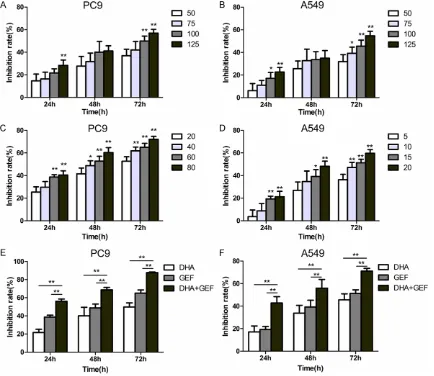
cells. DHA suppressed PC9 and A549 cell via-bility in a concentration- and time-dependent manner (Figure 1A and 1B). Following 72-h treatment with 125 µg/ml DHA, the rates of PC9 and A549 cell growth inhibition were 56.9 ± 3.3% and 54.8 ± 3.9%, respectively. Gefitinib significantly suppressed PC9 and A549 cell viability in a concentration- and time-depen-dent manner (Figure 1C and 1D), and had a much stronger inhibitory effect on the PC9 cells. Following 72-h treatment with high con-centrations of gefitinib (PC9 cells: 80 nmol/l; A549 cells: 20 µmol/l), the rates of PC9 and A549 cell growth inhibition were 72.1 ± 2.6% and 59.7 ± 3.2%, respectively. The combined gefitinib (60 nmol/l) and DHA (100 µg/ml) treat-ment had more potent inhibitory effects on the cell index as compared to gefitinib or DHA monotherapy. The combined therapy was supe-rior to monotherapy in inhibiting the growth of both PC9 and A549 cells (P < 0.01) (Figure 1E
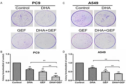
and 1F). Following 72-h gefitinib plus DHA Figure 2. DHA and gefitinib (GEF) inhibit colony formation of PC9 and A549 cell lines. And the combination treat-ment led to significant suppression of colony forming capacity. The two cell lines were divided into four treattreat-ment groups, and treated with DHA and/or GEF for 24 h. Both cell lines were treated with 100 µg/ml DHA; the PC9 and A549 cells were treated with 60 nmol/l and 15 µmol/l GEF, respectively. (A and C) Are representative sets of images from 3 reproducible independent experiments. Bars represent the mean ± SD of three separate experiments (B, D).
[image:4.612.90.523.69.358.2]ment, the rate of inhibition of PC9 cell growth was 87.6 ± 0.7%.
DHA and Gefitinib inhibited NSCLC cell colony
formation
A colony forming assay was used to examine the growth inhibition effect of DHA/Gefitinib on PC9/A549 cells. As is shown in Figure 2A and
2B, DHA or Gefitinib alone significantly sup-pressed the colony forming capacity of PC9 cells compared with control group (P < 0.01). Combined gefitinib and DHA treatment signifi-cantly decreased the colony formation number of PC9 cells as compared with gefitinib or DHA monotherapy (P < 0.01). Meanwhile, as depict-ed in Figure 2C and 2D, consistent results were gotten in A549 cell lines (P < 0.01). DHA com-bined with gefitinib significantly reduced A549 cell colony formation, which was also further than the cell line was treated with gefitinib or DHA alone. The results confirmed that DHA increased the gefitinib-induced inhibition of colony forming of NSCLC cells.
DHA enhanced gefitinib-induced apoptosis in
PC9 cells
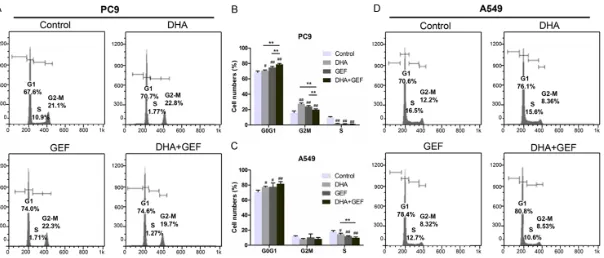
The effects of gefitinib and/or DHA on the induction of apoptosis in PC9 and A549 cells were detected by flow cytometry. Both gefitinib and DHA had slight apoptosis induction effects on the A549 cells, where the apoptosis index of the cells was increased, but without statistical significance as compared with the control group (P > 0.05) (Figure 3D and 3E). Gefitinib or DHA significantly induced apoptosis in the PC9 cells (P < 0.01). Combined gefitinib and DHA treatment significantly increased the per-centage of apoptotic cells as compared with gefitinib or DHA monotherapy (P < 0.01) (Figure 3A and 3B). Therefore, DHA increases the gefi-tinib-induced apoptosis induced in NSCLC cells with EGFR activating mutation.
Figure 3C and 3F depict the change in the nuclear morphology of NSCLC cells treated with DHA and/or gefitinib. The results
con-firmed that DHA increased the gefitinib-induced apoptosis in NSCLC cells, especially in NSCLC cells with EGFR activating mutation.
DHA enhanced gefitinib-induced G0/G1 ac -cumulation in PC9 cells and S-phase decrease in A549 cells
DHA increased the anti-tumor effect of gefitinib on NSCLC cells in vitro. Next, we detected the effects of DHA and/or gefitinib on the cell cycle. In the PC9 and A549 cells (Figure 4A and 4D), 48-h treatment with gefitinib and DHA mono-therapy both significantly increased the centage of G0/G1 cells and decreased the per-centage of S-phase cells (P < 0.05) as is shown in Figure 4B and 4C. Gefitinib and DHA induced G2/M accumulation in PC9 cells, but had no effect on the G2/M phase in A549 cells. Compared with monotherapy, combined gefi-tinib and DHA treatment increased the percent-age of G0/G1 PC9 cells and decreased the per-centage of S-phase A549 cells. The combined treatment increased the percentage of G2/M PC9 cells as compared to the control group, and decreased the percentage of G2/M PC9 cells as compared with monotherapy.
DHA enhanced gefitinib-induced inhibition of ERK1/2 phosphorylation in PC9 cells
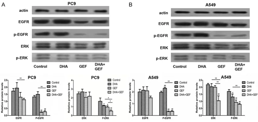
In the PC9 cells (Figure 5A), gefitinib inhibited EGFR and ERK1/2 phosphorylation in an obvi-ous manner. Gefitinib also inhibited p-EGFR and p-ERK1/2 expression in the A549 cells (Figure 5B), decreasing expression partially relative to the decrease in the PC9 cells. In the two cell lines, DHA upregulated EGFR phos-phorylation and downregulated p-ERK1/2 expression.
NSCLC was inhibited by DHA combined with Gefitinib
therapy was lower than that following mono-therapy (P < 0.05).
Discussion
As an important factor in the development of cancer, DHA has anti-tumor effects on many tumor types but has few adverse effects on most normal cells [13-15]. The epidemiological and laboratory data suggest that DHA also has anti-tumor effects on lung cancer cells [10, 12, 16]. In the present study, DHA inhibited PC9 and A549 cell proliferation in a dose- and time-dependent manner. In addition, DHA enhances the effectiveness of some chemical agents: When combined with many chemotherapeutic drugs, including paclitaxel, celecoxib, or 5-fluo-rouracil, DHA enhances colon cancer cell responsiveness to the drugs [17, 18]. Pola- varapu et al. found that DHA augments the growth inhibitory action of bleomycin on human neuroblastoma cells [19]. Moreover, DHA increases the anti-tumor effects of targeted therapy. In 2015, Zou et al. reported that DHA enhanced the effectiveness of the human EGFR 2 (HER2)-targeting drug trastuzumab by inhibiting the HER2 pathway [20]. In the pres-ent study, we aimed to find the influence of DHA on gefitinib-induced cytotoxic action on human lung adenocarcinoma cells.
The MTT assay results confirmed that both DHA and gefitinib suppress PC9 and A549 cell prolif-eration in a concentration- and time-dependent manner. The combined DHA and gefitinib treat-ment showed much greater inhibitory effects as compared with monotherapy in the NSCLC cells.
DHA induces apoptosis in lung cancer cells. The potential mechanism of action includes the proapoptotic effects caused by DHA, regulation of the expression of the dual phosphatase mitogen-activated protein kinase (MAPK) phos-phatase 1 (MKP-1), and the resulting modifica-tions in the phosphorylation state of MAPKs, especially ERK1/2 and p38 [21]. In the present study, flow cytometry showed that DHA en- hanced gefitinib-induced apoptosis in PC9 cells (P < 0.01), but increased apoptosis only slightly in the A549 cells (P > 0.05).
[image:8.612.92.520.71.274.2]In both the TKI-resistant A549 and TKI-sensitive PC9 cell lines, 48-h gefitinib treatment signifi-cantly increased the percentage of G0/G1 cells and decreased the percentage of S-phase cells; both cell lines followed the same trend after DHA treatment. The combined therapy had more significant effects on the cell cycle than monotherapy did. These synergistic effects contributed to the inhibition of NSCLC cell proliferation.
Figure 5. Western blot detection of the effect of DHA and gefitinib (GEF) on the protein expression of the EGFR and ERK signaling molecules in PC9 and A549 cells. (A) PC9 and (B) A549 cells were treated with gefitinib and/or DHA, and the data are expressed as the mean and SD of three separate experiments. #P < 0.05, ##P < 0.01 for test group
NSCLC was inhibited by DHA combined with Gefitinib
Gefitinib, an EGFR TKI, is one of the most repre-sentative targeting therapeutic drugs. Nume- rous studies have shown that gefitinib inhibits
EGFR-mutated NSCLC mainly through EGFR signaling [6, 22, 23]. Gefitinib blocks the ade-nosine triphosphate (ATP) binding pocket of EGFR, inhibiting autophosphorylation and the subsequent activation of the phosphatidylinosi-tol 3-kinase (PI3K)/AKT, MAPK/ERK, and Janus kinase/signal transducer and activator of tran-scription 3 (JAK/STAT3) downstream signaling that is essential for tumor migration, prolifera-tion, differentiaprolifera-tion, survival, and apoptosis [24, 25]. To investigate the molecular mecha-nisms responsible for the effects induced by the combined therapy along the EGFR signaling pathway, the expression of both the inactive (unphosphorylated) and activated (phosphory-lated) forms of EGFR and its downstream effec-tor ERK1/2 were investigated using western blotting. In the PC9 cells, gefitinib markedly reduced p-EGFR and p-ERK1/2 expression. In the A549 cells, which were used as a compara-tive cell line because they harbor wild-type
EGFR, gefitinib downregulated p-EGFR and p-ERK1/2 expression partially as compared with that in the PC9 cells. In both cell lines, DHA upregulated EGFR phosphorylation while down-regulating p-ERK1/2 expression. The results are consistent with that of previous studies [21, 26]. When the cells were treated with DHA, there were inconsistent changes in the EGFR and ERK1/2 phosphorylation levels. DHA induces membrane microdomain alterations, resulting in the exclusion of EGFR from caveo-lin-rich lipid raft fractions [26]. Lipid raft micro-domains play an important role, functioning as platforms that aggregate specific proteins, including EGFR, to facilitate cell signaling. Although it upregulated EGFR phosphorylation, we believe that DHA disrupted EGFR signaling and did not activate downstream signaling, as the EGFR protein was transferred out of the cell signal activity area. In 2004, Calviello et al. showed that n-3 PUFAs, including DHA, induced apoptosis by inhibiting ERK1 and ERK2 phos-phorylation in human colon cancer cells [27]. Serini et al. also found that DHA downregulated the phosphorylation state of MAPKs, especially ERK1/2, in lung cancer cells [21]. These results are in agreement with ours.
The level of ERK1/2 phosphorylation following combined therapy was lower than that of
mono-therapy, especially in the PC9 cells (P < 0.05), which suggests that the combined treatment affects the ERK1/2 pathway in NSCLC cells with EGFR mutation more effectively than DHA or gefitinib monotherapy. This result may be attributed to synergy between DHA and gefi-tinib. Nevertheless, we detected the combined effects of DHA and gefitinib only in vitro, there-fore further experiments in vivo are needed. In summary, we demonstrate that DHA enhanc-es the anti-tumor effects of gefitinib in NSCLC cells with EGFR activating mutation and TKI resistance by inducing apoptosis, regulating the tumor cell cycle, and inhibiting ERK1/2 pro-tein activation. Nutritional intervention with DHA is a promising approach for enhancing the therapeutic effect of gefitinib, or potentially reducing the drug dose. More research into the potential of using DHA supplementation is of critical importance.
Disclosure of conflict of interest
None.
Address correspondence to: Dr. Zhongmin Peng, Department of Thoracic Surgery, Shandong Pro- vincial Hospital Affiliated to Shandong University, 9677 Jing 10 Road, Ji’nan 250000, Shandong Province, PR China. Tel: +8615168887792; Fax: +86-531-87902348; E-mail: sdslyypzm@163.com
References
[1] Torre LA, Bray F, Siegel RL, Ferlay J, Lortet-Tieu-lent J and Jemal A. Global cancer statistics, 2012. CA Cancer J Clin 2015; 65: 87-108. [2] Crawford S. Is it time for a new paradigm for
systemic cancer treatment? Lessons from a century of cancer chemotherapy. Front Phar-macol 2013; 4: 68.
[3] Schiller JH, Harrington D, Belani CP, Langer C, Sandler A, Krook J, Zhu J, Johnson DH; Eastern Cooperative Oncology Group. Comparison of four chemotherapy regimens for advanced non-small-cell lung cancer. N Engl J Med 2002; 346: 92-98.
[5] Antonicelli A, Cafarotti S, Indini A, Galli A, Rus-so A, Cesario A, Lococo FM, RusRus-so P, Mainini AF, Bonifati LG, Nosotti M, Santambrogio L, Margaritora S, Granone PM and Dutly AE. EG-FR-targeted therapy for non-small cell lung cancer: focus on EGFR oncogenic mutation. Int J Med Sci 2013; 10: 320-330.
[6] Ono M and Kuwano M. Molecular mechanisms of epidermal growth factor receptor (EGFR) ac-tivation and response to gefitinib and other EGFR-targeting drugs. Clin Cancer Res 2006; 12: 7242-7251.
[7] de Goede J, Geleijnse JM, Boer JM, Kromhout D and Verschuren WM. Marine (n-3) fatty ac-ids, fish consumption, and the 10-year risk of fatal and nonfatal coronary heart disease in a large population of Dutch adults with low fish intake. J Nutr 2010; 140: 1023-1028.
[8] Sanders TA. Protective effects of dietary PUFA against chronic disease: evidence from epide-miological studies and intervention trials. Proc Nutr Soc 2014; 73: 73-79.
[9] Ali M, Heyob K and Rogers LK. DHA-mediated regulation of lung cancer cell migration is not directly associated with Gelsolin or Vimentin expression. Life Sci 2016; 155: 1-9.
[10] Yao QH, Zhang XC, Fu T, Gu JZ, Wang L, Wang Y, Lai YB, Wang YQ and Guo Y. omega-3 polyun-saturated fatty acids inhibit the proliferation of the lung adenocarcinoma cell line A549 in vi-tro. Mol Med Rep 2014; 9: 401-406.
[11] Xue M, Wang Q, Zhao J, Dong L, Ge Y, Hou L, Liu Y and Zheng Z. Docosahexaenoic acid in-hibited the Wnt/beta-catenin pathway and suppressed breast cancer cells in vitro and in vivo. J Nutr Biochem 2014; 25: 104-110. [12] Murphy RA, Mourtzakis M, Chu QS, Baracos
VE, Reiman T and Mazurak VC. Supplementa-tion with fish oil increases first-line chemother-apy efficacy in patients with advanced nons-mall cell lung cancer. Cancer 2011; 117: 3774-3780.
[13] Leslie MA, Abdelmagid SA, Perez K, Muller WJ and Ma DW. Mammary tumour development is dose-dependently inhibited by n-3 polyunsatu-rated fatty acids in the MMTV-neu(ndl)-YD5 transgenic mouse model. Lipids Health Dis 2014; 13: 96.
[14] Joubert AM, Panzer A, Joubert F, Lottering ML, Bianchi PC and Seegers JC. Comparative study of the effects of polyunsaturated fatty acids and their metabolites on cell growth and tyro-sine kinase activity in oesophageal carcinoma cells. Prostaglandins Leukot Essent Fatty Acids 1999; 61: 171-182.
[15] Ma YJ, Yu J, Xiao J and Cao BW. The consump-tion of omega-3 polyunsaturated Fatty acids improves clinical outcomes and prognosis in
pancreatic cancer patients: a systematic eval-uation. Nutr Cancer 2015; 67: 112-118. [16] Azrad M, Turgeon C and Demark-Wahnefried
W. Current evidence linking polyunsaturated Fatty acids with cancer risk and progression. Front Oncol 2013; 3: 224.
[17] Skender B, Hofmanova J, Slavik J, Jelinkova I, Machala M, Moyer MP, Kozubik A and Hyrslova Vaculova A. DHA-mediated enhancement of TRAIL-induced apoptosis in colon cancer cells is associated with engagement of mitochon-dria and specific alterations in sphingolipid metabolism. Biochim Biophys Acta 2014; 1841: 1308-1317.
[18] Bradley MO, Webb NL, Anthony FH, Devanesan P, Witman PA, Hemamalini S, Chander MC, Baker SD, He L, Horwitz SB and Swindell CS. Tumor targeting by covalent conjugation of a natural fatty acid to paclitaxel. Clin Cancer Res 2001; 7: 3229-3238.
[19] Polavarapu S, Mani AM, Gundala NK, Hari AD, Bathina S and Das UN. Effect of polyunsatu-rated Fatty acids and their metabolites on bleomycin-induced cytotoxic action on human neuroblastoma cells in vitro. PLoS One 2014; 9: e114766.
[20] Mason JK, Klaire S, Kharotia S, Wiggins AK and Thompson LU. α-linolenic acid and doco-sahexaenoic acid, alone and combined with trastuzumab, reduce HER2-overexpressing breast cancer cell growth but differentially reg-ulate HER2 signaling pathways. Lipids Health Dis 2015; 14: 91.
[21] Serini S, Trombino S, Oliva F, Piccioni E, Mone-go G, Resci F, Boninsegna A, Picci N, Ranelletti FO and Calviello G. Docosahexaenoic acid duces apoptosis in lung cancer cells by in-creasing MKP-1 and down-regulating p-ERK1/2 and p-p38 expression. Apoptosis 2008; 13: 1172-1183.
[22] Liu CY and Seen S. Gefitinib therapy for ad-vanced non-small-cell lung cancer. Ann Phar-macother 2003; 37: 1644-1653.
[23] Nurwidya F, Takahashi F and Takahashi K. Ge-fitinib in the treatment of nonsmall cell lung cancer with activating epidermal growth factor receptor mutation. J Nat Sci Biol Med 2016; 7: 119-123.
[24] Lemmon MA and Schlessinger J. Cell signaling by receptor tyrosine kinases. Cell 2010; 141: 1117-1134.
[25] Sordella R, Bell DW, Haber DA and Settleman J. Gefitinib-sensitizing EGFR mutations in lung cancer activate anti-apoptotic pathways. Sci-ence 2004; 305: 1163-1167.
dis-NSCLC was inhibited by DHA combined with Gefitinib
rupting its lipid raft association. Carcinogene-sis 2010; 31: 1523-1530.
[27] Calviello G, Di Nicuolo F, Gragnoli S, Piccioni E, Serini S, Maggiano N, Tringali G, Navarra P, Ranelletti FO and Palozza P. n-3 PUFAs reduce