Original Article
Treadmill exercise increases cystathionine
γ-lyase expression and decreases inflammation in
skeletal muscles of high-fat diet-induced obese rats
Dongdong Wu1, Zhen Lu1, Mengling Li1, Zhenxian Feng2, Ailian Liu2, Dongzhu Jin1, Ailing Ji1, Yanzhang Li1
1Henan University School of Medicine, Kaifeng 475004, Henan, China; 2Henan University School of Physical
Education, Kaifeng 475001, Henan, China
Received May 7, 2016; Accepted September 17, 2016; Epub December 15, 2016; Published December 30, 2016
Abstract: Obesity is a worldwide metabolic disorder that increases the risk of a number of human diseases. Physical
exercise has many beneficial effects on obesity and related disorders, such as dyslipidemia and hypertension. The present study investigated the effects of treadmill exercise on the generation of endogenous hydrogen sulfide (H2S)
and the level of inflammation in skeletal muscles of high fat diet (HFD)-induced obese rats. Here we showed that the concentration of H2S and the expression level of cystathionine γ-lyase (CSE) in skeletal muscles of HFD-fed rats were lower than those in skeletal muscles of low fat diet (LFD)-fed rats. However, treadmill running significantly increased the concentration of H2S and the expression of CSE at both the mRNA and protein level in skeletal muscles of HFD-fed rats. Furthermore, the expression of interleukin-6 (IL-6) in the HFD group was higher than that in the LFD group; whereas, treadmill running reduced the IL-6 levels in skeletal muscles of HFD-fed rats. Moreover, no significant
difference was observed in IL-15 expression between each group. In conclusion, treadmill exercise could enhance
the concentration of H2S by increasing CSE expression and decrease inflammation by reducing the level of IL-6 in skeletal muscles of HFD-fed rats.
Keywords: Treadmill running, hydrogen sulfide, cystathionine γ-lyase, inflammation, interleukin-6
Introduction
Obesity, which is an excessively high propor- tion of body fat, is a common metabolic disor-der affecting over 500 million people world- wide [1-3]. Obesity is highly associated with the development of a number of human diseases, including cardiovascular disorders, type 2 dia-betes, and non-alcoholic fatty liver disease [4]. Obesity is characterized by systemic, low-grade,
chronic inflammation, which can be partly at-tributed to an increase in the proinflammatory systemic milieu imposed by proinflammatory
cytokines [5, 6]. It has been demonstrated
that proinflammatory adipocytokines, leptin,
and C-reactive protein are elevated; however, adiponectin, the main insulin-sensitizing and
anti-inflammatory compound, is lower in
obe-sity [7]. A recent study showed that exercise
training significantly improved muscle endur
-ance and reduced markers of inflammation and
oxidative stress in skeletal muscles of aged men [8]. Whether exercise training could
ame-liorate inflammation in high-fat diet
(HFD)-in-duced obese rats needs further investigation.
Hydrogen sulfide (H2S) is a colorless, toxic gas with a characteristic rotten-egg odor and is now widely considered to be an endogenous gas-eous signaling molecule [9-11]. In mammals,
endogenous H2S is mainly generated from L- cysteine by the catalysis of two pyridoxal 5’- phosphate-dependent enzymes, cystathionine
γ-lyase (CSE) and cystathionine β-synthase
(CBS), and by the combined action of cysteine aminotransferase and 3-mercaptopyruvate
sul-fotransferase (3-MST) [11-13]. Species specific
differences have been observed with respect
to the expression of CBS, CSE, and 3-MST in
skeletal muscle [14]. A recent report suggest- ed that CSE and CBS expression could be detected in human skeletal muscle, whereas there was no expression of these enzymes in mouse skeletal muscle [15]. Another study
demonstrated that H2S-generating enzymes
levels in rat skeletal muscle [16]. However,
whether exercise training could modulate the
generation of endogenous H2S in HFD-induced
obese rats remains to be fully elucidated. In the present study, the effects of tread-
mill running on the levels of endogenous H2S
and inflammation in skeletal muscles of
HFD-induced obese rats were investigated. Our results showed that treadmill running increa- sed the expression of CSE and decreased
the level of IL-6 in skeletal muscles of
HFD-induced obese rats.
Materials and methods
Animals
All procedures for animal experiments were ap- proved by the Committee of Medical Ethics and
Welfare for Experimental Animals of Henan
University School of Medicine. Five-week-old
male SD rats were purchased from The
Experi-mental Animal Center of Zhengzhou University
(Henan, China). Rats were maintained in a
controlled environment (temperature 22 ± 3°C and humidity 55 ± 5%) on a 12-hour light/dark cycle with food and water ad libitum. The rats
were fed either a low fat diet (LFD, 10% kcal as fat, Medicience Ltd., Jiangsu, China) or high fat
diet (HFD, 45% kcal as fat, Medicience Ltd.,
Jiangsu, China) for a total of 14 weeks. After 8
weeks of feeding, the HFD-fed mice were ran
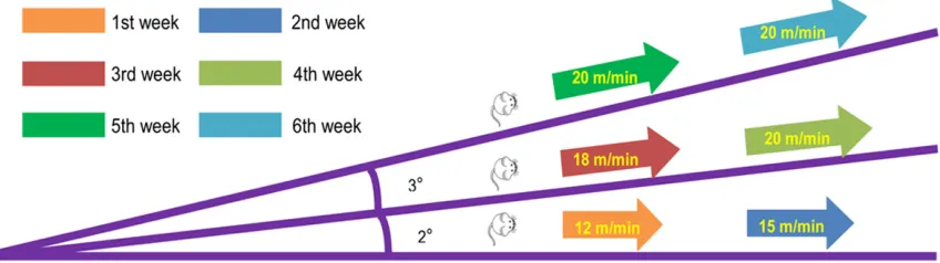
-domly assigned to the HFD group (n = 10) or the HFD with exercise training (EX) group (HFD + EX, n = 10). These mice were treated for an additional 6 weeks. EX consisted of running for
[image:2.612.95.519.78.197.2]40 minutes, 5 days per week, as described in
Figure 1. The LFD group (n = 10) continued with the LFD for the entire 6-week duration. The
body weight gain, food intake, and calorie con-sumption in rats were measured. At the end of the experiment, the rats were killed and the
gastrocnemius muscle was stored at -80°C for further study.
RNA extraction and RT-PCR
Total RNA was isolated from skeletal muscle using TRIzol reagent (Life Technologies,
Rock-ville, MD, USA), treated with DNase I (Roche Applied Science, Mannheim, Germany), and
purified using an RNA clean up kit (Cwbiotech,
Beijing, China). One microgram of total RNA was applied for cDNA synthesis using a cDNA reverse transcription kit (Cwbiotech, Beijing, China). Primers were designed with Primer Premier 5.0 (Premier Biosoft, Palo Alto, CA,
USA): CBS, forward 5’-CTCC-GGGAGAAGGGTT-TTGA-3’ and reverse 5’-CATGTTCCCGAGAG-TC-ACCAT-3’; CSE, forward 5’-GCTGAGAGCCTGG-GAGGATA-3’ and reverse 5’-TCACTGATCCCGA-GGGTAGCT-3’; 3-MST, forward 5’-CG-GCGCT-TCCAGGTAGTG-3’ and reverse 5’-CTGGTCAG-GAATTC-AGTGAATGG-3’; IL-6, forward TAT-GAACAGCGATGATGCAC-TG-3’ and reverse 5’-TTGCTCTGAATGACTCTGGCTT-3’; IL-15, forward 5’-GACAGTGACTTTCATCCCAGTT-3’ and rever-se 5’-CATT-CCTTGCAGCCAGAC-3’; and β-actin, forward 5’-ACCCGCGAGT-ACAACCTTCTT-3’ and reverse 5’-TATCGTCATCCATGGCGAACT-3’. The
PCR reactions were performed in a total
vol-ume of 25 μL using the following thermal
cycl-ing parameters: 95°C for 5 min, 35 cycles of 93°C for 30 s, 60°C for 45 s, and 72°C for 5
min. The mRNA expression levels of the test genes were normalized to β-actin levels.
Western blot analysis
For western blot analysis, frozen muscle sam-ples (50 mg) were powdered under liquid N2
and homogenized in 200 μL of ice-cold RIPA
tein assay kit (Beyotime Institute of Biotech-
nology, Shanghai, China). The extracted pro
-teins (40 μg) were separated on an SDS-PAGE
gel and transferred to a nitrocellulose mem-brane. After blocking, the membranes were incubated with an anti-CBS antibody (Santa Cruz Biotechnology, Santa Cruz, CA, USA), an-
ti-3-MST antibody (Santa Cruz Biotechnology,
Santa Cruz, CA, USA), anti-CSE antibody (Pro-
by using a commercial ELISA kit (Antibodies-online Inc., Atlanta, GA, USA) according to the manufacturer’s instructions.
Statistical analysis
All data were presented as the mean ±
stan-dard deviation (SD). The expression levels
of mRNA and protein were normalized to a calibrator which was considered as the ba-
sal sample. Significant differences between
groups were analyzed by two way analysis of variance using SPSS software version 19.0 (SPSS Inc., Chicago, IL, USA), followed by
Tukey’s test. A p-value of less than 0.05 was
considered to be statistically significant.
Results
Treadmill running reduces the body weight gain, food intake and calorie consumption in HFD-fed rats
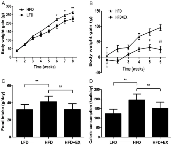
As shown in Figure 2A, in comparison to rats
[image:3.612.92.371.75.313.2]fed with a LFD for 8 weeks, HFD-fed rats exhib -ited increased body weight gain, which indicat-ed that an alimentary obesity model had been successfully established. It has been widely Figure 2. Effect of treadmill running on the body weight gain, food intake, and
calorie consumption in rats. A. The body weight gain of rats in the LFD and HFD groups. B. The body weight gain of rats in the HFD and HFD + EX groups.
C. Food intake. D. Calorie consumption. Values are presented as the mean ±
SD (n = 10); *P < 0.05, **P < 0.01 compared with the LFD group; #P < 0.05, ##P < 0.01 compared with the HFD group.
tein Tech Group, Chicago, IL, USA), and anti-β-actin (Pro-tein Tech Group, Chicago, IL, USA). The membranes were
thoroughly rinsed and then incubated with a horseradish peroxidase-conjugated sec-ondary antibody (Beyotime In- stitute of Biotechnology, Sh-
anghai, China). The reaction
was visualized using an en- hanced chemiluminescence
system (Thermo Fisher Scien-tific, Rockford, IL, USA). Immu-noblots were quantified by
densitometry using Quantity One software (Bio-Rad, CA, USA).
Measurement of H2S concen-tration
To investigate the effect of
treadmill running on the
pro-duction of H2S and release in skeletal muscles (50 mg), we performed enzyme-linked immunosorbent assay (ELISA)
Figure 3. Effect of treadmill running on H2S concen-tration in rat skeletal muscles. Values are presented
as the mean ± SD (n = 3); **P < 0.01 compared with the LFD group; ##P < 0.01 compared with the
[image:3.612.92.286.407.547.2]accepted that exercise training has beneficial
health effects on the prevention of obesity and related disorders, such as dyslipidemia and
hypertension [17-19]. The results showed that treadmill running for 6 weeks significantly re-duced the body weight gain in rats fed with HFD
(Figure 2B). Compared to the LFD-fed rats,
HFD-fed rats showed increased food intake
and calorie consumption, which were all rever- sed by tread mill running (Figure 2C and 2D).
Treadmill running increases the H2S concen-tration in skeletal muscles of HFD-fed rats
A recent study found that obesity in mice re-
duced the bioavailability of H2S and the
intra-cellular concentration of H2S was lower in
adi-pose tissue macrophages (ATMs) isolated from obese mice compared to ATMs from lean mice
[20]. Our results indicated that the concen-
tration of H2S in skeletal muscle of HFD-fed
rats was lower than that in skeletal muscles of
LFD-fed rats. However, after 6 weeks of tread
-mill running, the H2S concentration in skele-
tal muscles of HFD-fed rats was markedly
in-creased (Figure 3).
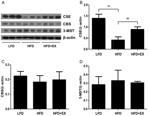
We further measured the protein expression of
CSE, CBS, and 3-MST in rat skeletal muscles. The results showed that the protein levels of CSE in the skeletal muscles of HFD-fed rats
were lower than those in skeletal muscles of LFD- fed rats (Figure 5A and 5B). After 6 weeks of treadmill running, the protein expression of
CSE in skeletal muscles of HFD-fed rats was significantly increased (Figure 5A and 5B).
Furthermore, there were no significant differ
-ences in the protein levels of CBS and 3-MST
between each group (Figure 5A, 5C and 5D).
Treadmill running reduces IL-6 gene expres-sion in skeletal muscles of HFD-fed rats
IL-6 plays an important role in the regulation of appetite, body composition, and energy ex- penditure [21, 22]. As shown in Figure 6A and
6C, the genetic expression of IL-6 in skeletal
muscles of HFD-fed rats was higher than that in skeletal muscles of LFD-fed rats. Treadmill run
-ning significantly reduced IL-6 gene expression in skeletal muscles of HFD-fed rats. IL-15 is
an anabolic factor that is highly expressed in skeletal muscles. Recent studies have shown Figure 4. Effect of treadmill running on the mRNA levels of CSE, CBS, and
3-MST in rat skeletal muscles. (A) The expression levels of CSE, CBS, and 3-MST were measured by RT-PCR. β-actin was used as an internal control.
Bar graphs showed the quantification of CSE (B), CBS (C), and 3-MST (D). Values are presented as the mean ± SD (n = 3); *P < 0.05, **P < 0.01 com-pared with the LFD group; ##P < 0.01 compared with the HFD group.
Treadmill running increases CSE gene expression in skel-etal muscles of HFD-fed rats
Considering that CSE, CBS,
and 3-MST are the main H2 S-generating enzymes in ma- mmals, we investigated the
genetic expression of H2 S-generating enzymes in rat
skeletal muscles. The results
suggested that the genetic expression of CSE, CBS, and
3-MST in skeletal muscles of HFD-fed rats were lower than
those in skeletal muscles of LFD-fed rats (Figure 4A-D), while treadmill running obser- vably increased CSE gene ex- pression in skeletal muscles
of HFD-fed rats (Figure 4A
and 4B).
that IL-15 is involved in skeletal muscle fiber
growth, glucose uptake, and hypertrophy [23-25]. Our results showed that there was no
sig-nificant difference in IL-15 gene expression
be-tween each group (Figure 6B and 6D).
H2S by increasing CSE expression in skeletal
muscles of HFD-fed rats.
Obesity is coupled to a low-grade, chronic in-
[image:5.612.92.375.75.295.2]flammatory state characterized by activation in Figure 5. Effect of treadmill running on the protein levels of CSE, CBS, and
3-MST in rat skeletal muscles. (A) The expression levels of CSE, CBS, and 3-MST were measured by western blot. β-actin was used as an internal con
-trol. Bar graphs show the quantification of CSE (B), CBS (C), and 3-MST (D). Values are presented as the mean ± SD (n = 3); **P < 0.01 compared with the LFD group; ##P < 0.01 compared with the HFD group.
Figure 6. Effect of treadmill running on the mRNA levels of IL-6 and IL-15 in
rat skeletal muscles. (A and B) The expression levels of IL-6 and IL-15 were measured by RT-PCR. β-actin was used as an internal control. Bar graphs
show the quantification of IL-6 (C) and IL-15 (D). Values are presented as the mean ± SD (n = 3); **P < 0.01 compared with the LFD group; #P < 0.05
compared with the HFD group.
Discussion
It is widely accepted that
CSE, CBS, and 3-MST are the three main H2S-generating
enzymes in mammals. These
enzymes could be detected in different organs and tis-sues, and their expression
levels are significantly differ -ent [26, 27]. CSE is mainly expressed in the cardiovas- cular system, while CBS is abundant in the central ner-vous system. Furthermore, 3-
[image:5.612.93.373.391.586.2]MST and CAT are apparently important for H2S production in the vasculature and brain
[9, 12, 28]. To explore the
ef-fect of treadmill running on the existence of the skeletal muscle-derived endogenous
H2S pathway in HFD-fed rats, we detected the H2S content
and the expression of H2 S-generating enzymes in rat skeletal muscles. Our results showed that CSE, CBS, and
3-MST could be detected at
the mRNA and protein levels in skeletal muscles of both
LFD- and HFD-fed rats, which is consistent with the
find-ings of a recent study [16].
Furthermore, the H2S concen-tration and the mRNA and protein levels of CSE in skele-
tal muscles of HFD-fed rats
were lower than those in skeletal muscles of LFD-fed rats, while treadmill running increased the concentration
of H2S and mRNA and protein levels of CSE in skeletal
mus-cles of HFD-fed rats. These
results indicate that CSE is
adipose tissue, immune cell infiltration, and systemic elevation of pro-inflammatory cyto -kines [5, 29, 30]. IL-6 is a novel adipokine that is expressed by the adipocytes and stromovas-cular matrix of visceral white adipose tissues involved in the development of obesity [18]. Recent studies have shown that the IL-6 con-centration in plasma is increased with weight gain and decreased upon weight loss [31, 32]. We observed that the IL-6 level in the
gastroc-nemius muscle of HFD-fed rats was higher
compared to the LFD group, suggesting that IL-6 plays an important role in
obesity-associat-ed inflammation. Recent findings indicate that
exercise training-induced adaptations in adi-pose tissue metabolism are mediated by IL-6, which is secreted from skeletal muscles during exercise [33-35]. In this study, we found that
treadmill running significantly reduced the IL-6 level in skeletal muscles of HFD-fed rats. These data demonstrate that the anti-inflammatory
effect of treadmill running could be achieved, at least in part, by reducing the expression of IL-6 in the gastrocnemius muscle of obese individuals.
IL-15 is a recently discovered pleiotropic
cyto-kine that belongs to the four α-helix bundle
family of cytokines that are known to enhance
natural killer cell activity and stimulate T cell
proliferation [25, 36]. A recent study showed that the expression level of IL-15 was lower in
the gastrocnemius muscle of HFD-fed rats than
in controls [25]. In contrast, another study
indi-cated that there was no significant difference
in the level of IL-15 protein in the gastrocne-mius muscle between Zucker diabetic fatty (ZDF) and ZDF lean control rats [23]. In our study, no remarkable difference was obser- ved in the gene expression of IL-15 in the
gas-trocnemius muscle between the LFD and HFD
groups. Recently, there has been controversy regarding the effects of exercise training on muscle IL-15 protein levels and mRNA expres-sion [37]. For instance, the mRNA expresexpres-sion of IL-15 in skeletal muscle has been shown to increase in response to acute resistance
exer-cise in fast skeletal muscles [38]. However,
another study suggested that chronic low fre-quency/low volume resistance training did not change the IL-15 mRNA level in plantaris mus-cles, despite the induction of hypertrophy [39]. Similarly, we found that treadmill running had no effect on the IL-15 mRNA level in the
gas-trocnemius muscle of HFD-fed rats. Taken to-gether, there was no significant difference in
the expression of IL-15 in the gastrocnemius
muscle between LFD and HFD-fed rats, and
treadmill running did not affect the expression level of IL-15 in the gastrocnemius muscle of
HFD-fed rats.
In conclusion, treadmill exercise could enhance
the concentration of H2S by increasing the ex- pression level of CSE and could decrease in-
flammation by reducing the expression of IL-6 in skeletal muscles of HFD-fed rats. Therefore,
treadmill exercise may possess therapeutic po-
tential in treating obesity-induced inflammation
in obese individuals.
Acknowledgements
This work was supported by the National
Na-tural Science Foundation of China under Grant U1504817, the Natural Science Foundation
of Education Department of Henan Province
under Grant 15A310017, and the Foundation
of Science & Technology Department of
He-nan Province under Grants 132300410012 and 162300410233.
Disclosure of conflict of interest
None.
Address correspondence to: Yanzhang Li and Ailing
Ji, Henan University School of Medicine, Kaifeng 475004, Henan, China. Tel: +86 371 23880585;
Fax: +86 371 23880585; E-mail: yanzhang206@ 163.com (YZL); [email protected] (ALJ)
References
[1] Sturm R and An R. Obesity and economic envi-ronments. CA Cancer J Clin 2014; 64: 337-350.
[2] Claussnitzer M, Dankel SN, Kim KH, Quon G, Meuleman W, Haugen C, Glunk V, Sousa IS,
Beaudry JL, Puviindran V, Abdennur NA, Liu J,
Svensson PA, Hsu YH, Drucker DJ, Mellgren G, Hui CC, Hauner H and Kellis M. FTO Obesity
Variant Circuitry and Adipocyte Browning in
Humans. N Engl J Med 2015; 373: 895-907.
[3] Fenske W, Athanasiou T, Harling L, Drechsler C, Darzi A and Ashrafian H. Obesity-related car
-diorenal disease: the benefits of bariatric sur -gery. Nat Rev Nephrol 2013; 9: 539-551. [4] Samuel VT and Shulman GI. Mechanisms for
[5] Gotoh K, Inoue M, Masaki T, Chiba S, Shimasaki T, Ando H, Fujiwara K, Katsuragi I, Kakuma T, Seike M, Sakata T and Yoshimatsu H. A novel anti-inflammatory role for spleen-derived inter
-leukin-10 in obesity-induced inflammation in
white adipose tissue and liver. Diabetes 2012; 61: 1994-2003.
[6] Arkan MC, Hevener AL, Greten FR, Maeda S, Li
ZW, Long JM, Wynshaw-Boris A, Poli G, Olefsky
J and Karin M. IKK-beta links inflammation to
obesity-induced insulin resistance. Nat Med 2005; 11: 191-198.
[7] Furukawa S, Fujita T, Shimabukuro M, Iwaki
M, Yamada Y, Nakajima Y, Nakayama O, Makishima M, Matsuda M and Shimomura I. Increased oxidative stress in obesity and its impact on metabolic syndrome. J Clin Invest 2004; 114: 1752-1761.
[8] Olesen J, Gliemann L, Bienso R, Schmidt J,
Hellsten Y and Pilegaard H. Exercise training,
but not resveratrol, improves metabolic and
inflammatory status in skeletal muscle of aged
men. J Physiol 2014; 592: 1873-1886. [9] Paul BD, Sbodio JI, Xu R, Vandiver MS, Cha
JY, Snowman AM and Snyder SH. Cystathio-nine gamma-lyase deficiency mediates neuro
-degeneration in Huntington’s disease. Nature
2014; 509: 96-100.
[10] Coletta C, Papapetropoulos A, Erdelyi K, Olah G, Modis K, Panopoulos P, Asimakopoulou
A, Gero D, Sharina I, Martin E and Szabo C.
Hydrogen sulfide and nitric oxide are mutually
dependent in the regulation of angiogenesis and endothelium-dependent vasorelaxation. Proc Natl Acad Sci U S A 2012; 109: 9161-9166.
[11] Wallace JL and Wang R. Hydrogen
sulfide-based therapeutics: exploiting a unique but ubiquitous gasotransmitter. Nat Rev Drug Discov 2015; 14: 329-345.
[12] Liu Y, Yang R, Liu X, Zhou Y, Qu C, Kikuiri T,
Wang S, Zandi E, Du J, Ambudkar IS and Shi
S. Hydrogen sulfide maintains mesenchymal
stem cell function and bone homeostasis via regulation of Ca(2+) channel sulfhydration. Cell Stem Cell 2014; 15: 66-78.
[13] Szabo C, Coletta C, Chao C, Modis K, Szczesny B, Papapetropoulos A and Hellmich MR. Tumor-derived hydrogen sulfide, produced by
cysta-thionine-beta-synthase, stimulates bioenerget-ics, cell proliferation, and angiogenesis in co-lon cancer. Proc Natl Acad Sci U S A 2013; 110: 12474-12479.
[14] Veeranki S and Tyagi SC. Role of hydrogen sul
-fide in skeletal muscle biology and metabo -lism. Nitric Oxide 2015; 46: 66-71.
[15] Chen NC, Yang F, Capecci LM, Gu Z, Schafer
AI, Durante W, Yang XF and Wang H. Regulation
of homocysteine metabolism and methylation in human and mouse tissues. FASEB J 2010; 24: 2804-2817.
[16] Du JT, LW, Yang JY, Tang CS, Li Q and Jin HF. Hydrogen sulfide is endogenously generated
in rat skeletal muscle and exerts a protective effect against oxidative stress. Chin Med J 2013; 126: 930-936.
[17] Pereira BC, da Rocha AL, Pauli JR, Ropelle
ER, de Souza CT, Cintra DE, Sant’Ana MR
and da Silva AS. Excessive eccentric exercise
leads to transitory hypothalamic
inflamma-tion, which may contribute to the low body weight gain and food intake in overtrained mice. Neuroscience 2015; 311: 231-242. [18] Gondim OS, de Camargo VT, Gutierrez FA,
Martins PF, Passos ME, Momesso CM, Santos
VC, Gorjao R, Pithon-Curi TC and Cury-Bo-aventura MF. Benefits of Regular Exercise on Inflammatory and Cardiovascular Risk
Markers in Normal Weight, Overweight and Obese Adults. PLoS One 2015; 10: e0140596. [19] Ito D, Cao P, Kakihana T, Sato E, Suda C,
Muroya Y, Ogawa Y, Hu G, Ishii T, Ito O, |Kohzuki M and Kiyomoto H. Chronic Running
Exercise Alleviates Early Progression of Nephropathy with Upregulation of Nitric Oxide Synthases and Suppression of Glycation in Zucker Diabetic Rats. PLoS One 2015; 10: e0138037.
[20] Velmurugan GV, Huang H, Sun H, Candela J, Jaiswal MK, Beaman KD, Yamashita M, Prakriya M and White C. Depletion of H2S
during obesity enhances store-operated Ca2+ entry in adipose tissue macrophages to in-crease cytokine production. Sci Signal 2015; 8: ra128.
[21] Ropelle ER, Flores MB, Cintra DE, Rocha GZ,
Pauli JR, Morari J, de Souza CT, Moraes JC,
Prada PO, Guadagnini D, Marin RM, Oliveira
AG, Augusto TM, Carvalho HF, Velloso LA, Saad
MJ and Carvalheira JB. IL-6 and IL-10
anti-in-flammatory activity links exercise to hypotha -lamic insulin and leptin sensitivity through
IKKbeta and ER stress inhibition. PLoS Biol
2010; 8.
[22] Wallenius V, Wallenius K, Ahren B, Rudling M, Carlsten H, Dickson SL, Ohlsson C and Jansson JO. Interleukin-6-deficient mice develop ma -ture-onset obesity. Nat Med 2002; 8: 75-79. [23] Kim HJ, Park JY, Oh SL, Kim YA, So B, Seong
JK and Song W. Effect of treadmill exercise
on interleukin-15 expression and glucose tol-erance in zucker diabetic Fatty rats. Diabetes Metab J 2013; 37: 358-364.
[24] Molanouri Shamsi M, Hassan ZM, Quinn LS,
Gharakhanlou R, Baghersad L and Mahdavi
resistance exercise in trained rats: effect of diabetes and skeletal muscle phenotype. Endocrine 2015; 49: 396-403.
[25] Yang HT, Luo LJ, Chen WJ, Zhao L, Tang CS, Qi
YF and Zhang J. IL-15 expression increased in response to treadmill running and inhibited endoplasmic reticulum stress in skeletal mus-cle in rats. Endocrine 2015; 48: 152-163. [26] Wu D, Si W, Wang M, Lv S, Ji A and Li Y.
Hydrogen sulfide in cancer: Friend or foe?
Nitric Oxide 2015; 50: 38-45.
[27] Wang R. Physiological implications of hydrogen
sulfide: a whiff exploration that blossomed.
Physiol Rev 2012; 92: 791-896.
[28] Wu D, Wang J, Li H, Xue M, Ji A and Li Y. Role of Hydrogen Sulfide in Ischemia-Reperfusion
Injury. Oxid Med Cell Longev 2015; 2015: 186908.
[29] Mauer J, Chaurasia B, Goldau J, Vogt MC, Ruud
J, Nguyen KD, Theurich S, Hausen AC, Schmitz J, Bronneke HS, Estevez E, Allen TL, Mesaros A,
Partridge L, Febbraio MA, Chawla A, Wunderlich
FT and Bruning JC. Signaling by IL-6
pro-motes alternative activation of macrophages to limit endotoxemia and obesity-associated resistance to insulin. Nat Immunol 2014; 15: 423-430.
[30] Lumeng CN and Saltiel AR. Inflammatory links
between obesity and metabolic disease. J Clin Invest 2011; 121: 2111-2117.
[31] Khan M and Joseph F. Adipose tissue and adi -pokines: the association with and application
of adipokines in obesity. Scientifica (Cairo)
2014; 2014: 328592.
[32] Smith MM and Minson CT. Obesity and adipo -kines: effects on sympathetic overactivity. J Physiol 2012; 590: 1787-1801.
[33] Knudsen JG, Bertholdt L, Joensen E, Lassen SB, Hidalgo J and Pilegaard H. Skeletal muscle
interleukin-6 regulates metabolic factors in
iWAT during HFD and exercise training. Obesity
(Silver Spring) 2015; 23: 1616-1624.
[34] Brandt C, Jakobsen AH, Adser H, Olesen J, Iversen N, Kristensen JM, Hojman P, Wojtas-zewski JF, Hidalgo J and Pilegaard H. IL-6 regu -lates exercise and training-induced adapta-tions in subcutaneous adipose tissue in mice. Acta Physiol (Oxf) 2012; 205: 224-235. [35] Knudsen JG, Murholm M, Carey AL, Bienso
RS, Basse AL, Allen TL, Hidalgo J, Kingwell BA, Febbraio MA, Hansen JB and Pilegaard H. Role
of IL-6 in exercise training- and cold-induced UCP1 expression in subcutaneous white adi-pose tissue. PLoS One 2014; 9: e84910. [36] Di Sabatino A, Calarota SA, Vidali F, Macdonald
TT and Corazza GR. Role of IL-15 in
immune-mediated and infectious diseases. Cytokine Growth Factor Rev 2011; 22: 19-33.
[37] Molanouri Shamsi M, Hassan ZH, Gharak-hanlou R, Quinn LS, Azadmanesh K, Baghersad
L, Isanejad A and Mahdavi M. Expression
of interleukin-15 and inflammatory cytokines in skeletal muscles of STZ-induced diabetic
rats: effect of resistance exercise training. Endocrine 2014; 46: 60-69.
[38] Nielsen AR, Mounier R, Plomgaard P, Mor-
tensen OH, Penkowa M, Speerschneider T, Pilegaard H and Pedersen BK. Expression of
interleukin-15 in human skeletal muscle ef-
fect of exercise and muscle fibre type
com-position. J Physiol 2007; 584: 305-312. [39] Zanchi NE, Lira FS, de Siqueira Filho MA, Rosa
JC, de Oliveira Carvalho CR, Seelaender M,
Santos RV and Lancha AH Jr. Chronic low
frequency/low volume resistance training
re-duces pro-inflammatory cytokine protein le-vels and TLR4 mRNA in rat skeletal muscle.