Original Article
Effect of butylphthalide in treating learning
disorders in rats with radiation brain injury
Chao-Wang An1, Jian-Min Li1, Yao Liu2, Ya-Ning Zhao2, Chang-Xiang Chen2, Jing Hao2, Cheng-Jing Xue1
1The Neurosurgery of Affiliated Hospital, 2Nursing and Rehabilitation College,North China University of Science and Technology, Tangshan 063000, China
Received April 7, 2016; Accepted February 23, 2017; Epub June 15, 2017; Published June 30, 2017
Abstract: Objective: We observed the effect of butylphthalide (NBP) in treating learning disorders in rats with radia-tion brain injury. Method: One hundred and twenty male Wistar rats were randomly divided into control group, model group, low-dose NBP group and high-dose NBP group. Radiation at a dose of 22 Gy was administered to the whole brain using linear accelerator to induce radiation brain injury. HE staining was performed to observe the morphologi-cal changes of hippocampal neurons. The expressions of extracellular regulated protein kinases 1/2 (ERK1/2) and growth associated protein-43 (GAP-43) were detected by immunohistochemistry and western blotting. MDA content was determined by thiobarbituric acid (TBA) colorimetry. SOD activity of the brain was measured by xanthine oxidase method. The learning capacity of rats was determined using the shuttle box paradigm. Result: Compared with the model group, the damage of nerve cells in the hippocampus was reduced and the MDA content was decreased of NBP group. The expression level of phospho-ERK1/2 and GAP-43 and activity of SOD were all higher in NBP group than in the model group; the shuttle box evaluation showed that NBP group animal active avoidance reaction was
significantly increased and passive avoidance latency decreased; the above changes were most significant in the
high dose NBP group. Conclusion: NBP has a good treatment effect in learning disorders caused by radiation brain injury in rats, which is related to enhanced activity of ERK1/2 and upregulation of GAP-43.
Keywords: Radiation injuries of the brain, learning, ERK1/2, GAP-43
Introduction
Radiation therapy is used as the first-line treat -ment for primary and secondary brain tumors and head and neck cancers. However, radiation brain injury (RBI) has become increasingly prev-alent due to radiation therapy [1] and mainly manifest as a decline in learning and memory capacities [2] in a dose-dependent manner. Hippocampus is the main site of cognition and learning [3], and RBI-induced learning disor-ders usually indicate structural and metabolic abnormalities of the hippocampus. At present, there are not available treatments against RBI. It is generally believed that cognitive deficit and neurogenic disorders caused by ionizing radia-tion play a role in RBI. Thus developing drugs that promote synaptic regeneration and recon-struction is the main concern. Growth associat-edprotein-43 (GAP-43) is related to nerve growth and considered the preferred molecular marker of neuronal regeneration and synaptic
the mechanism of therapeutic effect of NBP against RIB was discussed.
Materials and method
Grouping and modeling
One hundred and twenty adult male AD rats weighing 250 g-300 g were purchased from Beijing Huafukang Biotechnology Co., Ltd. (license No.: SCXK (Beijing) 2009-0004). The rats were randomly divided into control group, model group, high-dose RBI group and low-dose RBI group, with 30 rats in each group. For each group three time points were set up (7 d, 14 d, 28 d).
The rats in the control group were only anesthe-tized without radiation. The rats in the model group were conventionally anesthetized. The RBI model was induced by reference to litera-ture [9]. Brain irradiation using 6 MeV electrons was administered once with source-skin dis-tance of 100 cm, the absorbed dose rate of 250 Mu/min and total dose of 22 Gy.
The rats in the NBP groups received intragas-tric administration of NBP once daily starting 2 weeks before radiation. After radiation, NBP was continuously administered for different durations. According to literature [7], the high dose was set as 160 mg.kg-1 and the low dose 80 mg.kg-1.
Shuttle box learning
ZH-CSC shuttle box system was used. The rats were placed into the shuttle box at different time points and the test started from 9:00. After 5 min of adaptation, the stimulus (a tone) was given for 5 s, followed subsequently by an electric shock for 20 s. Another round of train-ing began after an interval of 10 s. If the rats succeeded in escaping to the safe zone within 5 s after the stimulus, it was considered active avoidance reaction and the present round of training stopped. Otherwise, 1.5 mA AC electric shock was given for 20 s. If the rats escaped to the safe zone after the second electric shock, it was considered passive avoidance reaction. If the rats still failed, the result was defined as negative for both active and passive avoidance reaction. Each rat received 30 electric shocks, and the passive avoidance latency (PAL) and the number of active avoidance reactions were
recorded. The percentage of active avoidance reactions to the total number of trainings was the active avoidance reaction rate (AARR). The higher the AARR and the lower the PAL, the stronger the learning capacity was.
Morphological observation
Five rats were selected from each group at each time point. Brain tissues were harvested after perfusion using 4% paraformaldehyde. The scope of resection was from optic chiasma to transverse cerebral fissure, followed by con -ventional paraffin embedding. The tissues were cut into coronal sections (thickness 5 μm), which were subjected to HE staining. For each rat, 4 slides were chosen and 4 fields of vision were randomly selected for each slide. The sec-tions were observed under the optical micro-scope. The number of surviving neurons in each field of vision (40 × 10) was determined, and the average percentage of surviving neu-rons in each field of vision was calculated (%) using Motic-6.0 image analysis system.
Immunohistochemical anlaysis of phospho-ERK1/2 and GAP-43
The expression of phospho-ERK1/2 and GAP-43 were detected by western blot
Western blot analysis included protein extrac-tion, electrophoresis, membrane transfer, and immune coloration. Respective 5 Five rats at each time point in each group were decapitat-ed under anesthesia. The hippocampal tissue was rapidly frozen in liquid nitrogen and homog-enized in ice-cold homogenization buffe. Homogenates were then centrifuged at 800 g for 5 min at 4°C. Then the supernatants were collected and protein concentrations were determined by the Kaumas Bradford method. Samples were stored at -80°C until required. Detection steps: 40 ug protein sample and the volume of the sample buffer were mixed, after boiling would be carried out twelve sodium dodecyl sulfate polyacrylamide gel electropho-resis (SDS-PAGE), and transfered film. The membrane was incubated with
phospho-ERK1/2 monoclonal antibody and GAP-43
monoclonal antibody (Zhongshan Beijing bio-logical company, both 1:2000) overnight at 4°C. The membrane was washed repeatedly with TBST and incubated with corresponding secondary antibody. The antibody was exposed with ECL solution and image analyzer was used to determine the optical density for quantitative analysis.
Determination of MDA content and SOD activ-ity
Five rats were randomly selected from each
tube-A of blank tube) × concentration of stan -dard ÷ protein content; SOD activity (U/mgprot) = (A of control tube-A of experimental tube)/(A of control tube) + 50% × (total volume of the reaction liquid/sample amount ml) ÷ protein content in the tissue.
Statistical analysis
The SPSS 17.0 software, was used for statisti-cal analysis. The measurement data was pre-sented as means ± standard deviation. Repeat measured ANOVA was used to multiple time points parameters. All data was presented as
P-values less than 0.05 was considered significant.
Result
Observation of the shuttle box
Compared with the control group, AARR of the model group decreased and PAL was prolonged (P<0.05). At 28 d after radiation, AARR and PAL did not show obvious recovery. As compared with the model group, NBP groups showed higher AARR and shorter PAL (P<0.05). Moreover, at 28 d after radiation, AARR and PAL of the NBP groups showed obvious recov-ery (Tables 1, 2). In order to further explain the role of NBP in the prevention and treatment, we further observed the changes in the morpholo-gy of neurons in the hippocampus.
Morphological changes of neurons
[image:3.612.89.377.81.147.2]Neurons of the control group were normal, with
Table 1. Comparison of AARR across the groups (_x±s, %)
Group 7 d 14 d 28 d
Control group 76.84±4.78 77.36±5.34 77.68±5.53 Model group 45.63±0.90* 32.71±0.65* 34.50±0.78* Low-dose NBP group 56.23±2.56*,Δ 44.66±2.79*,Δ 50.40±2.16*,Δ High-dose NBP group 64.71±5.65*,Δ,▲ 50.51±5.86*,Δ,▲ 69.98±6.26*,Δ,▲
*P<0.05 compared with the control group; ΔP<0.05 compared with the model
group; ▲P<0.05 compared with the low-dose NBP group.
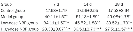
Table 2. Comparison of PAL across the groups
Group 7 d 14 d 28 d
Control group 17.68±1.79 17.56±2.55 17.53±3.64 Model group 40.11±1.57* 51.13±1.89* 49.08±1.78* Low-dose NBP group 34.11±1.57*,Δ 45.52±1.88*,Δ 39.52±1.79*,Δ High-dose NBP group 28.33±0.87*,Δ,▲ 36.53±2.70*,Δ,▲ 27.51±1.57*,Δ,▲
*P<0.05 compared with the control group; ΔP<0.05 compared with the model
group; ▲P<0.05 compared with the low-dose NBP group.
[image:3.612.91.373.201.269.2]Neurons of the model group showed degenera-tion and edema with obscure cell contour. Some neurons were apoptotic and the cell bod-ies shrank, presenting as polygonal or irregular shape. The morphological changes were less severe in NBP treatment groups, especially the
high-dose group (Figure 1). The above behav-ioral and morphological results indicated that NBP had a better control effect on the learning and memory impairment caused by radioactive brain injury. To further clarify whether the me- chanism of prevention and treatment of radia-Figure 1. Comparison of morphological changes of neurons across the groups (× 400). A: Control group; B: Model
[image:4.612.91.524.75.149.2]group at 14 d; C: Low-dose NBP group; D: High-dose NBP group.
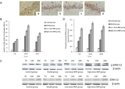
Figure 2. A: Immunohistochemical staining for phospho-ERK1/2 in each group (× 400). a: Control group; b: Model
[image:4.612.93.518.197.501.2]tion brain injury was related to the activation of ERK1/2 and the expression of GAP-43 in NBP. We further examined the changes of phospho-ERK1/2 and GAP-43 in the hippocampus.
Expressions of phospho-ERK1/2 and GAP-43
Immunohistochemical detection for phospho-ERK1/2 and GAP-43: Phospho-phospho-ERK1/2 and GAP-43 were mainly expressed in the nuclei, showing as small brown particles (Figures 2A,
3A). The control group had a few cells positive for phospho-ERK1/2. Compared with the con-trol group, the model group had more cells posi-tive for phospho-ERK1/2 at 7 d and 14 d; the number of positive cells decreased at 28 d, but it was still higher than that of the control group (P<0.05). Compared with the model group, the NBP treatment groups had higher number of cells positive for phospho-ERK1/2 at all time points, especially in the high-dose group (P< 0.05). The control group had a few cells
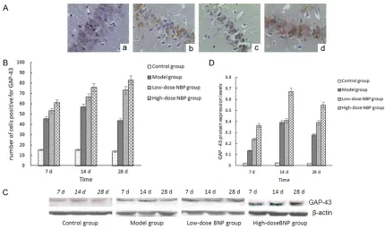
[image:5.612.91.521.68.324.2]posi-tive for GAP-43. Compared with the control group, the model group showed an obvious increase of cells positive for GAP-43 at 7 d and 14 d; the number of positive cells decreased at 28 d, but it was still higher than that of the con-trol group (P<0.05). Compared with the model group, NBP treatment groups had a significant increase of GAP-43-positive cells at each time point until 28 d, especially in the high-dose group (P<0.05) (Figures 2B, 3B). Western blot analysis forphospho-ERK1/2 and GAP-43: the IOD value of the protein bands after -actin was reflected by the value of the protein bands (Figures 2C, 3C). Compared with the control group, the expression of 7D in the model group, 14 d time and ERK1/2 phosphorylation levels of GAP-43 increased, 28 d decreased, but still higher than the control group (P<0.05); com-pared with the model group, the expression level of each time point in group NBP, the phos-phorylation of ERK1/2 and GAP-43 further Figure 3. A: Immunohistochemical staining for GAP-43 in each group (× 400). a: Control group; b: Model group at 14
increased, high expression continued to 28 d, especially changes in the high dose group (P<0.05, Figures 2D, 3D). The results showed that NBP could improve the brain radiation inju-ry in rats and promote the expression of hippo-campus ERK1/2 activity, GAP-43. In order to analyze the mechanism of the changes of the NBP regulation, we observed the changes of MDA and SOD activity in the intermediate metabolites of oxygen free radicals.
MDA content and SOD activity in each group
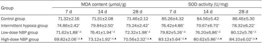
Compared with the control group, the model group showed a significant increase in MDA content and a reduction in SOD activity. MDA content further increased over time, while SOD activity declined (P<0.05). Compared with the model group, NBP treatment groups showed a significant reduction in MDA content and an increase in SOD activity (P<0.05), especially in the high-dose group (P<0.05, Table 3).
Discussion
RBI is a common complication after radiation therapy for craniofacial and cervical cancers. According to clinical reports, the probability of cognitive deficit in survivors receiving whole-brain radiation reaches 50%-90%. The decline in learning and memory capacity usually pres-ents as the symptom [10], in a dose-dependent manner. Study [3] shows that after radiation, the level of n-naeetyl aspartate (NAA) in the brain will decline, and ultrastructural damage of the brain usually occurs in a dose-dependent manner. Therefore, how to prevent the damage caused by ionizing radiation to patients’ learn-ing and memory capacity is a hot topic. Our results indicated that the rats of the NBP treat-ment groups suffered less severe learning dis-orders and the recovery of the learning capacity was faster. In the high-dose NBP group, AARR and PAL remained stable at 28 d, without show-ing continuous decline as in the model group. This indicated good preventive effect of NBP
against learning disorders in rats with RBI, which is consistent with the findings by Chen and Zhang [8, 12].
Radiation-induced impairment of learning and memory capacity is closely associated with neurogenic disorders. ERK1/2 is a key regula-tor of cell regeneration, differentiation and migration, which can be activated by several neurotrophic factors such as brain-derived neu-rotrophic factors and VEGF. As a result, nerve regeneration is promoted and the neuronal loss due to brain injuries is reduced. Conditional knockout of ERK1/2 would lead to failed axonal growth [13, 14], which suggested the key role played by ERK1/2 in nerve regeneration. GAP-43, an important axon growth factor, will be expressed intensively after brain injuries and react with intracellular signal molecule G pro-tein. This process will increase the reactivity of excitatory amino acid receptors and induce the release of calmodulin, thus promoting axonal sprouting and formation [15]. In recent years, Chinese scholars Huang and Liu et al. have confirmed that traditional Chinese medicine such as ShenxiongHuayu capsule and kidney-tonifying and brain-invigorating pills can en- hance neurogenesis and recovery of learning capacity after brain injury. The working mecha-nism is both associated with enhanced activity of ERK1/2 and upregulation of GPA-43 [16, 17]. Given the close connections of ERK1/2 signaling and GPA-43 to the learning capacity, we detected hippocampal expressions of phos-pho-ERK1/2 and GAP-43 in rats with RBI and both were significantly upregulated in a dose-dependent manner in the NBP groups; more-over, the high expressions persisted for some time. This indicated that NBP possibly worked by enhancing ERK1/2 activity and upregulating GAP-43.
[image:6.612.91.523.88.160.2]Ionizing radiation can cause oxidative stress to the brain, leading to neural damage, neu-rotransmitter release problems and cognitive
Table 3. Comparison of MDA content and SOD activity across the groups (_x±s)
Group MDA content (μmol/g) SOD activity (U/mg)
7 d 14 d 28 d 7 d 14 d 28 d
Control group 71.32±2.16 71.51±2.08 71.46±2.12 85.26±4.32 84.56±5.42 86.46±5.30
Intermittent hypoxia group 74.86±2.42* 79.84±2.50* 75.24±2.42* 76.42±4.86* 70.67±6.70* 78.32±6.22*
Low-dose NBP group 71.62±1.88*,Δ 76.41±1.94*,Δ 72.32±1.98*,Δ 79.82±5.26*,Δ 76.20±6.86*,Δ 80.12±5.76*,Δ
High-dose NBP group 69.82±2.06*,Δ,▲ 73.12±1.92*,Δ,▲ 70.56±2.32*,Δ,▲ 83.12±5.64*,Δ,▲ 80.62±5.86*,Δ,▲ 84.10±6.02*,Δ,▲
deficit [18, 19]. Huo [20] found that whole-brain radiation induced a reduction in the SOD level and an increase in the MDA level, which was accompanied by aggravated hippocampal neu-ronal damage and cerebral edema. Therefore, ionization-induced radiation injuries can be prevented by anti-oxidation. After NBP treat-ment in our study, the MDA level showed a sig-nificant reduction and SOD activity increased compared with the model group; furthermore, there was an improvement of learning and memory capacity. NBP can inhibit xanthine oxi-dase system, water solubility and lipid peroxi-dation system, thus reducing oxidative dam-age. Besides, NBP inhibits iron-dependent per-oxidation in the mitochondrial respiratory chain, thus preventing mitochondrial membrane dam-age caused by peroxidation and suppressing cascade reactions that amplify the oxidative stress [21-23]. Based on the above results, we believe that the strong anti-oxidative effect of NBP played a vital role. The latest research shows that anti-oxidation is crucial for activat-ing the signalactivat-ing pathway related to axonal regeneration. For example, Liu [24] reported that H2O2 pretreatment inhibited the oxidative stress-induced damage to PC12 cells by activat-ing ERK1/2 signalactivat-ing pathway, thus reducactivat-ing cell apoptosis. This represents a good evidence of the anti-oxidative effect of ERK1/2. Lu et al. [25] applied VitE to rats with cerebral ischemia and the results of induced axonal reconstruc-tion and repair were observed in the central nervous system, in addition to higher expres-sion of GAP-43 over a longer period of time. This means GAP-43 is involved in the resis-tance against oxidative stress in the presence of VitE. In this experiment, we detected the changes of MDA content and SOD activity in each group, both of which were significantly increased in a dose-dependent manner; more-over, the high expression and high activity per-sisted for some time. The MDA content after NBP treatment decreased significantly, also in a dose-dependent manner. We infer that the anti-oxidative effect of NBP in rats with RIB is probably related to enhanced ERK1/2 activity and upregulated GAP-43. This is the molecular mechanism of the neuroprotective effect of NBP against learning disorders in rats with RBI. Our experiment showed that NBP upregulated GAP-43 by activating ERK1/2 signaling path-way in the hippocampus, thus promoting
neuro-genesis and improving learning disorders in rats with RBI. NBP possesses multiple pharma-cological activities and its protective effect against ionizing radiation remains to be further investigated.
Disclosure of conflict of interest
None.
Address correspondence to: Dr. Min-Jian Li, The
Neurosurgery of Affiliated Hospital, North China
Uni-versity of Science and Technology, NO. 78 South Jianshe Road, Tangshan 063000, China. Tel: +86+15081978570; E-mail: [email protected]
References
[1] Tang Y, Luo D, Rong X, Shi X and Peng Y. Psychological disorders, cognitive dysfunction and quality of life in nasopharyngeal carcino-ma patients with radiation-induced brain inju-ry. PLoS One 2012; 7: e36529.
[2] He F, Wei X, Li JF, Li Y, Liao XW, Zeng Q and Niu
DL. Myelinbasicproteinreverts cognitive defi -cits of cranial irradiation injury by inducing hip-pocampal neurogenesis. Anatomy Research 2012; 34: 173-175.
[3] Ding WJ, Yang HH, Wang XF, Hu W, Lei H, Li CX, Fang F and Fang ZX. 1H-MR spectroscopy of the rat hippocampus after whole brain irradia-tion: an in vivo study. Chinese Journal of Radiological Medicine and Protection 2008; 28: 343-347.
[4] Takei H, Buckleair LW, Rivera A and Powell SZ. Brain tissue microarrays in neurodegenerative disease: validation of methodology and immu-nohistochemical study of growth-associated protein-43 and calretinin. J Pathol 2007; 57: 775-783.
[5] Zhao Y, Li J, Tang Q, Zhang P, Jing L, Chen C and Li S. Regulation of extracellular
signal-reg-ulated kinase 1/2 influences hippocampal
neuronal survival in a rat. Neural Regen Res 2014; 9: 749-56.
[6] Wang P, Wang LN, Liu YL, Li JM and Zhao YN. Effect of butylphthalide on brain edema and the expression of phosphorylated myosin light chain in peri-infarct tissue in focal cerebral ischemic rats. International Journal of Cere- brovascular Diseases 2014; 22: 376-38. [7] Zhao YN, Li JM, Zhang P, Chen CX and Li
SX. Protectove effects of dl-3butylphthalide against diffuse brain injury. Neural Regen Res 2013; 8: 2615-2624.
of Behavioral Medicine and Brain Science 2012; 21: 318-320.
[9] Fan XW, Guan SK and Wu KL. The effect of brain irradiation on mood and memory for rats. China Oncology 2014; 24: 841-849.
[10] Armstrong CL, Corn BW, Ruffer JE, Pruitt AA, Mollman JE and Phillips PC. Radiotherapeutie effects on hrain function: double dissociation of memory systems. Neuropsychiatry Neurop-sychol Behav Neurol 2000; 13: 101-111. [11] Greene-Shloessr D, Moore E and Robbins ME.
Molecular pathways: radiation-induced cogni-tive impairment. Clin Cancer Res 2013; 19: 2294-2300.
[12] Chen YZ, Zhang XX, Xiao L, Qi YH, Yang P, Tian Y and Bao SX. Effects of 1-3-n-Butylphthalide on the blood-brain barrier following whole brain irradiation in rats. Chinese Journal of Radiation Oncology 2012; 21: 392-3395. [13] Markus A, Zhong J and Snider WD. Raf and akt
mediate distinct aspects of sensory axon growth. Neuron 2002; 35: 65-76.
[14] Zhong J, Li X, McNamee C, Chen AP, Baccarini M and Snider WD. Raf kinase signaling func-tions in sensory neuron differentiation and axon growth in vivo. Nat Neurosci 2007; 10: 598-607.
[15] Rossini PM and Dal Forno G. Neuronal post-stroke plasticity in the adult. Restor Neurol Neurosci 2004; 22: 193-206.
[16] Huang HL, Li JM and Zhao YN. Effect of shenx-ionghuayu capsule on cerebral ischemia/re-perfusion injury and the expression of GAP43 in hippocampal CA1 of rats. Zhongguo Zhong Xi Yi Jie He Za Zhi 2014; 34: 185-190. [17] Liu YH, Li SW and Zheng QL. Effects of bushen
jiannao recipe on the content of acetylocholine and the hipppcampal ERK1 and ERK2 protein expressions of vascular dementia rats. Zhong- guo Zhong Xi Yi Jie He Za Zhi 2012; 32: 504-509.
[18] Zhang XX, Xiao L and Xu R. Pursuant to the adr
of radiation brain injury model, the influence of
cognitive dysfunction in rats. Chinese Journal of Radiological Medicine and Protection 2013; 33: 40-43.
[19] Jenrow KA, Brown SL, Lapanowski K, Naei H, Kolozsvary A and Kim JH. Selective inhibition
of microglia-mediated neuro-inflammation mit -igates radiation-induced cognitive impairment. Radiat Res 2013; 179: 549-556.
[20] Huo HM, Yang S, Chen LS, Lu HJ, Wang AD and Zhang LY. Hydrogen-rich saline alleviation on the oxidative stress and early-phase radiation-induced brain injury in rats. Chinese Journal of Radiological Medicine and Protection 2012; 32: 485-487.
[21] Dong GX and Feng YP. Butyl phthalide mito-chondrial ATPase on local cerebral ischemia reperfusion rats, the activity of antioxidant en-zyme and lipid peroxidation. Acta Academiae Medicinae Sinicae 2002; 24: 93-97.
[22] Huang RX, Li CX and Chen LY. Butyl phthalide on experimental effect of arterial thrombosis cerebral infarction. Chin J New Drugs 2005; 14: 985-988.
[23] Peng Y, Xu S, Chen G, Wang L, Feng Y and Wang X. 1-3-n-Butylphthalide improves cogni-tive impairment induced by chronic cerebral hypoperfusion in rats. J Pharmacol Exp Ther 2007; 321: 902-910.
[24] Liao XX, Wang YL, Guo RX, Zhang M, Yao QL, Li LQ, Chen PX and Feng JQ. ERK 1/2 mediates the cytoprotection of H2O2 preconditioning against oxidative injury in PC 12 cells. Chinese Pharmacological Bulletin 2008; 24: 1151-6. [25] Lu XH, Bao XQ, Peng WJ, Wang X, Zou CY and