Case Report
Use of the vastus medialis perforator flap
to treat defects around the knee
Liang Cheng, Tingxiang Chen, Zhijie Li
Department of Hand and Plastic Surgery, the Second Affiliated Hospital and Yuying Children’s Hospital of Wen-zhou Medical University, WenWen-zhou, China
Received March 31, 2017; Accepted July 2, 2017; Epub August 15, 2017; Published August 30, 2017
Abstract:Background and aim: Soft tissue reconstruction around the knee joint is usually complex for reconstruc-tive surgeons. Defects in this area are largely caused by trauma, chronic infection, tumor, and surgical infection.
This article presents the experience of using the vastus medialis perforator flap for reconstruction. Materials and methods: A total of 28 patients who had undergone reconstructive surgeries at the Second Affiliated Hospital and Yuying Children’s Hospital, Wenzhou Medical University, from December 2010 to December 2015 were enrolled. Age, sex, defect size, comorbidity, etiology, flap size, complication, follow-up, and postoperative range of knee joint were recorded and analyzed through a retrospective chart review. Results: All 28 flaps survived. In all, 22 of the
28 patients achieved full functional range of motion by the third month. Two patients had a 10° limitation in knee
extension and four cases had restricted knee flexion. Necrosis occurred in five cases; three were partial necrosis and two were superficial necrosis. Two patients were healed using conservative treatment, and the remaining three
cases were reconstructed with a full-thickness skin graft in two and directly sutured after debridement in one. All
patients were satisfied with their aesthetic and functional results. Conclusion: The vastus medialis perforator flap is
a reliable option for reconstructing soft tissue defects around the knee.
Keywords: Vastus medialis perforator flap, soft tissue defects around the knee joint, reconstructive
Introduction
Reconstructing soft-tissue defects around the knee remains a formidable task for reconstruc-tive surgeons. Common etiologies include de- fects resulting from trauma, tumor excision, poorly healing wounds, chronic infection, or orthopedic hardware needing sturdy coverage. The objectives of the surgery were to provide a settled soft-tissue cover, restore a favorable function to the joint [1, 2] and achieve an aes-thetically pleasing result. Various options can be employed, such as local fasciocutaneous flaps, free flaps, and muscle flaps; the chosen flap should have well-vascularized perfusion to ensure wound healing and facilitate any con-comitant orthopedic procedure [2]. Meticulous debridement, planning, and execution of a rea-sonable operative procedure are important in the management of a patient’s wounds. Out- lining a reconstructive plan requires
consider-ing the simplest technique likely to achieve wound closure with minimal donor-site morbid-ity [3-5]. If a soft-tissue defect around the knee requires flap coverage, the vastus medialis per -forator flap is a reliable, pliable, effective, and durable option. The use of this flap was first pio -neered by H. Zheng et al. in 2008 [6]. In this article, we reported our experience with perfo-rator flaps to cover knee defects.
Patients and methods
and 2 were falls. The defects occurred on one side in all patients. The defects were located on the superolateral aspects of the knee (N = 16), on the infrapatellar region of the knee (N = 3), and in the suprapatellar region or involving the knee (N = 9). Defect sizes ranged from 4 to 63 cm2 (average, 28.1 cm2). Three patients had
diabetes alone, and four had a combination of diabetes and hypertension or arteriopathy. There were no underlying diseases in 18 patients. Initial treatment began with aggres-sive debridement of bone and adjacent soft tis-sues and the removal of foreign materials and infected, non-viable, fibrotic, and granulation tissues. Before reconstructive procedures, patients received an average of two debride-ments. Subsequently, the vastus medialis per-forator flap was applied. All patients were evalu -ated for the viability of the flap and for function
-Table 1. Date of the patients Patient
NO. Age/Sex Etiology
Defect size
(cm) Comorbidities Flap size
(cm) Complication
Follow-up
(months) Knee joint function
1 56/M Traffic accident 8*6 HTN 9*7 No 10 Full range
2 67/M Traffic accident 7*4 None 8*5 No 7 Full range
3 47/M Skin ulcer 3*2 None 6*3 No 6 Full range
4 50/M Traffic accident 8*4 HTN 20*4 No 11 Full range
5 44/M Traffic accident 9*6 DM 10*6 Partial necrosis 14 Full range
6 62/F Traffic accident 5*2 DM/ART 8*3 No 14 Full range
7 60/M Skin ulcer 4*2 DM/ART 8*2 Partial necrosis 3 Limitation of knee flexion 30°
8 49/F Traffic accident 5*7 None 10*12 No 6 Full range
9 45/M Traffic accident 7*9 None 8*10 No 15 Full range
10 41/F Traffic accident 5*6 None 7*9 No 7 Full range
11 23/M Skin ulcer 6*8 None 8*10 Partial necrosis 24 Full range
12 26/M Traffic accident 6*8 None 8*10 No 9 Terminal 10° extention deficit 13 45/M Traffic accident 5*6 ART 7*9 No 21 Limitation of knee flexion 30° 14 49/F Traffic accident 2*5 None 4*15 Superficial necrosis 8 Full range
15 33/M Traffic accident 2*2 None 8*4 No 15 Full range
16 35/F Traffic accident 6*3 None 7*5 No 6 Full range
17 37/M Traffic accident 6*2 None 7*3 No 9 Full range
18 57/M Tumble 5*4 None 6*6 No 13 Full range
19 51/M Traffic accident 9*6 DM 10*7 No 7 Full range
20 50/M Skin ulcer 6*3 None 7*5 No 17 Full range
21 56/M Traffic accident 8*6 HTN/DM 9*7 No 10 Limitation of knee flexion 30°
22 28/F Traffic accident 5*3 None 6*4 No 12 Full range
23 56/F Traffic accident 6*3 HTN 7*5 No 7 Full range
24 24/M Skin ulcer 5*6 None 7*9 Superficial necrosis 4 Full range
25 61/F Traffic accident 5*3 None 6*3 No 11 Full range
26 66/M Tumble 6*4 HTN/DM 8*5 No 15 Terminal 10° extention deficit
27 37/M Traffic accident 9*6 None 10*7 No 7 Limitation of knee flexion 30°
28 28/F Traffic accident 4*2 None 8*3 No 12 Full range
[image:2.612.93.519.86.463.2]Note: M: male; F: female; HTN: hypertension; ART: arteriopathy; DM: diabetes mellitus.
Figure 1.The pedicle of the vastus medialis
perfora-tor flap during the transformation.
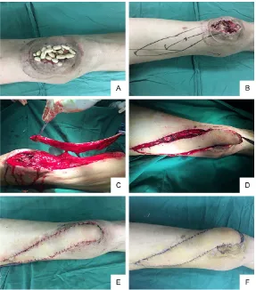
[image:2.612.91.289.496.646.2]Figure 2.A: The defect filled with the Antibiotic-PMMA bead on the anterior knee region; B: Planning of the flap; C: Elevated flap based on the pedicle supported by the perforator; D: The harvested flap insert into the defect; E:
Early postoperative result; F: 7 days after the operation.
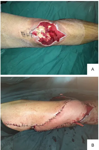
Figure 3.A: A circular defect on the left anterior knee region; B: Anterior view of late postoperative result.
al range of movement at the knee joint
com-pared to the contralateral knee joint. The fol- operative bandaging compromised vascular cir-culation and flap survival, so a simple window low-up period ranged from 3 to 24 months (mean, 10.7 months). A summary of the patients is shown in Table 1.
Surgical technique
[image:3.612.91.375.468.655.2]post-was made in the dressing to permit easy moni-toring in the ward. All cases received appropri-ate postoperative antibiotic therapy. All patients were followed up periodically until the wound site was healed and the donor had healed completely.
Results
A total of 28 vastus medialis perforator flaps were used in patients with soft tissue defects around the knee joint from May 2010 to September 2015. All flaps survived. The flaps were 16-120 cm2. In all, 22 patients achieved
full functional range of motion by the third month. Two patients had a 10° limitation in knee extension and four had restricted knee flexion. Most of the donor sites were closed pri -marily. Small superficial necrosis occurred in two cases, and both healed with conservative treatment. Partial necrosis was observed in three cases, two of which were covered with a splint thickness skin graft; the remaining one was directly sutured after debridement. No vas-cular compromise or other complications were
seen in the remaining patients. The length of hospital stay ranged from 7 to 80 days (aver-age, 33.36 days). Ultimately, all patients were satisfied with the functional results and could walk comfortably (Figures 2-4).
Discussion
[image:4.612.89.288.70.370.2]The knee region and proximal half of the lower leg have inherent characteristics that can make even a small defect a tremendous challenge because of exposed bones, tendons, metal fix -ation devices, or prostheses. Due to the lack of adequate soft tissue coverage or a stubborn infection, wound healing is markedly prolonged, leading to chronic wounds. Thin, pliable, dura-ble, and gliding coverage of the knee joint is a pre-condition for facilitating wound healing and any concomitant procedure. To restore the con-tour of the knee and preserve the function of the knee joint is the main goal of soft-tissue reconstruction [7]. Various options, such as local flaps, free flaps, and muscle flaps, have been used for reconstruction. The use of sev-eral muscle flaps has already been reported to repair soft tissue defects around the knee, including the vastus mediali [8], distally based vastus lateralis [9], sartorius [10] and gastroc-nemius [11]. A workhorse flap, the gastrocne -mius flap is famous for its easy operative tech -nique, better anti-infection capabilities, ade-quate three-dimensional defect coverage, and rich blood supply. The significant shortcomings of the gastrocnemius flap are unsatisfactory aesthetic outcomes and knee stiffness [12]. In addition, it is usually limited by defect size and location. Since Pontén [13] described the use of the fasciocutaneous flap in the lower leg in 1981, it has been an attractive choice for reconstructing defects around the knee joint [7], as it provides the closest match to the prim-itive tissue in complexion, volume, pliability, and tenacity [14]. However, local fasciocutane -ous flaps, such as the lateral or medial genicu -lar artery-island flap and the saphenous flap are usually associated with a sacrificed perfo -rator or sensory disturbances around the knee joint. Use of these flaps is also limited by exten -sive soft-tissue defects [15]. In addition, skin grafting is usually required at the donor site, and the application is usually restricted by deeply settled underlying pedicles. Free tissue transfer can be an excellent option if local flaps are unsuitable or cannot provide adequate cov-erage of the defect and if local tissue transfer is
unsuitable. Free flaps have been used in knee reconstruction extensively and efficiently with the advantage of sufficient bulkiness to fill the dead space and sufficient vascularity for the bone fragments. However, the choice of an appropriate recipient vessel is vital to the suc-cess and survival of the free flap. It is often dif -ficult to obtain an applicable recipient blood vessel in a patient due to damage from acci-dents or previous surgeries, and a free flap demands considerable microsurgical expertise and concentrated postoperative care facilities. In addition, it is a much more time-consuming procedure even in experienced hands and may not be available at most reconstructive cen-ters. It has also been reported that the vascu-lar thrombosis rate in patients who undergo free flap reconstruction of the knee is higher than in reconstruction of other parts of the limb [16].
The vastus medialis flap may be a promising option for reconstructing knee defects. Alth- ough vastus medialis flaps such as the muscle flap and perforator flap have been described several times in various forms in previous stud-ies, there have been few studies on the appli-cation of vastus medialis flaps around the knee, limited to a small number of patients (Table 2). We here present the largest series of cases with the use of vastus medialis perfora-tor flaps reported in the literature. A section of the quadriceps, the vastus medialis is located in the anteromedial aspect of the thigh beneath the sartorius and medial to the rectus femoris, where it plays an important role extending the knee joint. In 1981, Arnold et al. [8] first de-scribed the vastus medialis as a muscle flap for reconstructing the exposed knee joint in two patients. Arnold et al. reported that the vastus medialis was useful only for the upper part of the knee, because it had a relatively small arc of rotation and its blood supply was provided
[image:5.612.91.525.96.177.2]through several muscular branches of the superficial femoral artery. In addition, skin grafts were essential as the muscle does not have its own cutaneous territory. Although their first publication described the vastus medialis muscle without cutaneous territory, later ana-tomic studies and cadaveric dissections show- ed that the vastus medialis could be elevated as a myocutaneous flap. Tobin’s cadaveric study located a myocutaneous vascular supply to a large area of skin directly overlying the vas-tus medialis, suggesting that the vasvas-tus media-lis has its own cutaneous territory [17]. Kubota et al. [18] still upheld the absence of skin terri-tory in the vastus medialis muscle and reported the use in one case of a combination of the hemi V-Y flap and vastus medialis flap for the reconstruction of a knee with soft tissue loss and extensor rupture. In that study, the hemi V-Y flap moved as a rotating advancement flap and almost all of the blood supply was main-tained by continuity of the skin and subcutane-ous tissues. The controversy whether the vas-tus medialis muscle had its own skin territory continued until 2008, when Zheng et al. veri-fied that the vastus medialis had its own cuta -neous territory supplied by the musculocutane-ous perforators given off from the muscular arteries [6]. They demonstrated that the first musculocutaneous perforator was located around the mid-point of a surface projection line (located 9.4 ± 2.4 cm above the adductor tubercle and 4.1 ± 1.0 cm medial to the vertical line on the midpoint of the patella), which was traversed by the medial vastus medialis artery between a mid-point in the inguinal groove and the medial femoral condyle. In a few cases in a subsequent study [19], they introduced cover-age of soft-tissue defects around the knee using the pedicled vastus medialis perforator flap. We here present the largest series of cases with the use of vastus medialis perfora-tor flaps reported in the literature. A section of Table 2. List of the series regarding the application of the vastus medialis flaps reported in the litera -ture
Number
of cases Defect location Complication Additional Procedure Reference
2 Knee joint None Skin grafting (2) [8]
3 Knee joint None None [17]
1 Anterior knee None None [18]
6 Around the knee (5) Inferomedial aspect of the thigh (1)
Marginal necrosis (1) Sensitive antibiotic administration and frequent dressing change (1)
the quadriceps, the vastus medialis is located in the anteromedial aspect of the thigh beneath the sartorius and medial to the rectus femoris, where it plays an important role extending the knee joint. In 1981, Arnold et al. [8] first de-scribed the vastus medialis as a muscle flap for reconstructing the exposed knee joint in two patients. Arnold et al. reported that the vastus medialis was useful only for the upper part of the knee, because it had a relatively small arc of rotation and its blood supply was provided through several muscular branches of the superficial femoral artery. In addition, skin grafts were essential as the muscle does not have its own cutaneous territory. Although their first publication described the vastus medialis muscle without cutaneous territory, later ana-tomic studies and cadaveric dissections show- ed that the vastus medialis could be elevated as a myocutaneous flap. Tobin’s cadaveric study located a myocutaneous vascular supply to a large area of skin directly overlying the vas-tus medialis, suggesting that the vasvas-tus media-lis has its own cutaneous territory [17]. Kubota et al. [18] still upheld the absence of skin terri-tory in the vastus medialis muscle and reported the use in one case of a combination of the hemi V-Y flap and vastus medialis flap for the reconstruction of a knee with soft tissue loss and extensor rupture. In that study, the hemi V-Y flap moved as a rotating advancement flap and almost all of the blood supply was main-tained by continuity of the skin and subcutane-ous tissues. The controversy whether the vas-tus medialis muscle had its own skin territory continued until 2008, when Zheng et al. veri-fied that the vastus medialis had its own cuta -neous territory supplied by the musculocutane-ous perforators given off from the muscular arteries [6]. They demonstrated that the first musculocutaneous perforator was located around the mid-point of a surface projection line (located 9.4 ± 2.4 cm above the adductor tubercle and 4.1 ± 1.0 cm medial to the vertical line on the midpoint of the patella), which was traversed by the medial vastus medialis artery between a mid-point in the inguinal groove and the medial femoral condyle. In a few cases in a subsequent study [19], they introduced cover-age of soft-tissue defects around the knee using the pedicled vastus medialis perforator flap.
The perforator flap was originally described by Koshima in 1989 [20]. Attention has been
directed to improved methods of reconstruc-tion with progress on perforator techniques. The vastus medialis perforator flap is an alter -native solution for reconstructing soft tissue defects around the knee. The advantages of the vastus medialis perforator flap are: there is no need to sacrifice any main arteries or mus -cles; it is a time-efficient procedure with mini -mal donor site morbidity; the perforator site is relatively constant; the donor site and recipient area are in the same extremity, leading to less surgical injury; it is designed in a versatile form to give a better match to the defect, and the harvested area is bigger; advanced microsurgi-cal skills and vascular anastomosis are unnec-essary; the donor site is concealed and direct closure of the donor site is possible in most cases; the scar is above the knee, and the func-tion of the knee joint will not be affected; the recipient site has the most like-to-like tissues and can provide the most appropriate tissue with lower donor site morbidity and satisfactory aesthetic results; it can be harvested as a com-posite flap to reconstruct the extension mecha -nism of the knee; it can be harvested as a sen-sate flap; it can be rotated an appropriate degree based on the perforator pivot to cover a medium soft tissue defect; and the flap is thin and flexible. The main disadvantage of the vas -tus medialis perforator flap is that the femoral neurovascular bundle may become injured unless very close attention is paid during the dissection. In our series, the harvested flap was usually larger than the defect to obtain adequate distal perfusion of blood through the lower-resistance longitudinal nutrient vascular plexus of the cutaneous nerve made by a small modification to change the flap axis [21-23]. Thin and pliable coverage is required to ensure excellent mobility of the knee joint due to its particularities, flexion, and extension, and the vastus medialis perforator flap meets these standards. The use of this flap is a reliable and reproducible procedure providing low postop-erative morbidity, good daily function, and rela-tively satisfactory aesthetic results, without sacrificing any major vessels, nerves, or mus -cles. Hence, it is a suitable alternative for re-constructing medial soft tissue defects around the knee joint.
Acknowledgements
Disclosure of conflict of interest
None.
Address correspondence to: Dr. Zhijie Li, Department
of Hand and Plastic Surgery, The Second Affiliated Hospital and Yuying Children’s Hospital of Wenzhou Medical University, 109 Xue Yuan Xi Road, Wenzhou
325000, China. Tel: +86 13587969029; Fax: +86 0577-88816173; E-mail: 18767168160@163.com
References
[1] Nahabedian MY, Mont MA, Orlando JC, Dela-
nois RE and Hungerford DS. Operative man -agement and outcome of complex wounds fol-lowing total knee arthroplasty. Plast Reconstr Surg 1999; 104: 1688-1697.
[2] Gravvanis A and Britto JA. Venous
augmenta-tion of distally based pedicled ALT flap to re -construct the tibial tuberosity in a severely in-jured leg. Ann Plast Surg 2009; 62: 290-292. [3] Reddy V and Stevenson TR. Lower extremity
reconstruction. Plast Reconstr Surg 2008; 121: 1-7.
[4] Pan SC, Yu JC, Shieh SJ, Lee JW, Huang BM and Chiu HY. Distally based anterolateral thigh flap: an anatomic and clinical study. Plast
Reconstr Surg 2004; 114: 1768-1775. [5] Chen CY, Hsieh CH, Kuo YR, Jeng SF. An antero
-lateral thigh perforator flap from the ipsi-lateral
thigh for soft-tissue reconstruction around the knee. Plast Reconstr Surg 2007; 120: 470-473.
[6] Zheng H, Wang H, Zhang F and Yue S. Anatomic basis of perforator flaps of medial vastus mus -cle. Microsurgery 2008; 28: 61-64.
[7] Hallock GG. Local knee random fasciocutane
-ous flaps. Ann Plast Surg 1989; 23: 289-296.
[8] Arnold PG and Prunes-Carrillo F. Vastus
media-lis muscle flap for functional closure of the ex -posed knee joint. Plast Reconstr Surg 1981; 68: 69-72.
[9] Swartz WM, Ramasastry SS, Mcgill JR and
Noonan JD. Distally based vastus lateralis
muscle flap for coverage of wounds about the
knee. Plast Reconstr Surg 1987; 80: 255-265. [10] Petty CT and Hogue RJ Jr. Closure of an ex -posed knee joint by use of a sartorius muscle
flap: case report. Plast Reconstr Surg 1978;
62: 458-461.
[11] Moscona AR, Keret D and Reis ND. The
gas-trocnemius muscle flap in the correction of se
-vere flexion contracture of the knee. Arch
Orthop Trauma Surg 1982; 100: 139-142.
[12] Gravvanis AI, Iconomou TG, Panayotou PN and Tsoutsos DA. Medial gastrocnemius muscle
flap versus distally based anterolateral thigh flap: conservative or modern approach to the
exposed knee joint? Plast Reconstr Surg 2005; 116: 932-934.
[13] Pontén B. The fasciocutaneous flap: its use in
soft tissue defects of the lower leg. Br J Plast Surg 1981; 34: 215-220.
[14] Misra A and Niranjan NS. Fasciocutaneous
flaps based on fascial feeder and perforator
vessels for defects in the patellar and peri-patellar regions. Plast Reconstr Surg 2005; 115: 1625-1632.
[15] Liu TY, Jeng SF, Yang JC, Shih HS, Chen CC and Hsieh CH. Reconstruction of the skin defect of
the knee using a reverse anterolateral thigh
island flap: cases report. Ann Plast Surg 2010;
64: 198.
[16] Louer CR, Garcia RM, Earle SA, Hollenbeck ST, Erdmann D and Levin LS. Free flap reconstruc -tion of the knee: an outcome study of 34 cas-es. Ann Plast Surg 2015; 74: 57-63.
[17] Tobin GR. Vastus medialis myocutaneous and
myocutaneous-tendinous composite flaps.
Plast Reconstr Surg 1985; 75: 677-685. [18] Kubota Y, Tsubo K, Toh S and Ogawa T. Vastus
medialis muscle flap and hemi V-Y skin flap for
knee extensor and soft tissue reconstruction. Ann Plast Surg 2006; 56: 196-199.
[19] Zheng HP, Lin J, Zhuang YH and Zhang FH.
Convenient coverage of the soft-tissue defects around the knee by the pedicled vastus
media-lis perforator flap. J Plast Reconstr Aesthet
Surg 2012; 65: 1151-1157.
[20] Koshima I and Soeda S. Inferior epigastric
ar-tery skin flaps without rectus abdominis mus -cle. Br J Plast Surg 1989; 42: 645-648. [21] Chang SM and Hou CL. Chain-linked direction
-al vascular plexuses of the integument and
link-pattern vascularized flaps in distal extrem -ities. Plast Reconstr Surg 1998; 101: 2013-2015.
[22] Chang SM. The pedicle of neurocutaneous
is-land flaps. Plast Reconstr Surg 1996; 98:
374-376.