Original Article
Comparison of neurovascular relationships
between human pulp and rat pulp
Wei Zhang1*, Jie Zhang1,2*, Libo Zhao1, Yanjiao Jiang1, Xurui Hu1, Zhige Li1, Bin Liu1
1School of Stomatology, Lanzhou University, Lanzhou, China; 2School of Nuclear Science and Technology, Lanzhou University, Lanzhou, China. *Equal contributors.
Received October 14, 2016; Accepted March 1, 2017; Epub April 15, 2017; Published April 30, 2017
Abstract: The purpose of this study was to reveal the relationships of nerves and blood vessels between human and rat. Human premolar teeth, extracted for orthodontic reason, and rat maxilla and mandible were placed in 4%
para-formaldehyde. The presences of blood vessels (vascular endothelial growth factor VEGF), nerves (neurofilament heavy NFH) and myelinated nerve fiber (myelin basic protein MBP) were examined in the pulp by using immunohis -tochemistry (IHC). The pulp region was examined to determine the integral optical density (IOD) of blood vessels and
nerves by using fluorescence microscopy. Morphology of human pulp was largely consistent with that of rat molar pulp. Nerve fibers were seen to run along the blood vessels in both human and rat dental pulps. Myelinated nerve fiber was in the human peripheral area of the dental pulp, but appeared at central area of the rat molar pulp. In
addition, the distributions of human pulp nerves were appeared to vessel wall and perivascular space but the rat
pulp only existed in vessel wall. There were significant differences in blood vessels between human and rat teeth at
root and chamber (P<0.05), and the proportion of nerve fibers of human was significantly different from that of rat
teeth at root and chamber (P<0.05). The distribution and proportion of blood vessels and nerve fibers in rat molar teeth were significantly different from that of the human teeth, which indicated that the rat molar teeth could not a
valid model for human teeth research.
Keywords: Blood vessels, innervations, human pulp, rat pulp, suitable, animal model
Introduction
Dental pulp is a kind of highly vascularized and loose connective tissue, which contains cells, blood vessels and nerves. Dental pulp nerve
fibers and blood vessels, which provide nutri -ents and oxygen, remove waste material and
regulate inflammation, are in a state of dyna-mic balance. Dental pulp nerve fibers promote
angiogenesis of immune cells and reinforce pulpal defense system [1]. At the same time, the human dental pulp blood vessel is
con-trolled by nerve fibers [2]. Several studies [3-5]
have described distribution of blood vessels
and nerve fibers in human pulp, and nerve fiber bundles were along with vascular without branches in the root. However, nerve fibers
gave off branches in the crown, which were ac- companied by blood vessels in many instanc- es, forming a fan-shaped structure. In the pulp horns, nerve bundles and blood vessels pre-dominated formed a subodontoblastic plexus
and capillaries. Blood vessels and nerve fibers
not only play an important role in normal tooth function and self-repair, but also exert great
sig-nificance on dental pulp tissue regeneration
process [6-8].
in maturation [13]. Numerous immunoreactive
nerve fibers with varicosities were observed
along blood vessels in the center of the rat incisor pulp. In the periphery of the rat pulp,
many nerve fibers were seen in the subodonto -blastic region without extending into the pre-dentin or pre-dentin [14]. Previous study reported that the rat incisors simulating acute human pulpitis models were successful [15], but it is undeniable that rat incisor was absent from enamel at the pulp horns, nerve terminations in the dentine and subodontoblastic plexus. Fur- ther, rat incisor had special arrangement and development of the odontoblast [16]. In this case, the rat models simulating acute human pulpitis are still doubtful. Rat teeth were cap- ped with different adhesive resin systems and calcium hydroxide preparation different quan- tity of mineralized dentin formation, which can not explicitly verify whether the same story hap-pened in human dental pulp [11]. Further, the rat pulp demonstrated exceptional resilience and self-reparative capacity, which must be taken into account in the interpretation of ex- perimental results [17]. Hence, certain
speci-ficities of the model and intrinsic problems
have to be taken into consideration. Teeth of younger rat and older rat existed problem about technical aspects and physical chan-
ges. Cavities are more difficult to prepare in
younger rats, older rats have smaller pulps [18]. Although 6-week-old rats were the easi-
est experimental animal model, it difficult to
access the treatment area since the anato- mic position of the molar teeth posterior to a small diastema [18]. To our best knowledge, no study has reported/proved dental pulp of rat is consistent with that of human. Would the distribution, the number of nerves and blood vessels in rat dental pulp tissue be same to those of humans? Is the rat model suitable to study various human pulp diseases?
The aim of the present study was firstly to
undertake a semiquantitative assessment of the relationship of human pulpal blood vessels
and nerve fibers and those of rats by using an
immunocytochemcial approach. Secondly, we also provided experimental basis for dental pulp tissue engineering.
Materials and methods
Collection of samples
Thirty human premolar teeth were obtained from patients (15-40 years) who without
hyper-tension, heart disease and any other syste- mic diseases or contagious diseases because of orthodontic reasons. The patient’s consent was obtained. Teeth were extracted under local anesthesia without any signs of periodontal disease or caries at the Department of Ortho- dontics, Hospital of Stomatology, Lanzhou Uni-
versity. The tissue samples were fixed for 48 h
in 4% paraformaldehyde at 4°C. Surface enam-el and dentine of all human teeth were moved by high speed air turbine, making around the remaining dentin thickness of 2 mm.
20 male SD rats weighing 160~200 g were anesthetized by 10% chloral hydrate inject- ed intraperitoneally. Maxillae and mandibule were removed after cardiac perfusion with 4% paraformaldehyde. Incisors and molars of rat, which were completely peeled from maxillae
and mandibule, were fixed for in 4%
parafor-maldehyde 24 h at 4°C, and then rinsed with water for 1 h. The human and rat tooth were
decalcified for 3 months in 15% EDTA at room
temperature. EDTA were changed every two days and kept its volume 6 times more than that of all samples. The pulps of 15 chosen human teeth were carefully separated from hard structure with 11# surgical blade and tweezers, and then stored in 0.9% saline.
Standards of complete decalcification were
that all samples can be inserted with a 20#
k-file without resistance. Moreover, all samples
were pressed by tweezers and samples de- formed without resistance, and then slowly returned it.
Sample preparation
Fixed and decalcified samples were dehydrated
with the different concentration ethanol (70%, 75%, 85%, 95%, 100%) and treated with xy- lene. Then xylene was exchanged by molten
paraffin wax (56-58°C) with 2 changes, 1.5 h
each. Samples were embedded in fresh new
paraffin. Paraffin blocks were cut with micro
-tome to 3-5 μm thick sections and mounted
on glass slides. One part of slides was stain- ed with hematoxylin and eosin (H&E) and the other part of slides were submitted for immu- nohistochemical examination using the biotin-streptavidin system and tyramine signal am-
plification it. Blood vessels were labeled with
vascular endothelial growth factor (VEGF, 1:
1:50, Bioss, Beijing, China) and myelinated
nerve fibers were labeled with myelin basic pro -tein (BMP, 1:50, Bioss, Beijing China). Citrate buffer pH 6 were added into a pressure cooker with slides and heated up to keep boiling for 2 min. Repeated this procedure after slides cooled down at room temperature. The sec-tions were incubated for overnight with primary antibody at 4°C in blocking normal 1.5% goat serum. The secondary antibody (biotinylated goat anti-rabbit IgG) was applied for 30 min at 37°C and the slides were incubated with strep-tavidin-peroxidase complex. Slides were treat-ed with a color reagent DAB for visualizing of
Statistical analysis
Statistical analysis was performed using SPSS version 19.0 for Windows. All data were pre-sented as mean ± SE, and evaluated by Dun- nett-T3 test. The P values less than 0.05 were
considered statistically significant.
Results
Morphology of dental pulp between human and rat
Human molar was 2 cusps on the longitudinal section, but the frequency of cusp 4 was
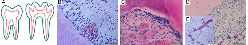
[image:3.612.93.525.74.153.2]pres-Figure 1. The contrast of human and rat dental histologic structure. Ideograph of human and rat tooth longitudinal section (A). In human dental pulp-dentin interface (B). In rat molar teeth pulp-dentin interface (C). In rat incisor teeth pulp-dentine interface (D). Inner of rat incisor teeth pulp (E). (B-E) is H&E staining. Scale bars: (B, C, E) 50 μm and (D) 100 μm.
Figure 2. Comparation between the width of human root pulp and rat root pulp.
positive signal and cellular nuclei were counterstained with hematoxylin.
Image analysis
Results were observed under
an immunofluorescence
mi-croscopy BX61-32FDIC-S08 (Olympus Japan). Images we- re processed for illustration purposes with Photoshop
CS5. Quantification of width
[image:3.612.93.371.226.494.2]ent in rat molar. Compared with human tooth, rat cusp parts of the enamel were platform-shaped, with dumbbell-shaped chamber, which showed elongated in middle and triangles on
both sides. The root and canal configuration of
rat was similar to human (Figure 1A). Diameter
of human premolar root pulp were not signifi -cantly different from that of rat (P>0.05), but
quantification of diameter of rat root pulp re-vealed a highly significant difference (P<0.05, Figure 2). Rat root were classified into wide root
and thin root based on pulp diameter. There
were no significant differences on microscopic
structure, cell morphology and pulp structure by the cross section of the dental pulp, the
rounded by irregular squares, and blood
ves-sels immersed within the fibrous tissue of the
center of the rat incisor pulp (Figure 1E).
Relationships between vasculature and the surrounding tissue
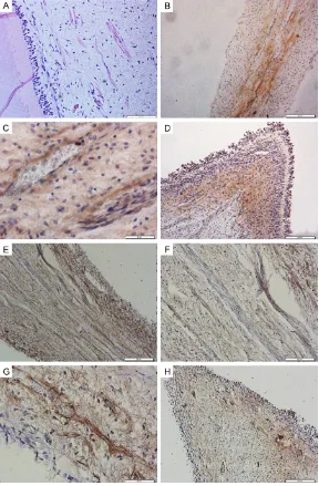
[image:4.612.89.524.71.200.2]Human pulp: In single-root teeth of human, there was the largest located blood vessel in the central portion of the pulp with some capil-laries running around (Figure 3A). In double-roots teeth, dental pulp displayed the exten-sive arrangement of medium-size blood ves- sels without large blood vessels immersed within two roots (Figure 3B). Reticular struc-
[image:4.612.92.373.272.484.2]Figure 3. Blood vessels in human dental pulps express VEGF (A, B, D, E) and centre of pulp (C) showing the blood vessels by H&E staining. The single-root teeth of human (A). The double-roots teeth of human (B). The inset shows a representative human premolar tooth with the root pulp area delimitated in red. Chambers (C, D). Horns (E). Scale bars: (D) 50 μm and (A-C, E) 200 μm.
Figure 4. The contrast of VEGF, NFH, MBP IOD in different part of human pulp.
peripheral one contained den-tin and predenden-tin, the bound-aries were not clear between a free-cell zone and a zone rich in cells in rat pulp alth- ough human pulp’s four-layer structure was clearly visible (Figure 1B, 1C). Rat incisor’s structure was completely dif-ferent from molars (Figure 1D). There was no obvious boundary in four-layer struc-ture of rat incisor. Morphology of odontoblast was irregular
in the central pulp with
fibr-ous connective tissue, and
fibroblasts and blood
ves-sels. The main direction of
the fiber in rat incisor was
sur-ture was formed by disorder blood vessels in the central area of the dental pulp (Figure 3C, 3D). Under the pulp horn regions, capillar-ies gathered passing through the odontoblast layer, when reaching the pulp horn, capillaries
single large-size blood vessel, while the blood vessels were sparse in the thin root (Figure 5E, 5F). There was significant difference between
[image:5.612.88.523.72.329.2]wide root and thin root (P<0.05) based on semi-quantitative analysis (Figure 6). In the rat pulp
[image:5.612.91.373.392.605.2]Figure 5. H&E staining (A-D) and immunocytochemical staining demonstrating VEGF-positive blood vessels in rat pulp (E-H). Wide root (A, E). Thin root (B, F), Large part of pulp chamber (C, G). Small part of rat pulp chamber (D, H). Scale bars: (A-D) 50 μm and (E-H) 200 μm.
Figure 6. The comparison IOD of VEGF, NFH, MBP between human root pulp and rat root pulp. H-Root: human root, R-Root: rat root.
gathered toward the horn (Figure 3E). At the same time, the greatest IOD of blood ves-sels were found in the pulp root region, compared with chamber and horn (P<0.05, Figure 4).
chamber, the vessels were mainly located in the near root instead of middle pulp chamber. Mild expression of VEGF was observed in the rat pulp horns and capillaries localized in the odontoblast layer. In large part of rat pulp chamber, the quantity of blood vessels was rich, but numbers of blood vessels were scarce in small part of rat pulp chamber (Figure 5G, 5H). The vascularity at large part of pulp
cham-ber was significantly greater than that of small
part of pulp chamber (P<0.05, Figure 7).
The relationships between human and rat nerve fibers and the surrounding tissues
Human pulp: Nerve fibers shape in human tooth was long fibrous shape observed from
classical histological examinations of the H&E stained. Neuron nucleus were fusiform (Figure 8A). In order to analyze the distributed
regular-ity of nerve fibers and myelinated nerve fibers, longitudinal paraffin sections were stained
us-ing NFH and MBP antibodies in human teeth and rat teeth. NFH-positive nerves were main- ly found at the blood vessel wall and perivas- cular region in human pulp (Figure 8C). Density
of nerve fibers was high in the
immunostain-ed in the mesial region of the root pulp (Figure 8B), while nerves ramified into a complex net
-work at chamber. Nerve fibers at the horn with
high intensity were located in the peripheral pulp and centre region of human pulp horn with
ber of myelinated nerve fiber and nerve fiber
in human pulp root was the most (Figure 4).
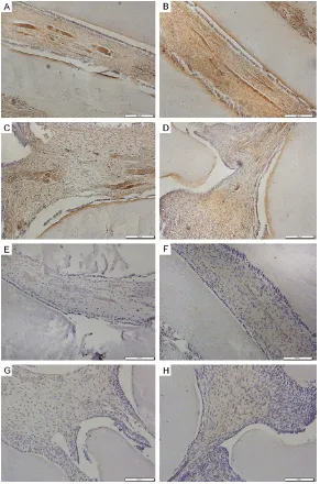
Rat pulp: The rat pulp was mainly innervated
by unmyelinated fibers. High levels of NFH
expression was found at the central portion of the root and near root rather than the mid- dle chamber by immunoreactivity of healthy rat
pulp. The distribution of rat pulp nerve fibers
was same as the distribution of blood vessels, mostly around the vessel wall (Figure 9A-D). Homogeneous MBP immunoreactivity of low intensity was evident at pulp and myelinated
fibers were often seen in close proximity to
vessels (Figure 9E-H). Expression of NFH and VEGF were found not only wide root, thin root, but also large part of chamber, small part of chamber, and IOD of NFH and VEGF were not different in different part of rat pulp (P>0.05, Figures 6, 7). NFH-expressing nerve fibers and MBP-expressing myelinated fibers were signifi -cantly different between wide root and thin root (P<0.05, Figure 6). There were significant
differences in expression of NFH and MBP be- tween large part of chamber and small part of chamber (P<0.05, Figure 7). In addition,
expres-sion of NFH and MBP which had
significant-ly differences can be observed in wide root and in thin root (P<0.05, Figure 6). Further, there
were significant differences in expression of
NFH and MBP in large part of chamber, while
there were no significant differences in expres
-Figure 7. The comparison IOD of VEGF, NFH, MBP between human chamber and rat chamber. H-Chamber: human chamber, R-Chamber: rat chamber.
slightly stained (Figure 8D).
Myelinated nerve fibers were
sparse in the peripheral of the pulp tissue obtained by MBP immunolabeling at pulp root (Figure 8E), the pulp chamber (Figure 8G) and pulp horn (Figure 8H). Large segmen- tal and nodular unmyelinated
nerve fibers were associated
with small myelinated nerve
fibers and unmyelinated fibers
were wrapped in myelin sh- eath with discontinuity (Figure 8F). Integral optical density
of nerve fibers was different
among root, chamber and horn of human teeth (P<0.05, Figure 4). There were signifi -cant differences in
myelinat-ed nerve fibers between root
num-sion of NFH and MBP in small part of chamber by electron microscopy (Figure 7).
Comparison with human and rat pulp: The inte-gral optical density of VEGF-positive blood
ves-sels did not reveal a significant difference
between human chamber and rat chamber (P>0.05, Figure 7). On the contrary, there
exist-ed significant differences between human root
and rat root (P<0.05, Figure 6). Myelinated
fibers expression within NFH of human cham
-ber was not significantly different from that of
Pulp sensitive fibers and blood vessels pene -trate through the apical foramen and gradual- ly fan out branches. Our data showed the ner- ves were always accompanied by blood ves- sels and not only presented in the blood ves-sels wall, but also proximity to the vesves-sels in human pulp. The dental pulp was mainly
inner-vated by unmyelinated nerve fibers, which were
widely distributed at the peripheral of pulp
tis-sue. Small myelinated nerve fibers were
close-ly associated with large unmyelinated nerve
fibers from our experiment. Myelinated nerve
Figure 8. Expression of NFH (B-D) in nerve fibers, and MBP (E-H) in myelinat
-ed fibers in human pulp as shown by IHC. H&E staining show-ed nerve fibers
(A). The double root (B, E). Chambers (C, F, G). Horns (D, H). Scale bars: (C, G) 50 μm, (F) 100 μm and (A, B, D, E, H) 200 μm.
and rat chamber (P>0.05, Figure 7), but the IOD of NFH
on human root were signifi -cant different from that of rat root pulp (P<0.05, Figure 6). MBP identified unmyelin
-ated fibers were not signifi -cantly different between hu- man chamber and rat cham-ber (P>0.05, Figure 7), and
there were not significant
differences in expression of MBP between human root and rat wide root (P>0.05),
while MBP revealed signifi -cant differences between hu- man root and rat thin root (P<0.05, Figure 6).
Discussion
The association between pul- pal nerves and the vascula-ture has been discussed in human teeth [3-5, 19-21]. However, it has not been described the relationship of
nerve fibers and pulpal blood
vessels in the rat tooth pulp.
We first described the distri -bution and number of nerve
[image:7.612.90.378.72.511.2]fibers with myelin sheath presented the seg -mental and incomplete shape, especially in the chamber. These phenomena were supported by other previous research [19-25]. Expression of protein gene product 9.5 (PGP9.5) was ob- served in the pulp roots, crowns and below the
odontoblast layer. Nerve fibers with few ramifi -cations were accompanied by larger vessels but branched into small bundles and passed through the odontoblast layer under the cell-rich zone [21]. NF-positive nerve bundles were distributed in the apical and crown, which were
tubules with low intensity, which could present interruptions [24]. However, not all nerve bun-dles accompanied arterioles in the pulp cham-ber and in the root canals and venules were not associated with nerve bundles [26]. Fur- thermore, our experiment showed there were
not significant differences in IOD of NFH and
VEGF in human pulp (root, chamber, horn, P>
0.05), but there were significant differences in
IOD of NFH and MBP in human pulp (root and chamber, P<0.05), indicating nerve fibers were
[image:8.612.89.380.71.511.2]mostly seen in close association with blood
Figure 9. Expression of NFH (A-D) in nerve fibers, and MBP (E-H) in myelin
-ated fibers in rat pulp as shown by IHC. Wide root (A, E). Thin root (B, F). Large
part of pulp chamber (C, G). Small part of pulp chamber (D, H). Scale bars: (A-H) 100 μm.
associated with blood vessels in many instances. In the cor-onal parts of the pulp, nerve
fibers fanned out, and
divid-ed into smaller branches that entered into the pulp den- tine interface. S100B label-ling was also detected in the region of apical, coronal and odontoblasts. NSE
immunore-active nerve fibers were pres -ent in mainly apical, coronal and odontoblastic parts of
the pulp [19]. β III-tubulin
im-munoreactivity was localized
to nerve fiber endings, which
were mostly seen in the pulp and predentin-dentin inter-face forming a terminal
net-work [22]. Most nerve fibers
including myelinated and un-
myelinated fibers within den -tal pulp expressed NFH and myelin sheaths were generally positive for MBP immunola-belling using confocal
micro-scopic and the nerve fibers
with myelin were prominent in coronal and peripheral re- gion, TH positive axons, in close proximity to blood ves-sels, were also observed [20].
vessels and the numbers of nerves and blood vessels could match well. Myelinated nerve
fibers were rare. The greatest numbers of nerve fibers, vascular and myelinated fibers were
found in the pulp root region because nerves and blood vessel entered the tooth via the api-cal foramina, ascended towards the coronal region and gave off branches en route. Vascular
and nerve fiber density were high at horns in human pulp. There were not significant differ -ences in IOD of VEGF, NFH and MBP in human pulp horns (P>0.05). We speculate it might be caused by their relatively small space, unclear borderline and measuring error.
The results of present study was in accordance with several previous studies [27, 28], which reported that rat dental pulp nerve was linked closely with the blood vessels, and the distribu-tion of nerve generally followed a similar pat-tern as blood vessels. In the rat root pulp, ner-
ve fiber bundles and blood vessels were
pa-rallel to the long axis of the tooth without ob-
vious branches, ramified in the coronal parts, and were present in an abundant fiber and vas -cular network. In addition, our study also dem-onstrated that the number between rat pulp blood vessels and nerves using
semi-quantita-tive analysis. There was significant difference
in IOD of VEGF, NFH and MBP expression in all parts of rat pulp, including wide root, thin root, large chamber and small chamber, suggesting
the number of nerve fibers and blood vessels
in wide root and large part of chamber was higher (P<0.05). The density of nerve fibers and blood vessels was not statistical significance
in rat root (P>0.05), while the density of nerve
fibers and myelinated fibers was statistical sig
-nificant (P<0.05), indicating that nerve fibers
accompanied vessels and the majority of the
nerve fibers in the rat root pulp were unmyelin
-ated fibers. Rat pulp express MBP, a protein commonly expressed myelinated fibers, was
in small numbers. Similarly, the IOD of nerve
fibers and blood vessels was not statistical significance in rat chamber (P>0.05), number
of nerve fibers and blood vessels was
basical-ly consistent. The fraction of NFH-expressing
nerve fibers was higher than MBP-expressing myelinated fibers in large part of chamber (P< 0.05). At variance with large part of chamber,
the proportion of nerve fibers was no signifi
-cantly difference from myelinated fibers in
small large part of chamber (P>0.05). Nerve
fibers were mainly unmyelinated fibers in large part of chamber and myelinated fibers in small
part of chamber showed a distinct distribution pattern on rat chamber, which may be proven that different innervation take place in different areas of the chamber. This hypothesis requires further experiments to testify.
The results from our HE staining experiment showed there were big structure differences between human teeth and rat incisor, but there was subtle difference from four microscopic zones of molar teeth between human and rat. Histological structure of rat molar teeth was unclear, although some authors suggested the biological responses of rats were consistent with the reaction of human due to their similar molar dental pulp tissue structure and anatom-ical structure of rat [29]. The majority of nerve
fibers in human dental pulp were presented in
central of human pulp. MBP was expressed in
the human myelinated nerve fibers presented
at the peripheral area. Both unmyelinated and
myelinated fibers in the human dental were
located blood vessels wall and perivascular
regions. However, nerve fibers and myelinated nerve fibers were located in central parts of
rat pulp, the expression of MBP in myelinated
nerve fibers accompanied by blood vessels, while unmyelinated nerve fibers in rat pulp
were mainly observed at the vascular walls. We also found that the number of blood vessels
and nerve fibers in the human root of pulp was
higher than those in rat root of pulp (P<0.05), while the number of blood vessels and nerves in the human chamber and rat chamber had no obvious difference (P>0.05), showed that radicular regions in human pulp were rich in blood supply and innervation, change of blood vessels and nerves was similar in human and rat pulp, suggesting different distribution and
number of nerve fibers may reflect the differ -ence in vascular supply nutrition for pulp
tis-sue, nerve responses may regulate blood flow, they influenced each other [2, 30]. In the
hu-man and rat pulp, distribution of myelinated
nerve fibers which were scarce was different.
In the rat, pituitary adenylate cyclase-activating polypeptide (PACAP)-immunoreactive (IR) nerve
fibers contained calcitonin gene-related pep -tide (CGRP)-IR and originated from the
trigemi-nal ganglion. PACAP-IR nerve fibers in the
from the superior cervical ganglion [31]. In the
rat incisor, nerve fibers did not form plexus in
the sub-odontoblast layer and not enter the dentin [32]. In the early stages of development, the sensory innervation pattern was different between rat incisor and molar pulp, but change in maturity, which might account for rat incisors are continuously growing [33]. Sensory nerve
fibers of the rat incisor pulp involved in the
vasoregulatory function, which made it a mo- del distinct from other teeth model for study- ing neurovascular interactions [14]. The
propor-tion of large myelinated fibers and unmyelinat
-ed fibers was significantly different between
molar and incisor within the parent axons. The
fraction of large myelinated fibers was signifi -cantly higher for the molar-than incisor-par- ent axons, while the fraction of unmyelinated
fibers was significantly higher for the
incisor-than molar-parent axons [34]. Current experi-mental samples were dominated by small ani-mals, which were a limitation in the study and recommended that it was necessary to use large mammals [35]. So, distribution and the
number of nerve fibers and blood vessels in
the human dental pulp were different from rat tooth pulp may be due to species differences in the oral environment, function and the state of engagement through tooth development. This suggests that the rat model for studying teeth is still controversial.
Regenerative endodontic treatment, which is the most perfect treatment of dental pulp ne- crosis, allows not only continuation of root de- velopment and apical closure, but also resto- ration of immune and sensory functions. How- ever, this clinical technology has not yet been approved. Further investigation is needed to
find a valid model to explore it [36]. The dental
pulp tissue regeneration of experimental ani-mal model is mainly rat [10-12, 37]. However, the present study showed that distribution and
number of blood vessels and nerve fibers in rat
molar teeth and the human teeth had a dramat-ic difference. In general, lower animals (rabbits, SD rats) had stronger regenerative capacity than higher animals, on account of the former with poor structural and functional organiza- tion and the latter with strong defense capabil-ity [38-40]. Therefore, rat molar teeth were not suitable for biological testing of human pulp tissue.
This study demonstrated that there was a
sig-nificant difference in the distribution and num
-ber of blood vessels and nerve fi-bers in rat
molar teeth and the human teeth by IHC, which supported why rats are not listed as suitable animals in ISO standard 7405, despite of simi-larity of their histological structure.
Acknowledgements
This work was supported by the Foundation of Stomatology College, Lanzhou University (Grant number: 20151204-2). Thanks to Hong Zhang (Chinese Academy of Science, China) for provid-ing experimental equipment, to Jie Li (Lanzhou University, China) for his support in obtaining rat teeth, and the authors also are grateful to Lihe Yao (First Hospital of Lanzhou University, China) for providing helpful advice that improved the manuscript.
Disclosure of conflict of interest
None.
Address correspondence to: Drs. Zhige Li and Bin Liu, School of Stomatology, Lanzhou University, 199 Donggang West Road, Lanzhou 730000, China. Tel: + 86 931 8915062; Fax: 931-8915051; E-mail: lizhg@lzu.edu.cn (ZGL); liubkq@lzu.edu.cn (BL)
References
[1] Nakashima M and Akamine A. The application of tissue engineering to regeneration of pulp and dentin in endodontics. J Endod 2005; 31: 711-718.
[2] Rodd HD and Boissonade FM. Immunocyto- chemical investigation of neurovascular rela-tionships in human tooth pulp. J Anat 2003; 202: 195-203.
[3] Dahl E and Mjör IA. The structure and distribu-tion of nerves in the pulp-dentin organ. Acta Odontol Scand 2009; 31: 349-356.
[4] Luthman J, Luthman D and Hokfelt T. Occurr- ence and distribution of different neurochemi-cal markers in the human dental pulp. Arch Oral Biol 1992; 37: 193-208.
[5] Zhu S, Yao J and Zhu Y. [The VIP-immunoreac-
tive nerve fibers in dental pulp-dentin complex
of human premolar]. Zhonghua Kou Qiang Yi Xue Za Zhi 2001; 36: 55-57.
[7] Mao JJ, Kim SG, Zhou J, Ye L, Cho S, Suzuki T, Fu SY, Yang R and Zhou X. Regenerative end-odontics: barriers and strategies for clinical translation. Dent Clin North Am 2012; 56: 639-649.
[8] Srisuwan T, Tilkorn DJ, Al-Benna S, Vashi A, Penington A, Messer HH, Abberton KM and Thompson EW. Survival of rat functional den- tal pulp cells in vascularized tissue engineer-ing chambers. Tissue Cell 2012; 44: 111-121. [9] Dammaschke T. Rat molar teeth as a study
model for direct pulp capping research in den-tistry. Lab Anim 2010; 44: 1-6.
[10] Lovschall H, Tummers M, Thesleff I, Fuchtbauer EM and Poulsen K. Activation of the Notch sig-naling pathway in response to pulp capping of rat molars. Eur J Oral Sci 2005; 113: 312-317. [11] Suzuki M, Taira Y, Kato C, Shinkai K and Katoh
Y. Histological evaluation of direct pulp cap-ping of rat pulp with experimentally developed low-viscosity adhesives containing reparative dentin-promoting agents. J Dent 2016; 44: 27-36.
[12] Zhang J, Liu X, Yu W, Zhang Y, Shi C, Ni S, Liu Q, Li X, Sun Y and Zheng C. Effects of human vascular endothelial growth factor on repara-tive dentin formation. Mol Med Rep 2015; 13: 705-712.
[13] Zerari-Mailly F, Braud A, Davido N, Toure B, Azerad J and Boucher Y. Glutamate control of
pulpal blood flow in the incisor dental pulp of
the rat. Eur J Oral Sci 2012; 120: 402-407. [14] Tabata S, Ozaki HS, Nakashima M and Uemu-
ra M. Blood vessels and nerve fibers in rat
incisor pulp. Immunoelectron microscopic ob-servation with anti-substance P antibody. Eur J Oral Sci 1998; 106 Suppl 1: 388-391. [15] Chidiac JJ, Rifai K, Hawwa NN, Massaad CA,
Jurjus AR, Jabbur SJ and Saade NE. Nociceptive behaviour induced by dental application of ir-ritants to rat incisors: a new model for tooth
inflammatory pain. Eur J Pain 2002; 6: 55-67.
[16] Kubota K, Katayama T, Hosaka K, Nagae K, Takada K, Iseki H, Shibanai S, Sato Y and Yonaga T. Structural and functional adaptation
of the pulpal nerve fibers in the rat incisor.
Anat Anz 1985; 160: 17-31.
[17] Goldberg M and Smith AJ. Cells and extracel-lular matrices of dentin and pulp: a biological basis for repair and tissue engineering. Crit Rev Oral Biol Med 2004; 15: 13-27.
[18] Six N, Lasfargues JJ and Goldberg M. Differ- ential repair responses in the coronal and ra-dicular areas of the exposed rat molar pulp in-duced by recombinant human bone morpho-genetic protein 7 (osteogenic protein 1). Arch Oral Biol 2002; 47: 177-187.
[19] Dourou V, Lyroudia K, Karayannopoulou G, Papadimitriou C and Molyvdas I. Compara-
tive evaluation of neural tissue antigens--
neurofilament protein (NF), peripherin (PRP), S100B protein (S100B), neuron-specific eno -lase (NSE) and chromogranin-A (CgA)--in both
normal and inflamed human mature dental
pulp. Acta Histochem 2006; 108: 343-350. [20] Henry MA, Luo S and Levinson SR. Unmye-
linated nerve fibers in the human dental pulp express markers for myelinated fibers and
show sodium channel accumulations. BMC Neurosci 2012; 13: 29.
[21] Tomaszewska JM, Miskowiak B, Matthews-Brzozowska T and Wierzbicki P. Characteristics
of dental pulp in human upper first
premo-lar teeth based on immunohistochemical and morphometric examinations. Folia Histochem Cytobiol 2013; 51: 149-155.
[22] Couve E, Osorio R and Schmachtenberg O. Re- actionary dentinogenesis and neuroimmune response in dental caries. J Dent Res 2014; 93: 788-793.
[23] Faria KM, Brandão TB, Ribeiro AC, Vasconcellos AF, de Carvalho IT, de Arruda FF, Castro Junior G, Gross VC, Almeida OP, Lopes MA, Santos-Silva AR. Micromorphology of the dental pulp is highly preserved in cancer patients who un-derwent head and neck radiotherapy. J Endod 2014; 40: 1553-1559.
[24] Manolea H, Vasile N, Opri M, Fronie A and Popescu MR. Immunohistochemical and elec-tron microscopy aspects of the nerve struc-tures from the dental pulp. Rom J Morphol Embryol 2014; 55: 147-152.
[25] Nair PN. Neural elements in dental pulp and dentin. Oral Surg Oral Med Oral Pathol Oral Radiol Endod 1995; 80: 710-719.
[26] Steiniger BS, Bubel S, Bockler W, Lampp K, Seiler A, Jablonski B, Guthe M and Stachniss
V. Immunostaining of pulpal nerve fibre bun -dle/arteriole associations in ground serial sec-tions of whole human teeth embedded in technovit(R) 9100. Cells Tissues Organs 2013; 198: 57-65.
[27] Sun Y, Tao R, Zhang M, Cao X, Wang H, Xue L and Wu M. Expression of calcitonin gene- related peptide in rat pulp and periodontal
tissues by indirect immunofluorescence
me-thod. Monoclon Antib Immunodiagn Immuno- ther 2013; 32: 404-408.
[28] Vandevskaradunovic V, Kvinnsland S and Kvin- nsland IH. Effect of experimental tooth
move-ment on nerve fibres immunoreactive to calci -tonin gene-related peptide, protein gene prod-uct 9.5, and blood vessel density and distribu-tion in rats. Eur J Orthod 1997; 19: 517-529. [29] Redlich M, Maly A, Aframian D, Shabat S, Ezov
molysis and thrombosis in rats*. J Oral Pathol Med 2004; 33: 424-429.
[30] Kvinnsland I and Heyeraas KJ. Effect of trau-matic occlusion on CGRP and SP
immunoreac-tive nerve fibre morphology in rat molar pulp
and periodontium. Histochemistry 1992; 97: 111-120.
[31] Ichikawa H and Sugimoto T. Pituitary adeny- late cyclase-activating
polypeptide-immunore-active nerve fibers in rat and human tooth
pulps. Brain Res 2003; 980: 288-292. [32] Byers MR. Dental sensory receptors. Int Rev
Neurobiol 1984; 25: 39-94.
[33] Nishikawa S. Developmental changes in pulpal sensory innervation of rat incisors and molars
shown on a single injection of the fluorescent
dye AM1-43. Anat Sci Int 2007; 82: 227-232. [34] Paik SK, Park KP, Lee SK, Ma SK, Cho YS,
Kim YK, Rhyu IJ, Ahn DK, Yoshida A and Bae YC. Light and electron microscopic analysis of the somata and parent axons innervating the rat upper molar and lower incisor pulp. Neuroscience 2009; 162: 1279-1286. [35] Wang YY, Zhao YM and Ge LH. [Isolation and
identification of Beagle dog dental pulp stem
cells]. Zhonghua Kou Qiang Yi Xue Za Zhi 2012; 47: 241-245.
[36] Zhujiang A and Kim SG. Regenerative end-odontic treatment of an immature necrotic mo-lar with arrested root development by using recombinant human platelet-derived growth factor: a case report. J Endod 2016; 42: 72-75.
[37] Zhang M, Kokabu S, Nakatomi C, Sugiyama G, Matsuo K and Jimi E. The distinct distributions of immunocompetent cells in rat dentin pulp after pulpotomy. Anat Rec (Hoboken) 2015; 298: 741-749.
[38] Hong L, Song D and Zeng Y. Comparison and
improvement in primary airway fibroblast cul -ture across different mammalian species. Cell Mol Biol (Noisy-le-grand) 2015; 61: 108-114. [39] Polezhayev LW. The loss and restoration of
re-generative capacity in the limbs of tailless Amphibia. Biol Rev Camb Philos Soc 1946; 21: 141-147.
[40] Seifert AW, Monaghan JR, Smith MD, Pasch B, Stier AC, Michonneau F and Maden M. The
in-fluence of fundamental traits on mechanisms