Original Article
Plastic wrap as a dressing material to treat stage III/IV
pressure ulcers in the inflammatory phase: a
randomized controlled trial
Jun Takahashi1, Kayoko Nakae1, Masaharu Miyagawa2, Osamu Yokota3,4, Yayoi Fujiki1, Masumi Ide1, Sumio Nishida1, Harusuke Aoki1, Takesuke Aoki1
1Department of Psychiatry, Minakuchi Hospital, Japan; 2Minamikusatsu Keyaki Clinic, Japan; 3Department of
Psychiatry, Kinoko Espoir Hospital, Japan; 4Department of Neuropsychiatry, Okayama University Graduate School
of Medicine, Dentistry and Pharmaceutical Sciences, Japan
Received October 23, 2016; Accepted January 3, 2017; Epub March 15, 2017; Published March 30, 2017
Abstract: The efficacy of plastic wrap (typically used for food) as dressing material to treat National Pressure Ulcer
Advisory Panel stage III/IV pressure ulcers was compared to standard treatment conforming to guidelines. This 12-week open-label randomized controlled trial was conducted in 10 wards of a hospital and 2 care facilities. Primary outcome was absolute surface area reduction. Secondary outcome was Pressure Sore Status Tool score reduction. Of the 142 participants enrolled, 74 were randomly allocated to plastic wrap dressing treatment and 68 to standard
treatment. Of these, 71 and 65, respectively, were analyzed. The plastic wrap dressing treatment reduced signifi -cantly more surface area than the standard treatment for 12 weeks (P < 0.0001). The mean surface area reduction with plastic wrap dressing treatment and standard treatment at 4, 8, and 12 week was 7.4 ± 7.1 and 4.7 ± 4.8 cm2,
P = 0.0103, (95% confidence interval of difference 0.7 to 4.8), 10.0 ± 9.3 and 5.8 ± 5.9 cm2, P = 0.0020, (1.6 to 6.9), and 11.1 ± 9.9 and 6.7 ± 7.1 cm2, P = 0.0032, (1.5 to 7.4), respectively. The median scores of Pressure Sore Status Tool score reduction in plastic wrap dressing treatment and standard treatment at 4, 8, 12 week was 13 and 9, P = 0.0076, 17 and 11, P = 0.0123, and 19 and 13, P = 0.0065, respectively. These differences were statistically
significant. The incidence of adverse events was comparable. These findings suggest that plastic wrap dressing
treatment was more effective than standard treatment to manage stage III/IV pressure ulcers.
Keywords: Dressing, food wrap, plastic wrap, pressure ulcer, randomized controlled trial, treatment
Introduction
Despite advances in various therapeutic meth-ods and materials, the development of a new treatment for severe pressure ulcers is impera-tive. Occlusive dressing technique (ODT) by modern dressing materials is a recommended standard treatment [1, 2], but has limitations in managing pressure ulcers with much exudate, necrotic tissue, and/or infection, even though the performance of dressing materials has improved. Some ointments with gauze dressing can manage more exudate than ODT, and can treat infected wounds. However, if wounds with massive exudate are treated, excess exudate which cannot be absorbed completely may increase pressure on the wounds, leading to deterioration or infection of the wounds. In
addition, if wounds with less exudate are treat-ed, both ointments and gauze dressing tend to let the wounds dry [1, 2]. The effect of negative pressure wound therapy compared with alter-natives for the treatment of pressure ulcers have not been well documented [3] although a few studies have suggested its effectiveness [4, 5]. In addition, this therapy is often impracti-cal in many geriatric hospitals and care facili-ties because of the associated costs and need for special equipment.
Plastic wrap dressing treatment may be one option to meet many kinds of treatment demands although this method is not widely known. Plastic wrap, typically used for foods, has been effectively used as a dressing
Panel (NPUAP) [6] stage II, III, and IV pressure
ulcers [7-9]. Because of effectiveness, ease of use, and low cost [8, 10], this treatment has been gradually accepted in many Japanese hospitals and care facilities.
The procedure using this treatment is easy and
simple [11, 12]: non-sterile plastic wrap is fixed
using non-woven tape. Because the adhesive power of this tape is weak, part of this dressing comes off easily when excess exudate accumu-lates under the dressing. Therefore, the exu-date is drained away from the wound, pressure on the wound reduces, and a moist environ-ment is maintained. Plastic wrap dressing treatment is consistent with the theory of wound bed preparation and moist wound heal-ing [13, 14]. By this mechanism of plastic wrap dressing, stage III/IV pressure ulcers that have more exudate and/or infection can be healed [7, 9, 11].
Although a few studies comparing the effective-ness of plastic wrap dressing to alternative treatments are available [10, 11, 15], no ran-domized controlled trial has been conducted to compare the effectiveness of plastic wrap dressing versus standard treatments in the management of stage III/IV pressure ulcers in
the inflammatory phase. The aim of this ran -domized controlled study is to determine differ-ence in reduction of surface area and the Pressure Sore Status Tool [PSST] [16] by plastic wrap dressing treatment versus standard treat-ment for stage III/IV pressure ulcers.
Methods
Trial design and participants
This 12-week prospective randomized con-trolled trial was conducted in 10 wards of a Japanese psychiatric and geriatric hospital and 2 care facilities between May 2005 and May 2015. This was an open-label design because masking either the participants or the doctors/ nurses to the intervention was impossible. This study was performed in accordance with the Declaration of Helsinki and its amendments. The Institutional Review Board approved this study protocol. Written informed consent was obtained from participants or their family members.
Patients aged 20 or older with NPUAP [6] stage III/IV pressure ulcers in inflammatory phase
were included. Eligible participants had pres-sure ulcers measuring 4 cm2 to 80 cm2 (from 2 to 4 in the item of Size on the PSST), and at least 50% of the surface area was covered by necrotic tissue (from 4 to 5 in the item of Necrotic tissue amount on the PSST). Wounds covered by hard, black eschar were included after surgical removal of the eschar. If a patient had multiple pressure ulcers, the largest one was selected for this study.
Participants were excluded if their skin ulcer was due to other causes, including peripheral arterial occlusive disease, skin cancer; if they had poorly controlled diabetes (HbA1c > 10% measured at registration); or they were treated with corticosteroids, immunosuppressants, cytotoxic agents, or radiotherapy. Pressure ulcers in the red proliferation phase were not included.
Baseline parameters included age, sex, mental disorder, systemic disease (including pneumo-nia, diabetes, and malignancy), hemoglobin and serum albumin, Braden Scale [17] score, surface area and location of the pressure ulcer, and PSST score. After obtaining these baseline characteristics, consenting participants were randomized to receive plastic wrap dressing treatment or standard treatment 1:1 by simple randomization. This was performed by the reg-istration center, which was independent of the hospital wards and care facilities.
Intervention
Procedure for plastic wrap dressing treatment [11, 12]
After cleansing with plenty of clean tap water or saline, the wound was covered with non-sterile plastic wrap of a suitable size. Non-woven
adhesive tape was used to fix four sides of the
wound may deteriorate, leading to serious wound infection and sepsis [18].
Devitalezed tissue was mainly removed by autolytic debridement promoted by the moist environment. Deep undermining or large wound cavities were not packed with dressings, oint-ments, or gels. In the early stage of treatment, plastic wrap dressing causes a foul odor and copious yellow exudate. However, these are not usually signs of infection or aggravation of the wound. With frequent dressing changes, the yellow exudate becomes translucent, and the amount of exudate and foul odor are reduced within a week without antibiotic agents. The fre-quency of dressing changes depends on the amount of exudate. In this study, dressings were exchanged at least 2 times per day. When an infected wound with much exudate was treated, the dressings were changed 3 or 4 times per day. If much exudate was drained around the wound, a thin paper diaper could be applied to absorb it and to protect the skin.
Procedure for standard treatment [19-22]
Wounds in the standard treatment group were treated with ointments and gauze dressing or ODT after cleansing with clean tap water or saline. If a wound had excess exudate, infec-tion, and/or deep undermining, and ODT could not be applied, various ointments and gauze dressing were used. Ointments used in this study were iodine-sugar, iodine-cadexomer paste, silver sulfadiazine, and enzymes. If oint-ments and gauze dressing were used, dress-ings were exchanged 2 or more times per day.
If a wound improved from the yellow inflamma -tory phase to the red proliferation phase and the amount of exudate was reduced, ODT was applied. The wounds were treated with hydro-colloid dressing, hydrocellular polyurethane
dressing, transparent film, and alginate dress -ing. These dressings were changed 2 or 3 times per week on average. When excess exudate accumulated under the dressing, the dressing was changed once or twice a day.
Common treatment procedures
If cellulitis or systemic infection developed in association with the pressure ulcer, systemic antibiotics were given. Surgical debridement or skin incision was undertaken when wound
infection was poorly controlled or necrotic tis-sue could not be removed by other debride-ment methods. Depending on the level of risk determined according to the Braden Scale, pre-vention protocols [22, 23] to improve moisture of skin, activity, and nutrition were implement-ed by a committee for prevention and manage-ment of pressure ulcers that was unrelated to this trial. Specialized mattresses, overlay, and seat cushions which met the needs of patients could be used for pressure redistribution.
Outcome measurement
The primary outcome measure was the abso-lute surface area reduction (SAR: baseline sur-face area - actual sursur-face area). Whether the differences between the treatments would affect SAR during the study period was investi-gated, and the SAR at 4, 8, and 12 week was compared for both treatments. The surface area was measured at baseline, and then every week via digital camera imaging by the same investigator, who was unaware of the treatment allocation. The area was calculated (length × width) using a ruler photographed with the wound.
Secondary outcomes included PSST score reduction at 4, 8, and 12 week compared to baseline (baseline PSST score - actual PSST score), and the incidence of adverse events including wound deterioration, local or systemic infection, hypergranulation, and maceration.
Using PSST, qualitative changes were evaluat -ed quantitatively, including depth of ulcer, amount of necrotic tissue and exudate, nature of granulation tissue, and state of skin around the wound. In this study, a doctor and nurse who were trained to treat and assess pressure ulcers marked the PSST score for all wounds separately at baseline and every 4 weeks. The evaluators were aware of the treatment alloca-tion. If the scores were different, they examined the wound together and marked the score again.
The difference in SAR for both groups during the study period was evaluated by a repeated measures analysis of variance (ANOVA) to investigate the effect of time and treatment. The comparisons of SAR at each observation point used the unpaired Student’s t-test and a
reduction was analyzed by the Mann-Whitney U
test with Bonferroni adjustment. Difference was assessed with a 2-sided test, with an alpha level of 0.05. For multiple comparisons, Bon- ferroni adjustment was applied to maintain an overall alpha level of 0.05 (each P-value <
0.0167 was accepted as significant).
Sample size and statistical analysis
We estimated that 140 participants were required to detect 6.0 ± 10.0 cm2 of difference
in SAR (mean ± standard deviation) between the 2 groups at 12 week, with alpha risk of 0.0167 (to maintain an overall alpha level of 0.05 by Bonferroni adjustment) and a study po- wer of 0.80, allowing for 10% loss to follow-up and unbalanced allocation. This estimate was based on the results of a previous study [11]. All analyses were performed using Stat View,
[image:4.612.94.519.82.515.2]version 5.0 (SAS Institute Inc. USA). The statisti -cal analyses were performed on an intention-to-treat basis. Data from all patients who Table 1. Baseline patient demographics and wound characteristics
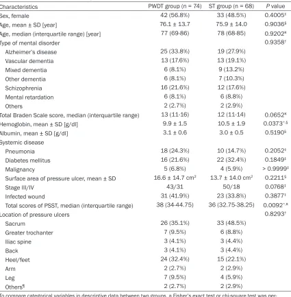
Characteristics PWDT group (n = 74) ST group (n = 68) P value
Sex, female 42 (56.8%) 33 (48.5%) 0.4005‡
Age, mean ± SD [year] 76.1 ± 13.7 75.9 ± 14.0 0.9036§
Age, median (interquartile range) [year] 77 (69-86) 78 (68-85) 0.9202ℵ
Type of mental disorder 0.9358†
Alzheimer’s disease 25 (33.8%) 19 (27.9%)
Vascular dementia 13 (17.6%) 13 (19.1%)
Mixed dementia 6 (8.1%) 9 (13.2%)
Other dementia 6 (8.1%) 7 (10.3%)
Schizophrenia 16 (21.6%) 12 (17.6%)
Mental retardation 6 (8.1%) 6 (8.8%)
Others 2 (2.7%) 2 (2.9%)
Total Braden Scale score, median (interquartile range) 13 (11-16) 12 (11-14) 0.0652ℵ Hemoglobin, mean ± SD [g/dl] 9.9 ± 1.5 10.5 ± 1.9 0.0373*,§
Albumin, mean ± SD [g/dl] 3.1 ± 0.6 3.0 ± 0.5 0.5190§
Systemic disease
Pneumonia 18 (24.3%) 10 (14.7%) 0.2052‡
Diabetes mellitus 16 (21.6%) 22 (32.4%) 0.1849‡
Malignancy 5 (6.8%) 4 (5.9%) > 0.9999‡
Surface area of pressure ulcer, mean ± SD 16.6 ± 14.7 cm2 13.7 ± 14.0 cm2 0.2211§
Stage III/IV 43/31 50/18 0.0768‡
Infected wound 31 (41.9%) 23 (33.8%) 0.3877‡
Total scores of PSST, median (interquartile range) 38 (34-44.75) 36 (32.75-38.25) 0.0092*,ℵ
Location of pressure ulcers 0.8293†
Sacrum 26 (35.1%) 33 (48.5%)
Greater trochanter 7 (9.5%) 6 (8.8%)
Iliac spine 3 (4.1%) 3 (4.4%)
Back 3 (4.1%) 3 (4.4%)
Heel/feet 24 (32.4%) 15 (22.1%)
Arm 2 (2.7%) 2 (2.9%)
Leg 7 (9.5%) 4 (5.9%)
Others¶ 2 (2.7%) 2 (2.9%)
Figure 1. Flow of participants though the trial.
underwent at least 1 post-baseline observation were in- cluded in the analyses, which were performed with the last observation carried forward (LOCF) method. If the values of SAR or reduction of PSST score were negative due to deterioration of the pressure ulcers, these were scored as zero, because wound exacer-bation would provide great negative outlier values.
Results
Between May 2005 and May 2015, 152 patients with skin ulcers were screened, and a total of 142 participants were randomized: 74 to plastic wrap dressing treatment and 68 to standard treatment. Baseline patient demograph-ics and wound characteristdemograph-ics are shown in Table 1. The 2 groups were similar in age, sex, type of mental disorder, Braden Scale score, preva-lence of systemic diseases, the surface area of the wound and location of pressure ulcer. PSST score in the plastic wrap dressing treatment group
were significantly greater than
in the standard treatment group (P = 0.0092).
The study flow of participants
through the trial is shown in Figure 1. Of these, 3 patients in the plastic wrap dressing group and 3 in the standard treatment group dropped out due to discharge, transfer to another hospital, or death
before the first observation.
The remaining 136 partici-pants (71 in the plastic wrap dressing treatment group and 65 in the standard treatment group) were included in the analysis. A total of 89 of the 136 participants completed the 12 weeks of the trial. Data from the remaining 47
partici-Figure 2. SAR of plastic wrap dressing and standard treatment groups (mean ± standard deviation). SAR in the plastic wrap dressing treatment group was
significantly greater than that in the standard treatment group during the en -tire study period. (P < 0.0001, repeated measures ANOVA). The comparisons of SAR at 4, 8, and 12 week were performed with an unpaired Student’s t-test and a P-value-based Bonferroni adjustment for multiple comparisons to
pants (25 in the plastic wrap dressing treat-ment group and 22 in the standard treattreat-ment group) were analyzed based on LOCF. During the study period, 20 participants in the plastic wrap dressing treatment group and 16 in the standard treatment group died of causes unre-lated to their pressure ulcers. All the data obtained are shown in Supplementary Table.
Primary outcomes
The SAR for both treatment groups is shown in Figure 2. The SAR in the plastic wrap dressing
in the plastic wrap dressing treatment group than in the standard treatment group. During the study period, the wound healed completely in 23/71 (32.4%) in the plastic wrap dressing treatment group and in 18/65 (27.7%) in the standard treatment group.
[image:6.612.91.377.97.151.2]Table 3 shows the incidence of adverse events. Deterioration of the wound, local wound infec-tion, and bacteremia/sepsis were observed approximately equally in both groups. More maceration developed in the plastic wrap Table 2. SAR of both treatments, each P value and 95% confi
-dence interval (CI) of difference at 4, 8 and 12 week
Weeks SAR in PWDT SAR in ST P value 95% CI of difference 4 7.4 ± 7.1 4.7 ± 4.8 0.0103* (0.7~4.8) 8 10.0 ± 9.3 5.8 ± 5.9 0.0020* (1.6~6.9) 12 11.1 ± 9.9 6.7 ± 7.1 0.0032* (1.5~7.4)
[image:6.612.93.375.210.485.2]PWDT = plastic wrap dressing treatment; ST = standard treatment; SAR = surface area reduction; *Statistically significant (P value based on unpaired Student’s t-test and Bonferroni adjustment to maintain an overall alpha level of 0.05).
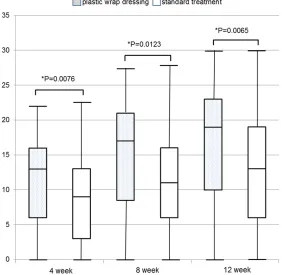
Figure 3. Reduction of PSST scores in plastic wrap dressing and standard treatment groups. Horizontal bar in each box represents the median.
Hori-zontal boundaries of the boxes represent the first and third quartile. Vertical
bars indicate the values of the 10th and 90th percentiles. *Statistically signifi -cant (P-value based on Mann-Whitney U test. To maintain an overall alpha
level of 0.05, Bonferroni adjustment was used).
treatment group was greater than in the standard treat-ment group during the entire study period. This difference
was significant (P < 0.0001).
The respective SAR values in the plastic wrap dressing treatment group at 4, 8, and 12 weeks were 7.4 ± 7.1, 10.0 ± 9.3, and 11.1 ± 9.9 cm2 (mean ± standard deviation). The corresponding values in the standard treatment group were 4.7 ± 4.8, 5.8 ± 5.9, and 6.7 ± 7.1 cm2. The plastic wrap dressing treatment re- duced the surface area of the pressure ulcers more than the standard treatment at each observation point, and all of these differences were
statis-tically significant. Each p
-val-ue and 95% confidence inter -val of difference is shown in Table 2.
Secondary outcomes
PSST score reductions in both treatment groups are shown in Figure 3. The median sco- res of the plastic wrap dress-ing treatment group at 4, 8, and 12 weeks were 13, 17, and 19, and the interquartile ranges were 6-16, 8.5-21, and 10-23, respectively. The corresponding values in the standard treatment group were 9, 11, and 13, and 3-13, 6-16, and 6-19. At every as- sessment point, the reduction in PSST score from the
dressing treatment group than in the standard treatment group, and hypergranulation devel-oped in the standard treatment group alone, although these differences were not
statisti-cally significant. Six of 7 cases of maceration in
the plastic wrap dressing treatment group and 2 of 2 in the standard treatment group devel-oped on the heel.
Discussion
This randomized controlled trial demonstrated
that stage III/IV pressure ulcers in the inflam -matory phase treated with plastic wrap
dress-ing showed significant improvement compared
to those receiving standard treatment. Plastic wrap dressing accelerated wound healing more than the standard treatment, in a comparison
of SAR. Significant differences in PSST score
reduction in both groups indicated that plastic wrap dressing treatment promoted qualitative improvement in pressure ulcers in addition to reducing the surface area. Baseline demo-graphics of patients in both groups were almost similar. Many participants were elderly and in poor general condition, and had severe system-ic disease. This study indsystem-icated that the plastsystem-ic wrap dressing was effective, even for severe wounds in patients in poor condition.
No statistical difference between groups was shown in the incidence of adverse events. The incidence of local wound infection and bactere-mia/sepsis in the plastic wrap dressing treat-ment was almost equal to that in the standard
treatment group. This finding agrees with prior
reports [10, 11, 15]. Plastic wrap dressing can manage bacterial bioburden on a wound with much exudate. Fewer wounds deteriorated with plastic wrap dressing treatment than with stan-dard treatment. Hypergranulation developed only in the standard treatment group, although
The characteristics of plastic wrap dressing treatment and standard treatment were com-pared. The greatest difference between the treatments is whether the wound is occluded completely. The plastic wrap dressing does not occlude the wound completely because of the weak adhesive power of non-woven adhesive tape used in this treatment, and the exudate is drained outside of the wound. Therefore, even if a wound with massive exudate is treated, plastic wrap dressing can maintain an ade-quate moist environment to facilitate wound healing, avoid increased pressure on the wound, and prevent wound infection despite maceration around the skin. Conversely, stan-dard treatment occludes completely, and dressing materials or ointments absorb the exudate. Excess exudate that is inadequately absorbed and is sealed under the dressing may make the wound overly wet, which prevents wound healing, causes infection and increased pressure, and aggravates the wound. This dif-ference may contribute to the superiority of plastic wrap dressing treatment in manage-ment of stage III/IV ulcers with much exudate, as shown by the present study and a previous non-randomized controlled study [11]. Con-
versely, differences in efficacy between the treatments were difficult to detect in the man -agement of mild pressure ulcers in prior stud-ies [10, 15]. When mild pressure ulcers with less exudate are treated, both the weak points of standard treatment and the strong points of plastic wrap dressing tend to be minimized, making the effectiveness of both treatments appear equal.
[image:7.612.92.323.86.186.2]In addition, plastic wrap as dressing material reduces the risk of aggravating the wound. Plastic wrap is smooth and slippery, and does Table 3. Incidence of adverse events
PWDT group
(n = 71) ST group (n = 65) P value Local wound infection 4 (5.6%) 5 (7.7%) 0.7366 Bacteremia/Sepsis 2 (2.8%) 2 (3.1%) > 0.9999 Maceration and odor 7 (9.9%) 2 (3.1%) 0.1689 Hypergranulation 0 (0.0%) 3 (4.6%) 0.1065 Deterioration of SAR 5 (7.0%) 10 (15.4%) 0.1709 Deterioration of PSST 3 (4.2%) 7 (10.8%) 0.1934
PWDT = plastic wrap dressing treatment; ST = standard treatment; Fisher’s exact test was performed.
not adhere to the surface of the wound and skin, unlike dressings used in standard treat-ment. Consequently, friction and shear stress as well as the damage caused by rubbing on the surface of the wound by dressing material do not occur with plastic wrap dressing treat-ment. Moreover, plastic wrap is so thin that local pressure on the wound can be avoided. Plastic wrap and non-woven adhesive tape are not just inexpensive alternative materials, but play important roles in this treatment due to their unique characteristics.
From the view of the burden in standard treat-ment, doctors and nurses who specialize in managing chronic wounds must select the most appropriate ointment or dressing for each case, depending on the depth of the wound and amount of exudate. Inappropriate selection of dressing or ointment may lead to a wet or dry environment, and can prevent wound healing. In addition, residual ointment or gel after inad-equate cleansing leads to wound infection. In particular, when pressure ulcers with deep undermining or large cavities are treated, much effort and time are needed to wash the wound. On the other hand, the procedure for plastic wrap dressing treatment need not change with the stage or condition of the pressure ulcer. Washing the wound is simple and easy, even if the wound has deep undermining or a large cavity. Technique and judgement requiring spe-cialization are not required. Frequent dressing changes and removal of necrotic tissue as needed can heal severe pressure ulcers. Since the procedure of plastic wrap dressing treat-ment is simple and easy, frequent dressing changes do not increase the workload of caregivers.
The limitations of this study must be consid-ered. First, this randomized controlled trial was not registered as an approved, publicly acces-sible clinical trial. Registration was uncommon when this trial was started in May 2005. Thus, this study does not meet all of the conditions set by the CONSORT 2010 checklist [24], although the CONSORT 2001 criteria [25] were fully met. Second, this trial could not be
double-blinded because of methodological difficulties.
Moreover, the PSST evaluators were aware of
the treatment allocation, because it was diffi -cult to obtain information on the wounds using only a digital camera image. Finally, even if
plastic wrap dressing treatment is superior, plastic wrap is not a medical material. More dis-cussions are needed about ethics, and further
investigations are necessary to confirm the
safety of plastic wrap dressing treatment. The incidence of adverse events was not statisti-cally different between groups; however, it is
possible that insufficient statistical power due to small sample size led to these findings. In
addition, long-term observation is needed. Conclusion
This study indicated that plastic wrap dressing treatment was more effective than standard treatment for stage III/IV pressure ulcers in the
inflammatory phase. The incomplete occlusion
of wounds by plastic wrap dressing plays an important role in managing copious exudate and maintaining an adequate environment for wound healing. Plastic wrap dressing can be useful in long-term care of patients with severe
pressure ulcers with respect to efficacy and
ease of use. More discussions about ethics are needed because plastic wrap is not a medical material.
Acknowledgements
This work was partially supported by a research grant from Minakuchi Hospital.
Disclosure of conflict of interest
None.
Address correspondence to: Jun Takahashi, Depart- ment of Psychiatry, Minakuchi Hospital, 2-2-43 Honmachi, Minakuchi-cho, 528-0031, Koka, Shiga, Japan. Tel: 1212; Fax: +81-748-62-1215; E-mail: [email protected]
References
[1] National Pressure Ulcer Advisory Panel, Euro
-pean Pressure Ulcer Advisory Panel and Pan Pacific Pressure Injury Alliance. Prevention and
treatment of pressure ulcers: quick reference guide. In: Emily Haesler, editor. Cambridge Me-dia: Osborne Park, Western Australia; 2014. [2] Guideline for prevention and management of
pressure ulcers. 4th edition. Jpn J PU 2015;
17: 487-557.
[4] de Laat EH, van den Boogaard MH, Spauwen PH, van Kuppevelt DH, van Goor H and Schoonhoven L. Faster wound healing with
topical negative pressure therapy in
difficult-to-heal wounds: a prospective randomized con-trolled trial. Ann Plast Surg 2011; 67: 626-631. [5] Dwivedi MK, Srivastava RN, Bhagat AK, Agar-wal R, Baghel K, Jain A and Raj S. Pressure ul-cer management in paraplegic patients with a novel negative pressure device: a randomised controlled trial. J Wound Care 2016; 25: 199-200, 202-204, 206-207.
[6] Pressure ulcers prevalence, cost and risk as-sessment: consensus development
confer-ence statement: The National Pressure Ulcer
Advisory Panel. Decubitus 1989; 2: 24-28. [7] Toriyabe S, Saito H and Sakurai K. Use of a
food wrap as a dressing material. Adv Skin Wound Care 1999; 12: 405-406.
[8] Okamura S, Urabe S, Hisatomi M, Iwasaki K
and Sakamoto M. The medical and economic effects of wrap-treatment for pressure ulcers.
Jpn J PU 2002; 4: 427-430.
[9] Oonisi S, Koike T, Shiogama K, Shimomura R
and Tsutsumi Y. Use of plastic wrap films for
treating pressure ulcers. Jpn J Med Pharm Sci 2006; 55: 561-567.
[10] Ueda T, Shimokubo S, Honda K, Kamikubo K,
Hanaki H, Torigoe T and Makisumi K. The eval-uation of plastic wrap treatment for pressure
ulcers. Jpn J PU 2006; 8: 551-559.
[11] Takahashi J, Yokota O, Fujisawa Y, Sasaki K, Ishizu H, Aoki T and Okawa M. An evaluation of
polyvinylidene film dressing for treatment of
pressure ulcers in older people. J Wound Care 2006; 15: 449-450, 452-454.
[12] Toriyabe S. Wrap Therapy for pressure ulcers [online]. Available at: http://www.geocities.jp/ pressure_ulcer/Accessed October 8, 2016. [13] Falanga V. The chronic wound: impaired
heal-ing and solutions in the context of wound bed preparation. Blood Cells Mol Dis 2004; 32: 88-94.
[14] Romanelli M and Mastronicola D. The role of wound-bed preparation in managing chronic pressure ulcers. J Wound Care 2002; 11: 305-310.
[15] Bito S, Mizuhara A, Oonishi S, Takeuchi K, Su-zuki M, Akiyama K, Kobayashi K and Matsuna-ga K. Randomised controlled trial evaluating
the efficacy of wrap therapy for wound healing acceleration in patients with NPUAP stage II
and III pressure ulcer. BMJ Open 2012; 2: e000371.
[16] Bates-Jensen BM, Vredevoe DL and Brecht ML. Validity and reliability of the Pressure Sore Sta-tus Tool. DecubiSta-tus 1992; 5: 20-28.
[17] Bergstrom N, Braden BJ, Laguzza A and Hol-man V. The Braden Scale for predicting pres-sure sore risk. Nurs Res 1987; 36: 205-210. [18] Moriyama Y. Severe complication associated
with inadequate wet dressing therapy: Benefits
and risk of so-called “Wrap Therapy”. Jpn Der-matol 2010; 120: 2187-2194.
[19] Pressure Ulcer Treatment. Clinical practice
guideline: quick reference guide for clinicians.
No. 15. Rockville, MD: U.S Department of
Health and Human Services. Public Health Service, Agency for Health Care Policy and Re-search. AHCRP Pub. No. 95-0653. Dec, 1994. [20] Japanese Society of Pressure Ulcers Evidence-Based Localized Pressure Ulcer Treatment
Guidelines. Tokyo: Shorinsya Co.; 2005. [21] European Pressure Ulcer Advisory Panel and
National Pressure Ulcer Advisory Panel. Treat -ment of pressure ulcers: Quick Reference
Guide. Washington DC: National Pressure Ul -cer Advisory Panel; 2009.
[22] Guideline for prevention and management of pressure ulcers. Tokyo: Shorinsya Co.; 2009. [23] Pressure ulcers in adults: prediction and
pre-vention. Quick reference guide for clinicians. AHCPR Publication No. 92-0050. Rockville, MD: Agency for Health Care Policy and
Re-search, Public Health Service, U.S. Depart -ment of Health and Human Services. May 1992.
[24] Schulz KF, Altman DG, Moher D; CONSORT Group. CONSORT 2010 statement: updated guidelines for reporting parallel group ran-domised trials. BMJ 2010; 340: c332. [25] Altman DG, Schulz KF, Moher D, Egger M,
Dav-idoff F, Elbourne D, Gotzsche PC, Lang T;