Original Article
The influence of different anesthesia
techniques on cognitive dysfunction in elderly patients
Yan-Bin Sun1*, Shu-Hong Sun2*, Da-Wei Liu3, Bing Bai4, Yan-Li Zhang5, You-Xin Guo6, Kui Jiang7
Departments of 1Anesthesiology, 3Vascular Surgery, 4Orthopaedics, 5Medicine, 6Otolaryngology, 7Radiology, Chengde Central Hospital, Chengde 067000, Hebei, China; 2Department of Gynaecology, Chengde Maternal and Child Care Hospital, Chengde 067000, Hebei, China. *Equal contributors.
Received November 25, 2015; Accepted February 13, 2016; Epub August 15, 2016; Published August 30, 2016
Abstract: This study aimed to investigate the influence of different anesthesia techniques on cognitive dysfunc
-tion in elderly patients after artificial femoral head replacement. One hundred and ninety-three elder patients with artificial femoral head replacement included in this study were randomly divided into general anesthesia (GA) and
epidural anesthesia (EA) group. Minimum mental state examination (MMSE) method was used to assess the
ner-vous and mental function in both subjects. Amyloid-beta (Aβ) protein levels in blood plasma were detected using enzyme-linked immunosorbent assay (ELISA) method. MMSE scoring and Aβ protein levels test were performed at the time of 24 hours preoperatively (T1), 24 hours postoperatively (T2) and 72 hours postoperatively (T3). The results showed that Amyloid beta protein levels in blood plasma were increased at the time of T2 compared with those at the time of T1 in GA group. Furthermore, MMSE scorings were upregulated, amyloid beta protein levels in blood plasma were downregulated and POCD incidence was decreased at the time of T3 compared with those at the time of T2 in GA group (P<0.05). Compared with GA group, MMSE scorings were elevated, amyloid beta protein
levels in blood plasma were reduced and POCD incidence was decreased in EA group at the time of T2 (P<0.05).
In conclusion, the general anesthesia method is more likely to exert influence on cognitive dysfunction compared to epidural anesthesia method in elderly patients after artificial femoral head replacement and general anesthesia
may induce cognitive impairment by upregulating amyloid beta protein expression.
Keywords: General anesthesia, epidural analgesia, postoperative cognitive dysfunction, minimum mental state examination, artificial femoral head replacement
Introduction
Postoperative cognitive dysfunction (POCD)
is a common complication characterized with mental derangement, anxiety and memory
impairment after surgery with general anaes -thesia in the elderly [1-3]. It has been reported
that the incidence of POCD is about 40.5% after surgery in the elderly [4]. Considerable data indicate that POCD might be caused
by anaesthesia [5, 6]. However, the exact
pathogenetic mechanism of POCD remains
undetermined.
It has been shown that amyloid precursor pro-tein (APP) plays an important role during the
occurrence of Alzheimer’s disease (AD) [7-9].
Considerable data indicate that dysregulation
of APP expression and beta-amyloid clearance are involved in the pathophysiology of AD [10-12]. Amyloid-beta (Aβ) protein is formed after
sequential cleavage of APP and serum Aβ pro -tein concentration test is used in the diagnosis
and prognostic evaluation of cognitive function
[13, 14].
The incidence and precise pathogenesis of POCD after artificial femoral head replacement in the elderly remains undetermined. Therefore, we aimed to study the incidence of POCD and serum Aβ protein concentration after artificial femoral head replacement with general anes -thesia (GA) and epidural anes-thesia (EA) in the elderly.
Materials and methods
Subjects
A total of 193 patients suffered from femoral neck fracture or femoral intertrochanter frac
-ment were recruited in the study. The ASA
clas-sification intended to describe pre-operative
physical status was II or III. All the included patients have no neuropsychiatric diseases, no vision and hearing disorders, and no antide-pressant and stabilizing agent used pre-opera-tion. The subjects were randomly divided into general anesthesia (GA) and epidural anesthe-sia (EA) group. Minimum mental state examina-tion (MMSE) method was used to assess the
nervous and mental function in both subjects.
Amyloid beta protein levels in blood plasma were detected using enzyme-linked immuno-sorbent assay (ELISA) method. Detailed
char-acteristics of included subjects were summa -rized in Table 1. This study was approved by the
ethical committee of Chengde
Central Hospital and every subject
provided an informed consent.
General anesthesia
After induction with Midazolam (0.1 mg/kg), fentanyl (3-5 μg/kg), propo
-fol (2 mg/kg), vecuronium bromide
(0.1 mg/kg), endotracheal intuba-tion and mechanical ventilaintuba-tion
were performed for the subjects
included in general anesthesia group. Then, the general anesthesia
was maintained with 1% to 3% vol isoflurane and the tidal volume was 8-10 mg/kg, respiratory frequency
was 10-12 time/min, and partial
pressure of carbon dioxide was
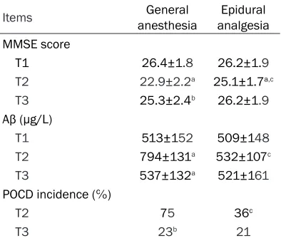
[image:2.612.90.350.84.287.2]maintained with 30-35 mmHg. Table 1. Characteristics of included subjects
Items anesthesia General analgesiaEpidural
Number of patients 98 95
ASA classification (II/III) 42/56 43/52a
Age (years) 67±7.2 68±8.4a
Body weight (kg) 58±5.4 59±7.2a
Gender (male/female) 49/49 48/47a
Years of education 16±3.8 15±4.1a
Operation time (min) 121±23 117±21a
Anesthesia time (min) 167±26 163±20a
Intraoperative hemorrhage quantity (ml) 339±101 385 ±121a
Blood transfusion amount (ml) 172±48 191±51a
Fluid infusion volume (ml) 1567±115 1497±120a
Urine output (ml) 301±105 324±118a
VAS 24 hours postoperatively 2.6±0.7 2.5±0.8a
aP>0.05 vs GA group.
Table 2. MMSE score, Aβ and POCD inci -dence comparison between two groups
Items anesthesia General analgesiaEpidural
MMSE score
T1 26.4±1.8 26.2±1.9 T2 22.9±2.2a 25.1±1.7a,c
T3 25.3±2.4b 26.2±1.9
Aβ (µg/L)
T1 513±152 509±148 T2 794±131a 532±107c
T3 537±132a 521±161
POCD incidence (℅)
T2 75 36c
T3 23b 21
aP<0.05 vs T1, bP<0.05 vs T2, cP<0.05 vs GA group.
Epidural anesthesia
The subjects were in the prone positions. L1-2 or L2-3 intervertebral spaces were the levels
of spinal puncture. Two milliliter ropivacaine (0.5%) was applied for anaesthetization. To keep the analgesic effect, fentanyl (0.001%) was injected intravenously with the speed of 2 ml/h for 48 hours.
Data collection and POCD assessment
The operation time, anesthesia time,
intraop-erative hemorrhage quantity, blood transfusion amount, fluid infusion volume, urine output and
visual analogue scale (VAS) 24 hours postop-eratively were recorded. Minimum mental state examination (MMSE) method was used to
assess the nervous and mental function in both subjects at the time of 24 hours preopera -tively (T1), 24 hours postopera-tively (T2) and
72 hours postoperatively (T3). POCD was diag -nosed when the MMSE score was less than
24 points or equal. Cognitive function was assessed by a trained and experienced profes
-sional without participating in the process of
anesthesia.
ELISA examination for Aβ
Aβ protein levels in blood plasma were detect -ed using enzyme-link-ed immunosorbent assay
(ELISA) method. Five milliliter fasting blood sample was drawn and centrifuged at 14000 rpm for 10 min. Then, the blood plasma was
[image:2.612.91.288.342.509.2]was assayed using specific ELISA kit (R&D systems) according to the manufacturer’s
instructions.
Statistical analysis
SPSS version 16.0 software was applied to
conduct the analyses and P value <0.05 was
considered statistically significant. The mea -surement data were shown as mean ±
stan-dard deviation. The differences of measure
-ment data were compared with the analysis of
variance test (among three groups) or t test
(between two groups). Differences test about count data was employed by the χ2 test. Correlations were assessed by Spearman’s
test.
Results
Patients characteristics
As shown in Table 1, there were no significant discrepancy about ASA classification, age, body weight, gender, years of education, operation
time, anesthesia time, intraoperative
hemor-rhage quantity, blood transfusion amount, fluid infusion volume, urine output and VAS 24 hours
postoperatively between GA group and EA group (P>0.05).
MMSE score, Aβ and POCD incidence in two groups
As shown in Table 2; Figures 1 and 2, there
were significant downregulation of MMSE scor
-ings at the time of T2 compared with those at
the time of T1 in GA group or EA group (P<0.05). Amyloid beta protein levels in blood plasma
were increased at the time of T2 compared with those at the time of T1 in GA group.
Furthermore, MMSE scorings were upregulat-ed, amyloid beta protein levels in blood plasma
were downregulated and POCD incidence was decreased at the time of T3 compared with those at the time of T2 in GA group (P<0.05). Compared with GA group, MMSE scorings were elevated, amyloid beta protein levels in blood
plasma were reduced and POCD incidence was decreased in EA group at the time of T2
(P<0.05). Moreover, amyloid beta protein levels were negatively correlated with MMSE scorings
in all patients at the time of T2 (r=0.769, P<0.001; n=193).
Discussion
There is ample evidence that POCD is a subtle disorder of thought processes which may influ -ence the short-term memory involving visual and verbal memory, attention, and visuospatial abstraction [15, 16]. Accumulating evidence
has shown that POCD incidence is associated
with age, trauma, anesthesia, medicine,
opera-tion type, a history of alcohol abuse, intraopera
-tive hemorrhage quantity, intraopera-tive blood
pressure, and concurrent brain diseases
preop-eratively [17-19]. In the present study, for better parallel we compared ASA classification, age, body weight, gender, years of education, opera -tion time, anesthesia time, intraoperative
hem-orrhage quantity, blood transfusion amount, fluid infusion volume, urine output and VAS 24
hours postoperatively between GA group and Figure 1. MMSE score comparison between GA
group and EA group. *P<0.05 vs T1, #P<0.05 vs T2, ※P<0.05 vs GA group.
Figure 2. Aβ incidence comparison between GA
group and EA group. *P<0.05 vs T1, ※P<0.05 vs GA
[image:3.612.93.285.71.232.2] [image:3.612.324.517.74.227.2]EA group and found that there were no signifi -cant discrepancy about them between the two groups.
The MMSE test is a 30-point questionnaire that
is used extensively in clinical and research set-tings to measure cognitive impairment [20]. It is used to estimate the severity and
progres-sion of cognitive impairment [21]. Our results showed that there were significant downregula
-tion of MMSE scorings at the time of T2 com
-pared with those at the time of T1 in GA group
or EA group. Compared with GA group, MMSE
scorings were elevated and POCD incidence was decreased in EA group at the time of T2. These findings indicate that epidural anesthe
-sia may be more beneficial than general anes
-thesia from the point of reducing POCD inci -dence. We speculate that the reasons resulting
in such change may be as follows. First, general anesthesia could affect central nervous sys -tem and induce cognitive impairment by regu-lating brain cholinergic system and memory
protein [22], however, the main acting site of
epidural anesthesia is the spinal cord instead
of central nervous system, thus central ner
-vous system is less affected by epidural anes -thesia. Second, epidural anesthesia may be
more helpful than general anesthesia in reduc
-ing stress response derived from the nervous system which is a key risk factor in the preva
-lence of postoperative delirium [23].
Further-more, MMSE scorings were upregulated and amyloid beta protein levels in blood plasma
were downregulated at the time of T3 com
-pared with those at the time of T2 in GA group
(P<0.05), which indicated that anesthetic drugs were gradually metabolized, the patients
grad-ually recovered and then their cognitive func -tion were gradually resumed until 72 hours postoperatively.
Considerable data indicate that Aβ protein is produced following sequential cleavage of the transmembrane protein APP by β- and γ-secretase and is released extracellularly in lengths ranging from approximately 37 to 43
amino acids [24-26]. Accumulating evidence
has shown that intrahippocampal injection of β-amyloid in rats induced the impairment of
learning and memory and the neuronal degen-eration in or around the injection sites [27, 28].
There is ample evidence that trauma,
anesthe-sia or surgery may induce the upregulation of IL-1β, IL-6 and TNF-α, which are capable of
aggravating cognitive function impairment by regulating the expression of Aβ protein, and inhibition of IL-1β, IL-6 or TNF-α may alleviating impairment of cognitive function [29-33]. The precise mechanisms whereby Aβ protein cause
neurotoxicity remain unknown. Jang et al. observed that beta-amyloid-induced apoptosis
in PC12 cells is associated with COX-2 up-regu
-lation through activation of NF-kappaB, which
is mediated by upstream kinases including ERK and p38 MAPK [34].
Liu et al. investigated the role of alpha7-nAChRs in the mediation of Aβ-induced neurotoxicity and found that alpha7-nAChRs are necessary for Aβ-induced neurotoxicity in hippocampal neurons because chronic Aβ significantly
increased LDH level in hippocampal cultures,
indicating a detrimental role of upregulated alpha7-nAChRs in the mediation of Aβ-induced
neurotoxicity [35]. Wang et al. demonstrated
that acteoside increased HO-1 expression through activation of ERK and PI3K/Akt signal
pathways, which may involve in the
neuropro-tection against Aβ-induced neurotoxicity [36]. Our results showed that amyloid beta protein
levels were negatively correlated with MMSE
scorings in all patients at the time of T2, indi -cating that amyloid beta protein levels were
a sensitive indicator of cognitive function.
Furthermore, amyloid beta protein levels in
blood plasma were increased at the time of T2 compared with those at the time of T1 in GA
group. Compared with GA group, amyloid beta protein levels in blood plasma were reduced
and POCD incidence was decreased in EA group at the time of T2. The results indicated
that general anesthesia may induce cognitive impairment by upregulating amyloid beta pro-tein expression.
In conclusion, the general anesthesia method
is more likely to exert influence on cognitive dysfunction compared to epidural anesthesia method in elderly patients after artificial femo -ral head replacement and gene-ral anesthesia may induce cognitive impairment by upregulat-ing amyloid beta protein expression.
Disclosure of conflict of interest
None.
Address correspondence to: Dr. Yan-Bin Sun, De-
pital, Chengde, Hebei, China. E-mail: [email protected]
References
[1] Silbert B, Evered L, Scott DA, McMahon S,
Choong P, Ames D, Maruff P and Jamrozik K.
Preexisting cognitive impairment is associated
with postoperative cognitive dysfunction after
hip joint replacement surgery. Anesthesiology 2015; 122: 1224-1234.
[2] Wang W, Wang Y, Wu H, Lei L, Xu S, Shen X, Guo X, Shen R, Xia X, Liu Y and Wang F.
Postoperative cognitive dysfunction: current
developments in mechanism and prevention. Med Sci Monit 2014; 20: 1908-1912.
[3] Lin GX, Wang T, Chen MH, Hu ZH and Ouyang
W. Serum high-mobility group box 1 protein
correlates with cognitive decline after gastro -intestinal surgery. Acta Anaesthesiol Scand 2014; 58: 668-674.
[4] Hansen MV. Chronobiology, cognitive function
and depressive symptoms in surgical patients. Dan Med J 2014; 61: B4914.
[5] Shi HJ, Xue XH, Wang YL, Zhang WS, Wang ZS
and Yu AL. Effects of different anesthesia methods on cognitive dysfunction after hip re -placement operation in elder patients. Int J Clin Exp Med 2015; 8: 3883-3888.
[6] Xu JH, Zhang TZ, Peng XF, Jin CJ, Zhou J and
Zhang YN. Effects of sevoflurane before car -diopulmonary bypass on cerebral oxygen bal-ance and early postoperative cognitive
dys-function. Neurol Sci 2013; 34: 2123-2129.
[7] Fu Y, Zhao D, Pan B, Wang J, Cui Y, Shi F, Wang C, Yin X, Zhou X and Yang L. Proteomic Analysis
of Protein Expression Throughout Disease Progression in a Mouse Model of Alzheimer’s
Disease. J Alzheimers Dis 2015; 47: 915-926. [8] Hahr JY. Physiology of the Alzheimer’s disease.
Med Hypotheses 2015; 85: 944-6.
[9] Kim HJ, Shin EJ, Lee BH, Choi SH, Jung SW, Cho IH, Hwang SH, Kim JY, Han JS, Chung C, Jang CG, Rhim H, Kim HC and Nah SY.
Oral Administration of Gintonin Attenuates
Cholinergic Impairments by Scopolamine,
Amyloid-beta Protein, and Mouse Model of Alzheimer’s Disease. Mol Cells 2015; 38:
796-805.
[10] Hata S. Molecular Pathogenesis of Sporadic Alzheimer’s Disease (AD) and Pharmaceutical Research to Develop a Biomarker for AD
Diagnosis. Yakugaku Zasshi 2015; 135: 1023-1027.
[11] Abramov AY, Ionov M, Pavlov E and Duchen MR. Membrane cholesterol content plays a
key role in the neurotoxicity of beta-amyloid: implications for Alzheimer’s disease. Aging
Cell 2011; 10: 595-603.
[12] Thal DR. Clearance of amyloid beta-protein and its role in the spreading of Alzheimer’s dis -ease pathology. Front Aging Neurosci 2015; 7: 25.
[13] Kimoto A, Kasanuki K, Kumagai R, Shibata N, Ichimiya Y and Arai H. Serum insulin-like
growth factor-I and amyloid beta protein in Alzheimer’s disease: relationship with cogni
-tive function. Psychogeriatrics 2015; [Epub ahead of print].
[14] de Souto Barreto P, Andrieu S, Payoux P, Demougeot L, Rolland Y, Vellas B; Multido- main Alzheimer Preventive Trial/Data Sharing Alzheimer Study Group. Physical Activity and Amyloid-beta Brain Levels in Elderly Adults with Intact Cognition and Mild Cognitive Impairment. J Am Geriatr Soc 2015; 63: 1634-1639. [15] Sorrell JM. Postoperative cognitive dysfunction
in older adults: a call for nursing involvement. J
Psychosoc Nurs Ment Health Serv 2014; 52: 17-20.
[16] Valentin LS, Pietrobon R, Aguiar Junior W, Rios
RP, Stahlberg MG, Menezes IV, Osternack-Pinto K and Carmona MJ. Definition and appli
-cation of neuropsychological test battery to evaluate postoperative cognitive dysfunction.
Einstein (Sao Paulo) 2015; 13: 20-26. [17] Shoair OA, Grasso Ii MP, Lahaye LA, Daniel R,
Biddle CJ and Slattum PW. Incidence and risk
factors for postoperative cognitive dysfunction
in older adults undergoing major noncardiac surgery: A prospective study. J Anaesthesiol Clin Pharmacol 2015; 31: 30-36.
[18] Gvozdenovic L and Antanaskovic A. History of alcohol abuse after major non-cardiac surgery and postoperative cognitive dysfunction. Eur J
Intern Med 2015; 26: e51.
[19] Liu C and Han JG. Advances in the
mecha-nisms and early warning indicators of the post
-operative cognitive dysfunction after the extra -corporeal circulation. Zhongguo Yi Xue Ke Xue Yuan Xue Bao 2015; 37: 101-107.
[20] Koekkoek PS, Rutten GE, van den Berg E, van Sonsbeek S, Gorter KJ, Kappelle LJ and Biessels GJ. The “Test Your Memory” test
per-forms better than the MMSE in a population without known cognitive dysfunction. J Neurol
Sci 2013; 328: 92-97.
[21] Larner AJ. Mini-mental Parkinson (MMP) as a dementia screening test: comparison with the Mini-Mental State Examination (MMSE). Curr Aging Sci 2012; 5: 136-139.
[22] Perouansky M. Liaisons dangereuses? General anaesthetics and long-term toxicity in the CNS. Eur J Anaesthesiol 2007; 24: 107-115. [23] Smith PJ, Attix DK, Weldon BC, Greene NH and
Monk TG. Executive function and depression as independent risk factors for postoperative
[24] Ripoli C, Cocco S, Li Puma DD, Piacentini R,
Mastrodonato A, Scala F, Puzzo D, D’Ascenzo
M and Grassi C. Intracellular accumulation
of amyloid-beta (Abeta) protein plays a major role in Abeta-induced alterations of glutama -tergic synaptic transmission and plasticity. J Neurosci 2014; 34: 12893-12903.
[25] Namekawa Y, Baba H, Maeshima H, Nakano Y, Satomura E, Takebayashi N, Nomoto H, Suzuki
T and Arai H. Heterogeneity of elderly depres
-sion: increased risk of Alzheimer’s disease
and Abeta protein metabolism. Prog Neuro- psychopharmacol Biol Psychiatry 2013; 43: 203-208.
[26] Gunderson WA, Hernandez-Guzman J, Karr JW, Sun L, Szalai VA and Warncke K. Local
struc-ture and global patterning of Cu2+ binding in fibrillar amyloid-beta [Abeta(1-40)] protein. J
Am Chem Soc 2012; 134: 18330-18337. [27] Altobelli GG, Cimini D, Esposito G, Iuvone T and
Cimini V. Analysis of calretinin early expression in the rat hippocampus after beta amyloid
(1-42) peptide injection. Brain Res 2015; 1610: 89-97.
[28] James D, Kang S and Park S. Injection of
beta-amyloid into the hippocampus induces meta-bolic disturbances and involuntary weight loss
which may be early indicators of Alzheimer’s
disease. Aging Clin Exp Res 2014; 26: 93-98. [29] Ma Y, Cheng Q, Wang E, Li L and Zhang X.
Inhibiting tumor necrosis factor-alpha signal -ing attenuates postoperative cognitive
dys-function in aged rats. Mol Med Rep 2015; 12:
3095-3100.
[30] Jiang P, Ling Q, Liu H and Tu W. Intracisternal
administration of an interleukin-6 receptor an -tagonist attenuates surgery-induced cognitive
impairment by inhibition of neuroinflammatory
responses in aged rats. Exp Ther Med 2015; 9: 982-986.
[31] Peng L, Xu L and Ouyang W. Role of peripheral inflammatory markers in postoperative cogni
-tive dysfunction (POCD): a meta-analysis. PLoS One 2013; 8: e79624.
[32] Cao L, Li L, Lin D and Zuo Z. Isoflurane induces
learning impairment that is mediated by
inter-leukin 1beta in rodents. PLoS One 2012; 7:
e51431.
[33] Cibelli M, Fidalgo AR, Terrando N, Ma D, Monaco C, Feldmann M, Takata M, Lever IJ, Nanchahal J, Fanselow MS and Maze M. Role
of interleukin-1beta in postoperative cognitive dysfunction. Ann Neurol 2010; 68: 360-368.
[34] Jang JH and Surh YJ. Beta-amyloid-induced apoptosis is associated with cyclooxygenase-2 up-regulation via the mitogen-activated pro- tein kinase-NF-kappaB signaling pathway. Free Radic Biol Med 2005; 38: 1604-1613. [35] Liu Q, Xie X, Emadi S, Sierks MR and Wu J. A
novel nicotinic mechanism underlies beta-am-yloid-induced neurotoxicity. Neuropharmaco- logy 2015; 97: 457-463.
[36] Wang HQ, Xu YX and Zhu CQ. Upregulation of
heme oxygenase-1 by acteoside through ERK
and PI3 K/Akt pathway confer neuroprotection