Original Article
miroRNA-31 affects the expression of
asthma-related cytokines via regulation of CD44
Ling Li1*, Yu Hui1*, Chaofeng Xing3, Yun Guo1, Qian Wang1, Jin Shu1, Jun Qian1, Guoping Zhou2
1Department of Pediatrics, Wuxi City People’s Hospital Respiratory Department Affiliated with Nanjing Medical
University, Wuxi 214023, Jiangsu Province, China; 2Department of Pediatrics, The First Affiliated Hospital of
Nan-jing Medical University, NanNan-jing 210029, Jiangsu Province, China; 3Department of Respiratory Medicine, Nanjing
Children’s Hospital Affiliated with Nanjing Medical University, Nanjing 210008, Jiangsu Province, China. *Equal
contributors.
Received August 5, 2016; Accepted September 27, 2016; Epub November 15, 2016; Published November 30, 2016
Abstract: MiroRNAs (miRNAs) play a crucial role in inflammatory development and the progression of asthma. In this study, we aimed to identify miRNAs that regulate gene expression of CD44, an asthma-related inflammatory fac -tor, by targeting CD44 promoter elements and to analyse the role of miRNAs in asthma. Bioinformatic analysis was performed to predict miRNAs that potentially regulate gene expression of CD44 by binding to the CD44 promoter. The expression of these miRNAs was detected in epithelial cells and plasma in both asthma patients and healthy controls. We then transfected the relevant miRNA mimic/inhibitor into human bronchial epithelial cells; measured the expression of CD44 using real-time quantitative polymerase chain reaction (qPCR), immunoblotting (Western blot) and cellular immunofluorescence; and detected asthma-related cytokine (IL-6, IL-8 and ICAM) expression lev -els by ELISA. We first identified that miR-31 expression was enhanced in the epithelial cells and plasma of asthma patients. In vitro, our data indicated that overexpression of miR-31 induced the expression level of CD44 and en-hanced asthma-related cytokines in BEAS-2B cells, while knockdown of endogenous miR-31 decreased CD44 and asthma-related cytokine levels. Further studies demonstrated that miR-31 regulated the progression of asthma by directly binding the promoter region of the CD44 gene and that re-suppression and restoration of CD44 expression reversed the effects of miR-31 on expression of asthma-related cytokines. Taken together, our findings indicate that miR-31 may play a valuable role in the asthma-related inflammatory response and may be a promising interven -tional therapeutic target for asthma.
Keywords: microRNA-31, asthma, cytokines, CD44
Introduction
Asthma is a chronic airway inflammatory dis -ease that seriously threatens human health
and is characterized by airway inflammation,
exaggerated bronchial airway
hyperresponsive-ness (AHR), and variable airflow obstruction in
response to inhaled antigens [1, 2]. According to the World Health Organization (WHO), it is estimated that there are approximately 150-200 million asthma patients around the world [3]. Although great advances have been made
in studies of immunologic and inflammatory
mechanisms of asthma, its molecular
mecha-nism has yet to be definitively characterized.
miroRNAs (miRNAs) are endogenous non-cod-ing small RNA molecules with a length of 21 to
24 nucleotides [4, 5] that play an important role in the processes of cell differentiation, prolifer-ation and apoptosis and regulate the body’s growth, development and disease development
process [6]. Studies have confirmed that miR -NAs can inhibit mRNA translation or lead to mRNA degradation by binding to complemen-tary sequences in the 3’-untranslated region (3’-UTR), 5’-UTR or open reading frame of target genes [7-9]. In addition, miRNAs have been proven to mediate gene activation via binding to the target gene promoter region [10, 11]. Despite deeper progress in the understanding of miRNA biological functions, regulatory mech-anisms still need to be elucidated.
CD44, a transmembrane glycoprotein, belongs
is involved in activating lymphocytes,
facilitat-ing specific adhesion between cells and sub -strates, increasing airway reactivity, promoting
the accumulation of inflammatory cells and
sti-mulating a variety of cell proliferation activities
[12]. Our previous study identified that the
ex-pression of CD44 is high in asthmatic rat lung tissue [13]. However, the relationship between CD44 and asthma is not clear, and a speci-
fic miRNA for the expression and regulation of
CD44 may play a critical role. To explore wheth-er miRNAs play a role in CD44-mediated airway
inflammation, the levels of endogenous miR -NAs were measured inunaffected controls and asthma patients. We then transfected miR-31 mimic/inhibitor into BEAS-2B cells to inves- tigate the relationship between miR-31 and
inflammatory mechanisms of asthma. The
re-sults reveal that miR-31 playa potentially
sig-nificant role in regulating asthma by the direct
regulation of CD44. Materials and methods Patients and tissue samples
Patients with asthma (n = 10) and healthy con-trols (n = 10) were recruited from the Depart- ment of Respiratory Medicine, Nanjing Child-
ren’s Hospital affiliated with Nanjing Medical
University between 2013 and 2014. Diagnos-
es of asthma were verified by a respiratory phy -sician in our hospital according to the diagnos-tic criteria of asthma. Healthy control subjects had no respiratory symptoms. None of the
subjects had ever smoked or received inhaled or oral corticosteroids or leukotriene antago
-nists. For each subject, blood and bronchial
epithelial brushing samples were collected. All samples were obtained with informed consent, and the study protocol was approved by the Ethics Committee of Nanjing Medical University. Cell culture
The BEAS-2B cell line was a gift from
paediat-rics professor Zhou Guoping of the Fourth
School of Clinical Medicine of Nanjing Medical University. The cells were grown in Dulbecco’s
modified Eagle’s medium (DMEM, Invitrogen,
Carlsbad, CA, USA) with 10% foetal bovine serum (Invitrogen, Carlsbad, CA, USA) at 37°C and 5% CO2 in a humidified chamber.
Cell transfection
BEAS-2B cells were plated in 6-well and 24- well plates and incubated overnight until the
cells were 50-70% confluent. miRNAs and
CD44 siRNA were purchased from Dharmacon
(Austin, TX, USA) and ThermoFisher
(Shang-hai, China). The cells were transfected with a miRNA mimic (50 nM) or siRNA (25 nM) us- ing LipofectamineTM 2000 reagent (Invitrogen, Carlsbad, CA) according to the manufacturer’s instructions. The cells were counted and
pho-tos were taken under a green fluorescent
in-verted microscope to detect BEAS-2B cells
with a FAM label after transfection.
Real-time quantitative polymerase chain reac -tion (qPCR)
qPCR was performed to verify the expression of miRNAs and CD44 in BEAS-2B cells. After
transfection for 48 h, RNA was extracted from
BEAS-2B cells by TriZOL. qPCR was perform- ed using the SYBR PCR Master Mix (Applied
Biosystems, Foster City, CA, USA) on an ABI 7500 Fast system (Applied Biosystems).
Pri-mers were synthesized from Changzhou Bo Hong Biological Engineering Co., LTD (Jiangsu, China). The miRNA primer sequences were as follows: miR-21, 5’-TAGCTTATCAGACTGATGTT- GA-3’; miR-31, 5’-CGGAGGCAAGATGCTGCATA- GCT-3’; miR-141, 5’-ATCTTTACCAGACAGTGTTA- TT-3’; U6 was used as an internal reference. Primers for CD44 analysis were as follows: CD44 forward, 5’-GAGAGCTGGCCAAGTCTTCA- 3’, reverse, 5’-GTCAAGCTGTGCTTCCAGAGTTA-
3’; β-actin was selected as an internal refer -ence. The reactions were performed using SYBR Premix Ex TaqTM II (Qiagen, Austin, TX), with conditions as follows: pre-degeneration at 95°C for 30 sec and 40 cycles of denatu- ration at 95°C for 5 sec and annealing/ex- tension at 60°C for 34 sec. The expression lev-els of miR-21, miR-31, miR-141 and CD44 mRNA were calculated by the 2-ΔΔCt method.
Western blots
Protein was extracted by RIPA lysis according to the instructions. The protein concentration was detected using the BCA Kit (Pierce, IL, USA),
and 20 μg of each protein sample was sepa -rated by 10% SDS-polyacrylamide gel electro-phoresis (PAGE) and transferred onto nitrocel-lulose (NC) membranes. The membranes were
then blocked with 5% skim milk at room tem -perature for 1 h and washed in TBST for 5 min three times. Primary antibodies to CD44 (Cell Signaling Technology, Boston, USA, Catalogue
anti-human monoclonal antibody) and β-actin (Cell
Signaling Technology, Boston, USA, Catalogue
NO. #3700; final dilution, 1:1,000; mouse
anti-human monoclonal antibody) were incubated at 4°C overnight. The HRP-labelled goat anti-mouse IgG (Cell Signaling Technology, Bos-
ton, USA, Catalogue NO. #7072; final dilution,
1:2,000; mouse human monoclonal anti-body) was incubated at room temperature for 1 h. Then, the bands were visualized with an enhanced chemiluminescence detection rea-gent usingthe ChemiDoc XRS system (Bio-Rad, Hercules, CA, USA).
Immunofluorescence
The cells were washed with PBS and fixed in
4% paraformaldehyde. Membranes were per-meabilized with 0.2% Triton, and cells were
blocked in normal fluid (1% BSA, 1% goat
se-rum) for 30 min at room temperature. Subse- quently, cells were washed with PBS and incu-bated with the primary antibody to CD44 (Cell Signaling Technology, Boston, USA, Catalogue
NO. #3570; final dilution, 1:1,000; mouse
anti-human monoclonal antibody) at 4°C in a hu- mid chamber overnight. This was followed by
incubation with FITC-labelled goat anti-mouse
IgG for 1 h at room temperature and then by
analysis using a confocal laser fluorescent microscope (Zeiss 710; Carl Zeiss, Oberchoken,
Germany).
ELISA analysis
Culture supernatant was collected and
ana-lysed according to the ELISA kit instructions.
The specimens and standard, respectively, were added to IL-6 monoclonal antibody-coat-ed enzyme panels at different concentrations
(100 μl/hole) and were incubated for 2 h at
room temperature, followed by incubation with the HRP-labelled anti IL-6 monoclonal anti-
body (100 μl/hole) for 1 h at room temperature.
Then, the plate was washed four times and a chromogenic agent was added; then, the plates were incubated away from light at room temperature for 10-30 min. The reaction was stopped with stop solution. The optical density value was measured by microplate
reader (Varioskan Flash 3001, Thermo, USA). Detection of the IL-8 and ICAM levels were conducted according to the kit instructions as
described above.
Plasmid construction
To construct a luciferase reporter vector, the
1-kb transcriptional start region of CD44, as
well as the mutant sequence of CD44, was synthesized by PCR. The primers used con-tained the following restriction sites: CD44 forward, 5’-AACTCCCCACCCCTCACTCCC-3’, re- verse, 5’-CCTCGCCCA AAACTGCGCGC-3’; and Mut CD44 forward, 5’-TCCTGTGAAACCAAGA- GATCGGGCTC-3’, reverse, 5’-GAGCCCGATCTC- TTGGTTTCACAGGA-3’. The PCR product was
cloned into the SpeΙ and HindΙΙΙ restriction
sites downstream of the luciferase open read-ing frame in the pMIR-REPORT vector (Ambion, Carlsbad, CA, USA). The CD44-overexpressing
plasmid was amplified by PCR with the follow -ing primers: forward, 5’-ATGGACAAGTTTTGGT- GGCAC-3’ and reverse, 5’-TTACACCCCAATCTT- CATGTCC’. The PCR amplicons of CD44 were cloned into the T vector (Promega, Madison, WI, USA).
Luciferase reporter assays
For the luciferase assay, BEAS-2B cells were grown to 70-80% confluence in 24-well plates and co-transfected with a firefly luciferase
re-porter vector containing the seed sequence or its mutant sequence and miRNA mimics or inhibitors (50 nM) using LipofectamineTM 2000 reagent (Invitrogen, Carlsbad, CA, USA) accord-ing to the manufacturer’s instructions. The
luciferase activity assay was performed 48 h
after co-transfection using the Dual Lucifer- ase Assay System (Promega, Madison, WI, USA), and the values were normalized with Renilla luciferase activity.
Statistical analysis
Statistical analyses were carried out using SPSS 15.0 software (SPSS, Inc., Chicago, IL, USA). All data are shown as the mean ±
stand-ard deviation (SD). The statistical
significan-ce between groups was analysed using Stu- dent’s t-test. P < 0.05 for the difference was
considered statistically significant.
Results
The expression of miR-31 is up-regulated in asthma patients
Figure 1. Enhanced miR-31 expression in asthma patients. A: The sequence of the CD44 promoter transcription start site was analysedvia bioinformatics using the miRNA target prediction software programs miRBase, TargetScan and RegRNA. B, C: Plasma and epithelial cell miR-31 expression was determined by quantitative PCR in subjects with asthma and in healthy controls. Data are expressed as the mean ± SD. *, P < 0.05, n = 10.
[image:4.612.92.520.412.682.2]Figure 3. miR-31 targets CD44 by directly binding the transcriptional start region. A: Potential miR-31 target se-quences in the transcriptional start region of CD44 are shown (solid lines indicate matching base pairs and crosses represent non-matching base pairs). B: The effect of miRNA mimic or inhibitor on luciferase intensity controlled by the wild-type or mutant fragment was determined with a luciferase assay. Data are expressed as the mean ± SD. *, P < 0.05, n = 5.
promoter transcription start region and con-ducted a bioinformatic analysis using the miRNA target prediction software programs miRBase, TargetScan and RegRNA, screening miRNAs that may regulate the CD44 promoter. Three miRNAs (miR-31, miR-21 and miR-141) were predicted to bind to the CD44 promoter region (Figure 1A). We detected the express- ion level of miRNAs by qPCR in the plasma and epithelial cells of asthma patients. Com- pared with the control group, the expression level of miR-31 was obviously higher in asthma patients (Figure 1B and 1C). However, there were no significant differences observed in the miR-21 and miR-141 levels.
miR-31 regulates CD44 expression in BEAS-2B cells
To demonstrate whether miR-31 regulated CD44 expression in BEAS-2B cells, we
up-regu-sion increased CD44 expresup-regu-sion, while the inhi-bition of miR-31 expression inhibits CD44 pro-tein expression in BEAS-2B cells (Figure 2D). miR-31 directly regulates CD44 gene expres-sion via targeting its promoter region
To verify whether CD44 is a direct target of miR-31 in BEAS-2B cells, we cloned wild-type and mutant CD44 seed fragments into a luciferase reporter gene system (Figure 3A). Wild-type or mutant binding sequence constructs were co-transfected with miR-31 or control mimic/inhib-itor into BEAS-2B cells, followed by measure-ment of luciferase activity. The luciferase reporter assay indicated that miR-31 expres-sion led to activation of the wild-type seed
sequence, whereas knockdown of miR-31
decreased wild-type luciferase activity. In con-trast, it had no effect on the luciferase intensity controlled by the mutant sequence (Figure 3B). Representative images of CD44 immunofluorescence in BEAS-2B cells (scale bar = 50 μm). Relative expression of CD44 was calculated based on fluorescence intensity. Data are expressed as the mean ± SD. *, P < 0.05, n = 5.
Table 1. The levels of IL-6, IL-8 and ICAM (pg/ml)
Group IL-6 IL-8 ICAM
Control mimic 5.52±0.25 5.13±0.25 2.48±0.21 miR-31 mimic 6.63±0.84* 6.45±0.67* 3.56±0.25*
miR-21 mimic 5.53±0.90 5.05±0.27 2.10±0.27 miR-141 mimic 5.50±0.25 4.60±0.82 2.55±0.51 Control inhibitor 5.41±0.35 5.10±0.87 2.55±0.33 miR-31 inhibitor 4.35±0.75# 4.10±0.50# 1.97±0.40#
miR-21 inhibitor 5.20±0.76 5.60±0.60 2.71±0.36 miR-141 inhibitor 5.59±0.36 4.98±0.90 2.60±0.37
*, P < 0.05 vs. Control mimic group; #, P < 0.05 vs. Control inhibitor
group.
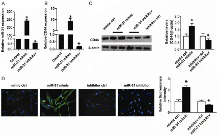
lated and down-regulated miR-31 expres-sion in BEAS-2B cells by miR-31 mimic/ inhibitor transfection. Compared with the control group, the expression of miR-31 was obviously increased after transfection with the mimic and was decreased after transfection with the inhibitor (Figure 2A). qPCR and western blot results demon-strated that the CD44 mRNA and protein
expression levels were significantly elevat -ed by miR-31 mimic transfection, but
sig-nificantly reduced by miR-31 inhibitor
transfection (Figure 2B and 2C). Im-
munofluorescence analysis also showed
[image:5.612.93.323.339.459.2]overexpres-miR-31 elevates asthma-related cytokine ex -pression by targeting CD44
To explore the effect of miR-31 on asthma, we detected the expression of asthma-related cy-
tokines by ELISA after miRNA
transfect-ion. ELISA showed that overexpression of
miR-31 increased the levels of IL-6, IL-8 and ICAM. However, inhibition of miR-31 significantly reduced the IL-6, IL-8 and ICAM levels. The expression of IL-6, IL-8 and ICAM did not change
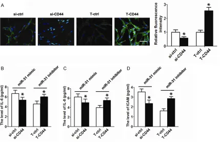
in the presence of the miR-21 or miR-141 mimic/inhibitor (Table 1). Moreover, we re-sup-pressed and restored the CD44 expression in BEAS-2B cells by transfecting CD44 siRNA and
a recombinant plasmid.
Immunofluorescen-ce analysis revealed decreased expression of the CD44 protein after CD44 siRNA transfec-tion, while increased CD44 protein expression was found after CD44 recombinant plasmid transfection (Figure 4A). Functionally, inhibition
of CD44 expression abrogated the effect of
miR-31, resulting in significantly attenuated lev
-els of asthma-related cytokines. In contrast, restoration of CD44 expression re-induced
asthma-related cytokine expression (Figure 4B-D).
Discussion
Current studies imply that the mechanism of asthma primarilyoccurs through chronic airway
inflammation and airway remodelling, in which chronic airway inflammation is related to eosin
-ophil, mast cell and lymphocyte infiltration and the release of inflammatory factors [14, 15].
A previous study suggests that CD44 partici-pates in eosinophil and lymphocyte activation, increases airway reactivity, promotes a variety of cell proliferation activities, and is closely as- sociated with the onset of asthma [16]. In ad- dition, excessive expression of CD44 in lung tissue is closely related to the process of as- thma, and participates in the onset of asthma at an early stage [17]. CD44 expression is also associated with eosinophil and lymphoid cell
chemotaxis and infiltration, resulting in the in-flammation of airway tissue in asthma [18, 19].
[image:6.612.94.523.71.348.2]In the diagnosis and treatment of disease, miR-NA-mediated target gene transcriptional activa-tion also has great advantages in target
speci-ficity and the flexibility of target genes. Recent
studies have focused on the regulatory mecha-nism of miRNAs in asthma. Some studies indi-cated that miRNAs mediate target gene tran-scriptional activation by binding to the promoter region of the target gene (by partially comple-mentary pairing) in the nucleus [20, 21], sug-gesting that the recognition of gene promoters targeted by miRNAs may be a natural and gen-eral mechanism for gene transcriptional regula-tion [22]. Although miRNA binding sequence-sare highly conserved in different species, even with good homologues, a bioinformatically-pre-dicted target may not be the real target. In this study, we used bioinformatics software to
ana-lyse 1 kb upstreamof the CD44 transcriptional start site and identified three miRNAs (miR-31,
miR-21, miR-141) that were highly
complemen-tary with the CD44 promoter region. Further-more, we then identified that miR-31 expres -sion was enhanced in the plasma and epithe- lial cells of asthma patients relative to unaf-fected people.
In vitro, we demonstrated that overexpress- ion of a miR-31 mimic in BEAS-2B cells led to high expression of CD44 and asthma-related
cytokines (IL-6, IL-8 and ICAM). However, knock -down of miR-31 expression in BEAS-2B cells decreased the CD44 and asthma-related cy-
tokine levels. In addition, we confirmed that
CD44 was a direct target gene of miR-31, and we found that miR-31 positively regulated CD44 expression by directly targeting the pro-moter region of the CD44 gene in BEAS-2B
cells. Further studies will verify whether miR-31
plays a regulatory role in an animal asthma model in vivo. The role of miRNA-mediated gene transcriptional activation in the pathogen-esis of asthma is still in its infancy, and many problems still need to be solved.
In summary, our results are the first to indicate
that miR-31 affects the expression of
asthma-related cytokines by up-regulation of CD44. It is
expected that miR-31 will become a new target for diagnosis and treatment of asthma in the future.
Disclosure of conflict of interest
None.
Address correspondence to: Guoping Zhou, Depart- ment of Pediatrics, The First Affiliated Hospital, Nanjing Medical University, 300 Guang Zhou Road, Nanjing 210029, Jiangsu Province, China. Tel: +86-025-86863443; Fax: +86-025-86862670; E-mail: gpzhou2003@126.com; Jun Qian, Wuxi City People’s Hospital Respiratory Department Affiliated with Nanjing Medical University, 299 Qing Yang Road, Wuxi 214023, Jiangsu Province, China. Tel: +86-51085350616; Fax: +86-51085350613; E-mail: qian@wuxiph.com
References
[1] Hejazi ME, Modarresi-Ghazani F and Entezari-Maleki T. A review of Vitamin D effects on com -mon respiratory diseases: Asthma, chronic obstructive pulmonary disease, and tuberculo-sis. J Res Pharm Pract 2016; 5: 7-15.
[2] Rebane A and Akdis CA. MicroRNAs in allergy and asthma. Curr Allergy Asthma Rep 2014; 14: 424.
[3] Tarlo SM. Update on work-exacerbated asth -ma. Int J Occup Med Environ Health 2016; 29: 369-374.
[4] Lagos-Quintana M, Rauhut R, Lendeckel W and Tuschl T. Identification of novel genes cod -ing for small expressed RNAs. Science 2001; 294: 853-858.
[5] Lau NC, Lim LP, Weinstein EG and Bartel DP. An abundant class of tiny RNAs with probable regulatory roles in Caenorhabditis elegans. Science 2001; 294: 858-862.
[6] Carrington JC and Ambros V. Role of microR-NAs in plant and animal development. Science 2003; 301: 336-338.
[7] Lai EC. Micro RNAs are complementary to 3’ UTR sequence motifs that mediate negative post-transcriptional regulation. Nat Genet 2002; 30: 363-364.
[8] Moretti F, Thermann R and Hentze MW. Me-chanism of translational regulation by miR-2 from sites in the 5’ untranslated region or the open reading frame. RNA 2010; 16: 2493-2502.
[9] Qin W, Shi Y, Zhao B, Yao C, Jin L, Ma J and Jin Y. miR-24 regulates apoptosis by targeting the open reading frame (ORF) region of FAF1 in cancer cells. PLoS One 2010; 5: e9429. [10] Place RF, Li LC, Pookot D, Noonan EJ and
Dahiya R. MicroRNA-373 induces expression of genes with complementary promoter se-quences. Proc Natl Acad Sci U S A 2008; 105: 1608-1613.
plications in cancer. Nucleic Acids Res 2012; 40: 1695-1707.
[12] Rothenberg ME. CD44-a sticky target for asth -ma. J Clin Invest 2003; 111: 1460-1462. [13] Li L, Yang L, Tang H and Jin R. [Role of CD44
on airway inflammatory response in rats with asthma]. Zhongguo Dang Dai Er Ke Za Zhi 2009; 11: 142-145.
[14] Webley WC and Aldridge KL. Infectious asth- ma triggers: time to revise the hygiene hypo- thesis? Trends Microbiol 2015; 23: 389-391. [15] Plantier L, Pradel A and Delclaux C. [Mech-
anisms of non-specific airway hyperrespon -siveness: Methacholine-induced alterations in airway architecture]. Rev Mal Respir 2016; [Epub ahead of print].
[16] Tai HY, Tam MF, Chou H, Perng DW and Shen HD. Pen ch 13 major fungal allergen decreas-es CD44 exprdecreas-ession in human bronchial epi-thelial cells. Int Arch Allergy Immunol 2010; 153: 367-371.
[17] Yang C, Liang H, Zhao H and Jiang X. CD44 variant isoforms are specifically expressed on peripheral blood lymphocytes from asthmatic patients. Exp Ther Med 2012; 4: 79-83.
[18] Hart SP, Rossi AG, Haslett C and Dransfield I. Characterization of the effects of cross-link -ing of macrophage CD44 associated with in-creased phagocytosis of apoptotic PMN. PLoS One 2012; 7: e33142.
[19] Ascon M, Ascon DB, Liu M, Cheadle C, Sarkar C, Racusen L, Hassoun HT and Rabb H. Renal ischemia-reperfusion leads to long term infil -tration of activated and effector-memory T lym-phocytes. Kidney Int 2009; 75: 526-535. [20] Huang V and Li LC. miRNA goes nuclear. RNA
Biol 2012; 9: 269-273.
[21] Huang YP, Qiu LZ and Zhou GP. MicroRNA-939 down-regulates CD2-associated protein by tar-geting promoter in HEK-293T cells. Ren Fail 2016; 38: 508-513.