• No results found

Original Article Aggressive mandibularaneurysmal bone cyst misdiagnosed as simple bone cyst: a case report and literature review

N/A
N/A
Protected

Academic year: 2020

Share "Original Article Aggressive mandibularaneurysmal bone cyst misdiagnosed as simple bone cyst: a case report and literature review"

Copied!
6
0
0

Loading.... (view fulltext now)

Full text

Figure

Figure 1. Images depicting the primary lesion. A: A panoramic radiograph show-ing swelling of the left condyle and coracoid
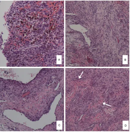
Figure 2. Pathological examination of the recurrent lesion. A and B: Specimen in January 2013 showing spindle cells and multinucleated giant cells and “vascular” form in the lesion
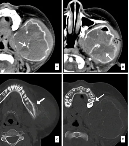
Figure 3. Images depicting of the lesion (June 2013). A: Wave-like pattern of cortical bone and calcified foci (arrow)
Figure 4. June 2013 A: Surgical specimen showing a cystic appearance. B: Cytology smear showing numerous blood cells with scattered giant cells (Original magnification, 200×).

References

Related documents